


BIOL 203 Final Exam Updated Study Guide
Previous (Old) Material
Scientists and contributions
oLeeuwenhoek, Pasteur, Koch, Semmelweis, Flemming
oKoch’s Postulates
1. Leeuwenhoek: described live microorganisms using microscope
2. Pasteur: showed that microorganisms are present in the air, S-shaped flask (kept
microbes out but let air (Golden age of micro began with pasteurs work), showed that
microbes are responsible for fermentation, pasteurization (application of high heat for a
short time) (germ theory of disease: Pasteur believed that another disease was cause by
a protozoan)
3. Koch: proved that a bacterium causes anthrax-kochs postulates
4. Kochs postulates-proves that a specific microbe causes a specific disease
-Microorganism must be found in all organism suffering from disease but not in a
healthy one
-must be isolated and grown in pure culture
-Should cause disease when introduced to healthy organism
-organism must be reisolated and then proven to be the causative agent
5. Semmelweis: advocated hand washing to prevent transmission of pleural fever
6.Fleming: discovered the 1st antibiotic (Penicillin), observed that the penicillium fungus
made an antibiotic that killed S. aureus
END CHAP 1
Microscopy/viewing
oBrightfield light microscopy, darkfield microscopy, transmission vs scanning electron
microscopy
1. Brightfield: dark object visible against light background, light reflected off
specimen does NOT enter objective lens
2. Darkfield: light object visible against dark background, light reflected off
specimen DOES enter lens
3. Transmission electron (TEM): ultrathin of specimens, light PASSES through
specimen then through electromagnetic lens to a screen or film (specimen may
be stained with heavy metal salts
4. Scanning electron (SEM): electron gun produces beam of electrons that SCANS
surface of whole specimen, secondary electrons emitted from the specimen
produce an image
oStaining techniques: Gram stain, endospore stain, capsule stain, acid-fast stain
1. Gram: differential, positive- are killed by penicillin and detergents, negative- are
more resistant to antibiotics
§Crystal violet: primary stain, purple for both positive and negative
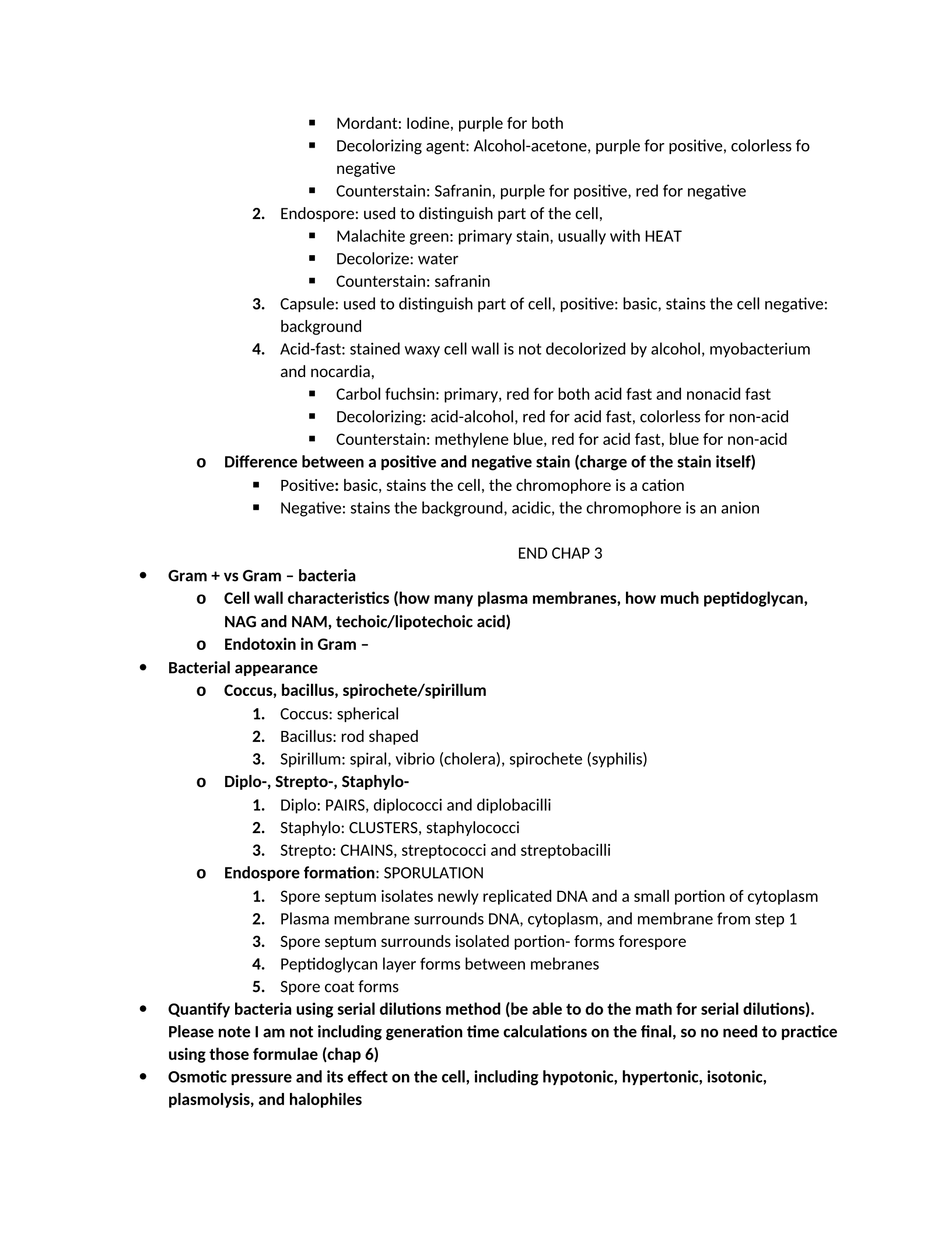
§Mordant: Iodine, purple for both
§Decolorizing agent: Alcohol-acetone, purple for positive, colorless fo
negative
§Counterstain: Safranin, purple for positive, red for negative
2. Endospore: used to distinguish part of the cell,
§Malachite green: primary stain, usually with HEAT
§Decolorize: water
§Counterstain: safranin
3. Capsule: used to distinguish part of cell, positive: basic, stains the cell negative:
background
4. Acid-fast: stained waxy cell wall is not decolorized by alcohol, myobacterium
and nocardia,
§Carbol fuchsin: primary, red for both acid fast and nonacid fast
§Decolorizing: acid-alcohol, red for acid fast, colorless for non-acid
§Counterstain: methylene blue, red for acid fast, blue for non-acid
oDifference between a positive and negative stain (charge of the stain itself)
§Positive: basic, stains the cell, the chromophore is a cation
§Negative: stains the background, acidic, the chromophore is an anion
END CHAP 3
Gram + vs Gram – bacteria
oCell wall characteristics (how many plasma membranes, how much peptidoglycan,
NAG and NAM, techoic/lipotechoic acid)
oEndotoxin in Gram –
Bacterial appearance
oCoccus, bacillus, spirochete/spirillum
1. Coccus: spherical
2. Bacillus: rod shaped
3. Spirillum: spiral, vibrio (cholera), spirochete (syphilis)
oDiplo-, Strepto-, Staphylo-
1. Diplo: PAIRS, diplococci and diplobacilli
2. Staphylo: CLUSTERS, staphylococci
3. Strepto: CHAINS, streptococci and streptobacilli
oEndospore formation: SPORULATION
1. Spore septum isolates newly replicated DNA and a small portion of cytoplasm
2. Plasma membrane surrounds DNA, cytoplasm, and membrane from step 1
3. Spore septum surrounds isolated portion- forms forespore
4. Peptidoglycan layer forms between mebranes
5. Spore coat forms
Quantify bacteria using serial dilutions method (be able to do the math for serial dilutions).
Please note I am not including generation time calculations on the final, so no need to practice
using those formulae (chap 6)
Osmotic pressure and its effect on the cell, including hypotonic, hypertonic, isotonic,
plasmolysis, and halophiles
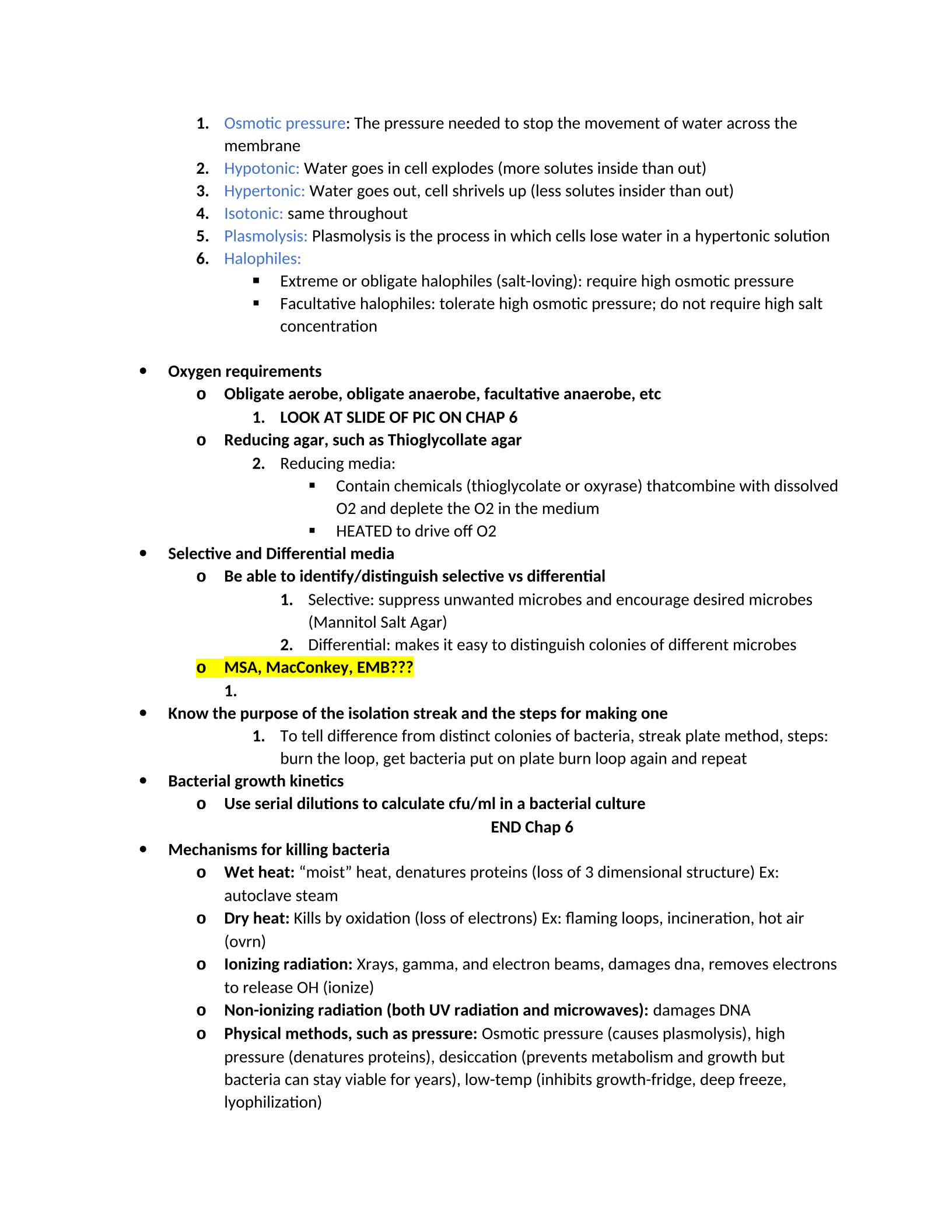
1. Osmotic pressure: The pressure needed to stop the movement of water across the
membrane
2. Hypotonic: Water goes in cell explodes (more solutes inside than out)
3. Hypertonic: Water goes out, cell shrivels up (less solutes insider than out)
4. Isotonic: same throughout
5. Plasmolysis: Plasmolysis is the process in which cells lose water in a hypertonic solution
6. Halophiles:
§Extreme or obligate halophiles (salt-loving): require high osmotic pressure
§Facultative halophiles: tolerate high osmotic pressure; do not require high salt
concentration
Oxygen requirements
oObligate aerobe, obligate anaerobe, facultative anaerobe, etc
1. LOOK AT SLIDE OF PIC ON CHAP 6
oReducing agar, such as Thioglycollate agar
2. Reducing media:
§Contain chemicals (thioglycolate or oxyrase) thatcombine with dissolved
O2 and deplete the O2 in the medium
§HEATED to drive off O2
Selective and Differential media
oBe able to identify/distinguish selective vs differential
1. Selective: suppress unwanted microbes and encourage desired microbes
(Mannitol Salt Agar)
2. Differential: makes it easy to distinguish colonies of different microbes
oMSA, MacConkey, EMB???
1.
Know the purpose of the isolation streak and the steps for making one
1. To tell difference from distinct colonies of bacteria, streak plate method, steps:
burn the loop, get bacteria put on plate burn loop again and repeat
Bacterial growth kinetics
oUse serial dilutions to calculate cfu/ml in a bacterial culture
END Chap 6
Mechanisms for killing bacteria
oWet heat: “moist” heat, denatures proteins (loss of 3 dimensional structure) Ex:
autoclave steam
oDry heat: Kills by oxidation (loss of electrons) Ex: flaming loops, incineration, hot air
(ovrn)
oIonizing radiation: Xrays, gamma, and electron beams, damages dna, removes electrons
to release OH (ionize)
oNon-ionizing radiation (both UV radiation and microwaves): damages DNA
oPhysical methods, such as pressure: Osmotic pressure (causes plasmolysis), high
pressure (denatures proteins), desiccation (prevents metabolism and growth but
bacteria can stay viable for years), low-temp (inhibits growth-fridge, deep freeze,
lyophilization)
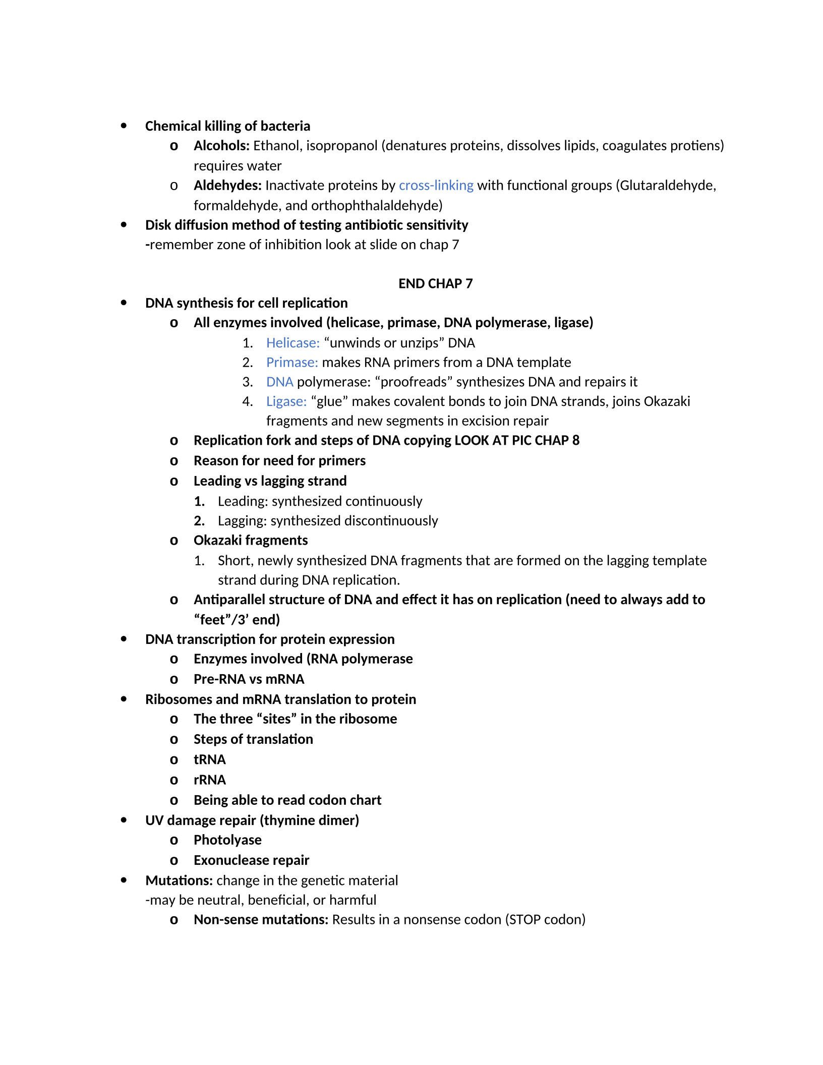
Chemical killing of bacteria
oAlcohols: Ethanol, isopropanol (denatures proteins, dissolves lipids, coagulates protiens)
requires water
oAldehydes: Inactivate proteins by cross-linking with functional groups (Glutaraldehyde,
formaldehyde, and orthophthalaldehyde)
Disk diffusion method of testing antibiotic sensitivity
-remember zone of inhibition look at slide on chap 7
END CHAP 7
DNA synthesis for cell replication
oAll enzymes involved (helicase, primase, DNA polymerase, ligase)
1. Helicase: “unwinds or unzips” DNA
2. Primase: makes RNA primers from a DNA template
3. DNA polymerase: “proofreads” synthesizes DNA and repairs it
4. Ligase: “glue” makes covalent bonds to join DNA strands, joins Okazaki
fragments and new segments in excision repair
oReplication fork and steps of DNA copying LOOK AT PIC CHAP 8
oReason for need for primers
oLeading vs lagging strand
1. Leading: synthesized continuously
2. Lagging: synthesized discontinuously
oOkazaki fragments
1. Short, newly synthesized DNA fragments that are formed on the lagging template
strand during DNA replication.
oAntiparallel structure of DNA and effect it has on replication (need to always add to
“feet”/3’ end)
DNA transcription for protein expression
oEnzymes involved (RNA polymerase
oPre-RNA vs mRNA
Ribosomes and mRNA translation to protein
oThe three “sites” in the ribosome
oSteps of translation
otRNA
orRNA
oBeing able to read codon chart
UV damage repair (thymine dimer)
oPhotolyase
oExonuclease repair
Mutations: change in the genetic material
-may be neutral, beneficial, or harmful
oNon-sense mutations: Results in a nonsense codon (STOP codon)
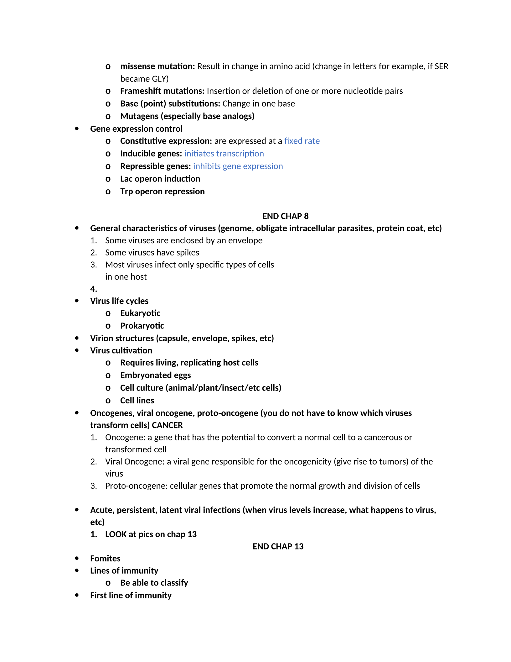
omissense mutation: Result in change in amino acid (change in letters for example, if SER
became GLY)
oFrameshift mutations: Insertion or deletion of one or more nucleotide pairs
oBase (point) substitutions: Change in one base
oMutagens (especially base analogs)
Gene expression control
oConstitutive expression: are expressed at a fixed rate
oInducible genes: initiates transcription
oRepressible genes: inhibits gene expression
oLac operon induction
oTrp operon repression
END CHAP 8
General characteristics of viruses (genome, obligate intracellular parasites, protein coat, etc)
1. Some viruses are enclosed by an envelope
2. Some viruses have spikes
3. Most viruses infect only specific types of cells
in one host
4.
Virus life cycles
oEukaryotic
oProkaryotic
Virion structures (capsule, envelope, spikes, etc)
Virus cultivation
oRequires living, replicating host cells
oEmbryonated eggs
oCell culture (animal/plant/insect/etc cells)
oCell lines
Oncogenes, viral oncogene, proto-oncogene (you do not have to know which viruses
transform cells) CANCER
1. Oncogene: a gene that has the potential to convert a normal cell to a cancerous or
transformed cell
2. Viral Oncogene: a viral gene responsible for the oncogenicity (give rise to tumors) of the
virus
3. Proto-oncogene: cellular genes that promote the normal growth and division of cells
Acute, persistent, latent viral infections (when virus levels increase, what happens to virus,
etc)
1. LOOK at pics on chap 13
END CHAP 13
Fomites
Lines of immunity
oBe able to classify
First line of immunity
oSkin (salty, sebum, keratinized)
oMucus
oCiliary escalator
oNormal microbiota
Inflammation- Four Cardinal Signs, vasodilation, cytokines and chemokines
Fever- transferrins, interferons, high temperature, shivering, what happens during crisis/shock
Immunological terms- susceptibility, innate immunity, adaptive immunity, Toll-like receptors
(TLR’s), pathogen associated molecular patterns (PAMP’s), chemotaxis, etc
Cytokines- TNF, IL-1, IFN-α, IFN-β, IFN-γ
Second (macrophages, Dendritic cells, granulocytes, etc) and third lines of immune defense (B
cells, T cells, etc)
Lymphatic system- lymph, lymph nodes, etc
Phagocytosis- what is it, what cells do it, how does this relate to adaptive immunity (more in
Ch 17)
Complement- series of proteins already in blood but are inactive, activated, steps of cascade
to formation of C9 pore. Note: only need to know only classical and lectin pathways of
complement activation (not alternate method of complement activation)
oClassical pathway- initiated by antibody after it binds to bacteria or virus infected cell
oLectin pathway- initiated by lectins (MBL) binding to foreign sugar residues on
bacterial cells
Need to know effects of complement activation (opsonization, inflammation, cytolysis), and
how they are mediated (C3b mediating opsonization, C3a and C5a initiating inflammation, C3b
initiating membrane attack complex+ the factors involved in that: C5b-C9)
Four cardinal signs of inflammation
1. Redness
2. Swellng
New Material
Chapter 17- Adaptive Immunity
Humoral vs Cellular immunity
B cells, Helper T cells, Th1, Th2, Cytotoxic T cells, Tc, macrophages, antigen presenting cells
(APC’s), exogenous antigens, endogenous antigens, apoptosis, major histocompatibility
complex (MHC)
Classes of antibodies- just IgM, IgG, IgA, IgE and what they are able to do
Cytokines- IL-1, IL-2, TNFα, Type I IFN, Type II IFN
Chemokines- tell cells where to go- recruit more immune cells to site of infection
Antigen, hapten, epitope, clonal deletion, plasma cells, memory cells
Antibody activity- opsonization, agglutination, neutralization, complement fixing, antibody-
dependent cell-mediated cytotoxicity (ADCC)
Activation of B cells (T cell independent, T cell dependent) and T cells (helper and cytotoxic)
Killing cells/parasites with granzymes and perforin
Regulatory T cells (T reg)
Cytokines and cytokine storm
Major histocompatibility complexes (MHCs)
Primary response vs secondary response
Antibody titer
Naturally acquired active immunity, naturally acquired passive immunity, artificially acquired
active immunity, artificially acquired passive immunity
Powered by TCPDF (www.tcpdf.org)

