


BMET 221: PATHOPHYSIOLOGY
ALTERATIONS IN WATER MOVEMENT
Edema
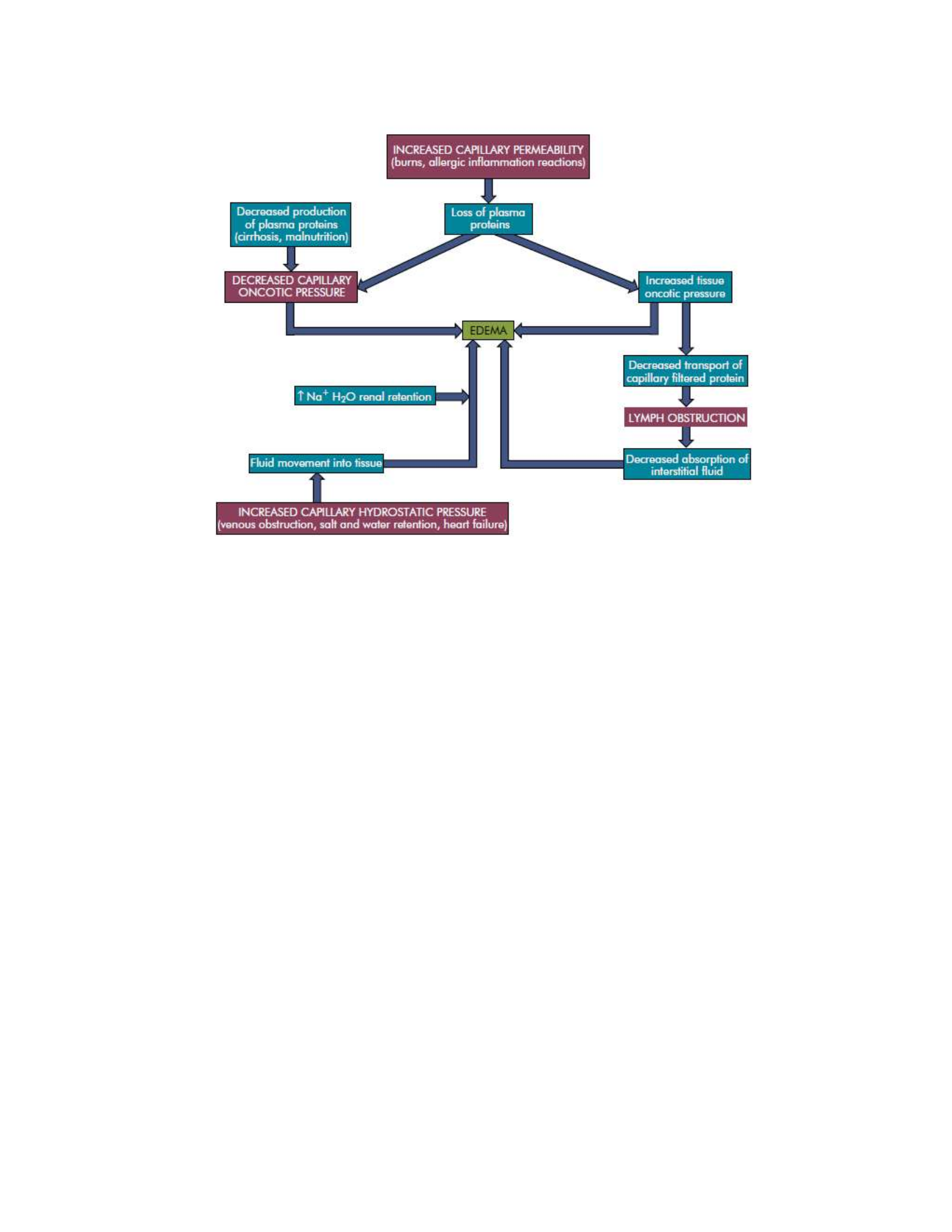
Edema is excessive accumulation of fluid within the interstitial spaces. The forces favoring fluid
movement from the capillaries or lymphatic channels into the tissues are increased capillary
hydrostatic pressure, decreased plasma oncotic pressure, increased capillary membrane
permeability, and lymphatic channel obstruction.
Pathophysiology
Hydrostatic pressure increases as a result of venous obstruction or salt and water retention. Venous
obstruction causes hydrostatic pressure to increase behind the obstruction, pushing fluid from the
capillaries into the interstitial spaces. Thrombophlebitis (inflammation of veins), hepatic
obstruction, tight clothing around the extremities, and prolonged standing are common causes of
venous obstruction. Congestive heart failure, renal failure, and cirrhosis of the liver are associated
with excessive salt and water retention, which cause plasma volume overload, increased capillary
hydrostatic pressure, and edema. Lost or diminished plasma albumin production (e.g., from liver
disease or protein malnutrition) contributes to decreased plasma oncotic pressure. Plasma proteins
are lost in glomerular diseases of the kidney, serous drainage from open wounds, hemorrhage,
burns, and cirrhosis of the liver. The decreased oncotic attraction of fluid within the capillary
causes filtered capillary fluid to remain in the interstitial space, resulting in edema. Capillaries
become more permeable with inflammation and immune responses, especially with trauma such
as burns or crushing injuries, neoplastic disease, and allergic reactions. Proteins escape from the
vascular space and produce edema through decreased capillary oncotic pressure and interstitial
fluid protein accumulation. The lymphatic system normally absorbs interstitial fluid and a small
amount of proteins. When lymphatic channels are blocked or surgically removed, proteins and
fluid accumulate in the interstitial space, causing lymphedema. For example, lymphedema of the
arm or leg occurs after surgical removal of axillary or femoral lymph nodes, respectively, for
treatment of carcinoma. Inflammation or tumors may cause lymphatic obstruction, leading to
edema of the involved tissues.
Evaluation and Treatment
Specific conditions causing edema require diagnosis. Edema may be treated symptomatically until
the underlying disorder is corrected. Supportive measures include elevating edematous limbs,
using compression stockings, avoiding prolonged standing, restricting salt intake, and taking
diuretics.
BMET 221: PATHOPHYSIOLOGY
ALTERATIONS OF HORMONAL REGULATION
Mechanisms of Hormonal Alterations
Significantly elevated or significantly depressed hormone levels may result from various causes.
Dysfunction of an endocrine gland may involve its failure to produce adequate amounts of
biologically free or active hormone, or a gland may synthesize or release too much hormone.
Feedback systems that recognize the need for a particular hormone may fail to function properly
or may respond to inappropriate signals. Once hormones are released into the circulation, they may
be degraded at an altered rate or be inactivated before reaching the target cell by antibodies that
function as circulating hormone inhibitors. Other causes of decreased hormone delivery to the
target cell include an inadequate blood supply to the gland or target tissues or an insufficient
amount of the appropriate carrier proteins in the serum. Ectopic sources of hormones (hormones
produced by non-endocrine tissues) may cause abnormally elevated hormone levels without the
benefit of the normal feedback system for hormone control; in this case, the ectopic hormone
production is said to be autonomous. Target cells may not respond appropriately to hormonal
stimulation for a number of reasons. The following are the two general types of target cell
insensitivity to hormones:
1. Cell surface receptor–associated disorders. These disorders have been identified primarily in
water-soluble hormones, such as insulin. They may involve a decrease in the number of receptors,
leading to decreased or defective hormone-receptor binding; impaired receptor function, resulting
in insensitivity to the hormone; presence of antibodies against specific receptors that either reduce
available binding sites or mimic hormone action, suppressing or exaggerating, respectively, the

BMET 221: PATHOPHYSIOLOGY
target cell response; or unusual expression of receptor function, for example, tumor cells with
abnormal receptor activity.
2. Intracellular disorders. These disorders involve acquired defects in post-receptor signaling
cascades or inadequate synthesis of a second messenger, such as cyclic adenosine monophosphate
(cAMP), needed to transduce the hormonal signal into intracellular events. The target cell for
water-soluble hormones may have a faulty response to hormone-receptor binding and thus fail to
generate the required second messenger, or the cell may respond abnormally to the second
messenger if levels of intracellular enzymes or proteins are altered. As a result, the target cell fails
to express the usual hormonal effect.
Pathogenic mechanisms affecting target cell response for lipid-soluble hormones are recognized
less often than those affecting water soluble hormones. When they do occur, the mechanisms are
similar to those for water-soluble hormones, including changes in the number and binding affinity
of intracellular receptors or altered generation of new messenger ribonucleic acid (RNA) and
substrates for new protein synthesis.
Diabetes Mellitus
Type 1 Diabetes Mellitus
Type 1 diabetes mellitus is the most common pediatric chronic disease. Two distinct types of type
1 diabetes have been identified: autoimmune and non-immune. Autoimmune type 1 diabetes is
called type 1A. Nonimmune type 1 diabetes is far less common than immune. It occurs secondary
to other diseases, such as pancreatitis, or to a more fulminant disorder termed idiopathic (type 1B)
diabetes. Type 1B diabetes occurs mostly in people of Asian or African descent and affected
individuals have varying degrees of insulin deficiency.
Pathophysiology
Type 1A diabetes mellitus is a slowly progressive autoimmune T cell–mediated disease that
destroys beta cells of the pancreas. Destruction of beta cells is related to genetic susceptibility and
environmental factors. The strongest genetic association is with histocompatibility leukocyte
antigen (HLA) class II alleles HLADQ and HLA-DR. The HLA-DR marker is associated with other
autoimmune disorders, such as celiac, Graves, Hashimoto, and Addison diseases. Environmental
factors that have been implicated include exposure to certain drugs, foods, and viruses. These gene-
environment interactions result in the formation of autoantigens that are expressed on the surface
of pancreatic beta cells and circulate in the bloodstream and lymphatics. Cellular immunity (T
cytotoxic cells and macrophages) and humoral immunity (autoantibodies) are stimulated, resulting
in beta-cell destruction and apoptosis. Over time, insulin synthesis declines and hyperglycemia
develops. For insulin synthesis to decline enough such that hyperglycemia occurs, 80% to 90% of
the insulin- secreting beta cells of the islet of Langerhans must be destroyed. Insulin normally
suppresses secretion of glucagon and, thus, hypo-insulinemia leads to a marked increase in
glucagon secretion. Glucagon, a hormone produced by the alpha cells of the islets, acts in the liver
to increase blood glucose level by stimulating glycogenolysis and gluconeogenesis. In addition to
the decline in insulin secretion, there is decreased secretion of amylin, another beta-cell hormone.

BMET 221: PATHOPHYSIOLOGY
One of the critical actions of amylin is to suppress glucagon release from the alpha cells. Thus
both alpha-cell and beta-cell functions are abnormal and both a lack of insulin and a relative excess
of glucagon contribute to hyperglycemia in type 1 diabetes.
Type 2 Diabetes Mellitus
Type 2 diabetes mellitus (non–insulin-dependent diabetes mellitus) is much more common than
type 1 and has been rising in incidence since 1940. A genetic-environmental interaction appears
to be responsible for type 2 diabetes. The most well-recognized risk factors are age, obesity,
hypertension, physical inactivity, and family history. The metabolic syndrome is a constellation of
disorders (central obesity, dyslipidemia, prehypertension, and an elevated fasting blood glucose
level) that together confer a high risk of developing type 2 diabetes and associated cardiovascular
complications.
Pathophysiology
Many genes have been identified that are associated with type 2 diabetes, including those that code
for beta-cell mass, beta-cell function (ability to sense blood glucose levels, insulin synthesis, and
insulin secretion), proinsulin and insulin molecular structures, insulin receptors, hepatic synthesis
of glucose, glucagon synthesis, and cellular responsiveness to insulin stimulation. These genetic
abnormalities combined with environmental influences, such as obesity, result in the basic
pathophysiologic mechanisms of type 2 diabetes: insulin resistance and decreased insulin secretion
by beta cells.
Insulin resistance: is defined as a suboptimal response of insulin sensitive tissues (especially
liver, muscle, and adipose tissue) to insulin and is associated with obesity. Cellular insulin
resistance and obesity are present in 60% to 80% of those with type 2 diabetes. Obesity contributes
to the development of insulin resistance and diabetes through several important mechanisms:
1. Adipokines (leptin and adiponectin) are hormones produced in adipose tissue. Obesity results
in increased serum levels of leptin and decreased levels of adiponectin. These changes are
associated with inflammation and decreased insulin sensitivity.
2. Elevated levels of serum free fatty acids (FFAs) and intracellular deposits of triglycerides and
cholesterol are also found in obese individuals. These changes interfere with intracellular insulin
signaling and thus decrease tissue responses to insulin and contribute to beta-cell apoptosis, a
process known as lipotoxicity.
3. Inflammatory cytokines (tumor necrosis factor-alpha [TNF-α], interleukin-1-beta [IL-1β], and
interleukin-6 [IL-6]) are released from intra-abdominal adipocytes or adipocyte-associated
mononuclear cells; they induce insulin resistance and are cytotoxic to beta cells.
4. Obesity is correlated with hyperinsulinemia and decreased insulin receptor density.
Compensatory hyperinsulinemia prevents the clinical appearance of diabetes for many years.
Eventually, however, beta-cell dysfunction develops and leads to a relative deficiency of insulin

BMET 221: PATHOPHYSIOLOGY
activity. The islet dysfunction is caused by a combination of a decrease in beta-cell mass and a
reduction in normal beta-cell function. A progressive decrease in the weight and number of beta
cells occurs and many of the remaining cells develop “exhaustion” from increased demand for
insulin biosynthesis. Glucagon concentration is increased in type 2 diabetes because pancreatic
alpha cells become less responsive to glucose inhibition, resulting in an increase in glucagon
secretion. These abnormally high levels of glucagon increase blood glucose level by stimulating
glycogenolysis and gluconeogenesis. As was discussed under type 1 diabetes, type 2 diabetes also
is associated with a deficiency in amylin, further increasing glucagon levels. Drugs aimed at
improving amylin function are being evaluated. Hormones released from the gastrointestinal (GI)
tract play a role in insulin resistance, beta-cell function, and diabetes. Ghrelin is a peptide
produced in the stomach and pancreatic islets that stimulates growth hormone release. Decreased
levels of circulating ghrelin have been associated with insulin resistance and increased fasting
insulin levels. The incretins are a class of peptides that are released from the GI tract in response
to food intake and function to increase the sensitivity of beta cells to circulating glucose levels,
thus improving insulin responsiveness to meals. Incretins also suppress glucagon secretion, delay
gastric emptying, suppress appetite, reduce beta-cell apoptosis, and induce pancreatic acinar cells
to differentiate into new beta cells. Between meals they are inactivated by the enzyme dipeptidyl
peptidase IV (DPP-IV). Incretin analogs and DPP-IV inhibitors are being used for the treatment
of type 2 diabetes.
ALTERATIONS OF CARDIOVASCULAR FUNCTION
Diseases of the Arteries
Hypertension
Hypertension is consistent elevation of systemic arterial blood pressure. The chance of developing
primary hypertension increases with age. Although hypertension is usually considered an adult
health problem, it is important to remember that hypertension does occur in children and is being
diagnosed with increasing frequency. All stages of hypertension are associated with increased risk
for target organ disease events, such as myocardial infarction, kidney disease, and stroke; thus both
stage I and stage II hypertension need effective long-term therapy.
Primary Hypertension
Primary hypertension is the result of an extremely complicated interaction of genetics and the
environment mediated by a host of neuro-humoral effects. Multiple pathophysiologic mechanisms
mediate these effects, including the sympathetic nervous system (SNS), the renin-angiotensin-
aldosterone system (RAAS), and natriuretic peptides. Inflammation, endothelial dysfunction,
obesity-related hormones, and insulin resistance also contribute to both increased peripheral
resistance and increased blood volume. Increased vascular volume is related to a decrease in renal
excretion of salt, often referred to as a shift in the pressure-natriuresis relationship. This means
that for a given blood pressure, individuals with hypertension tend to secrete less salt in their urine.
The sympathetic nervous system has been implicated in both the development and the maintenance
of elevated blood pressure and plays a role in hypertensive end-organ damage. Increased SNS
activity causes increased heart rate and systemic vasoconstriction, thus raising the blood pressure.

BMET 221: PATHOPHYSIOLOGY
Additional mechanisms of SNS-induced hypertension include structural changes in blood vessels
(vascular remodeling), renal sodium retention (shift in natriuresis curve), insulin resistance,
increased renin and angiotensin levels, and pro-coagulant effects. In hypertensive individuals,
over-activity of the RAAS contributes to salt and water retention and increased vascular resistance.
High levels of angiotensin II contribute to endothelial dysfunction, insulin resistance, and platelet
aggregation. Further, angiotensin II mediates arteriolar remodeling, which is structural change in
the vessel wall that results in permanent increases in peripheral resistance. Angiotensin II is
associated with end-organ effects of hypertension, including atherosclerosis, renal disease, and
cardiac hypertrophy. Finally, aldosterone not only contributes to sodium retention by the kidney
but also has other deleterious effects on the cardiovascular system. Medications, such as
angiotensin-converting enzyme (ACE) inhibitors and angiotensin receptor blockers (ARBs),
oppose the activity of the RAAS and are effective in reducing blood pressure and protecting against
target organ damage.
Aneurysm
An aneurysm is a localized dilation or outpouching of a vessel wall or cardiac chamber. True
aneurysms involve all three layers of the arterial wall and are best described as a weakening of
the vessel wall. Most are fusiform and circumferential, whereas saccular aneurysms are basically
spherical in shape. False aneurysm is an extravascular hematoma that communicates with the
intravascular space. A common cause of this type of lesion is a leak between a vascular graft and
a natural artery. The aorta is particularly susceptible to aneurysm formation because of constant
stress on the vessel wall and the absence of penetrating vasa vasorum in the media layer. Three
fourths of all aneurysms occur in the abdominal aorta. Atherosclerosis is the most common cause
of arterial aneurysms because plaque formation erodes the vessel wall and contributes to
inflammation and release of proteinases that can further weaken the vessel. Hypertension also
contributes to aneurysm formation by increasing wall stress. Collagen-vascular disorders (e.g.,
Marfan syndrome), syphilis, and other infections that affect arterial walls also can cause
aneurysms. Cardiac aneurysms most commonly form after myocardial infarction when
intraventricular tension stretches the noncontracting infarcted muscle. The stretching produces
infarct expansion, a weak and thin layer of necrotic muscle, and fibrous tissue that bulges with
each systole. Clinical manifestations depend upon where the aneurysm is located. Aortic
aneurysms often are asymptomatic until they rupture, and then cause severe pain and hypotension.
Thoracic aortic aneurysms can cause dysphagia (difficulty swallowing) and dyspnea
(breathlessness). An aneurysm that impairs flow to an extremity causes symptoms of ischemia.
Cerebral aneurysms, which often occur in the circle of Willis, are associated with signs and
symptoms of increased intracranial pressure. Signs and symptoms of stroke occur when cerebral
aneurysms leak. Aneurysms in the heart present with dysrhythmias, heart failure, and embolism of
clots to the brain or other vital organs. Aortic aneurysms can be complicated by the acute aortic
syndromes, which include aortic dissection, hemorrhage into the vessel wall, or vessel rupture.
Dissection of the layers of the arterial wall occurs when there is a tear in the intima and blood
enters the wall of the artery. Dissections can involve any part of the aorta (ascending, arch, or

BMET 221: PATHOPHYSIOLOGY
descending) and can disrupt flow through arterial branches, thus creating a surgical emergency.
The diagnosis of an aneurysm is usually confirmed by ultrasonography, computed tomography,
magnetic resonance imaging, or angiography. Medical treatment is indicated for slow-growing
aortic aneurysms, particularly in early stages, and includes cessation of smoking, reduction of
blood pressure and blood volume, and implementation of beta-adrenergic blockade. For those
aneurysms that are dilating rapidly or have become large, surgical treatment is indicated and
usually includes replacement with a prosthetic graft. New endovascular surgical techniques make
aneurysm repair possible for more individuals.
Embolism
Embolism is the obstruction of a vessel by an embolus—a bolus of matter circulating in the
bloodstream. The embolus may consist of a dislodged thrombus; an air bubble; an aggregate of
amniotic fluid; an aggregate of fat, bacteria, or cancer cells; or a foreign substance. An embolus
travels in the bloodstream until it reaches a vessel through which it cannot fit. No matter how tiny
it is, an embolus will eventually lodge in a systemic or pulmonary vessel determined by its source.
Pulmonary emboli originate on the venous side (mostly from the deep veins of the legs) of the
systemic circulation or in the right heart; arterial emboli most commonly originate in the left heart
and are associated with thrombi after myocardial infarction, valvular disease, left heart failure,
endocarditis, and dysrhythmias. Embolism causes ischemia or infarction in tissues distal to the
obstruction, causing organ dysfunction and pain. Infarction and subsequent necrosis of a central
organ are life-threatening. For example, occlusion of a coronary artery will cause a myocardial
infarction, whereas occlusion of a cerebral artery causes a stroke.
ALTERATIONS OF HEMATOLOGIC FUNCTION
Anemia is a reduction in the total number of circulating erythrocytes or a decrease in the quality
or quantity of hemoglobin. The causes of anemia are (1) altered production of erythrocytes, (2)
blood loss, (3) increased erythrocyte destruction, or (4) a combination of all three.
Anemias are classified by their causes (e.g., anemia of chronic disease) or by the changes that
affect the size, shape, or substance of the erythrocyte. The most common classification of anemias
is based on the changes that affect the cell’s size and hemoglobin content. Terms used to identify
anemias reflect these characteristics. Terms that end with cytic refer to cell size, and those that end
with chromic refer to hemoglobin content. Additional terms describing erythrocytes found in some
anemias are anisocytosis (assuming various sizes) and poikilocytosis (assuming various shapes).
Macrocytic-Normochromic Anemias
The macrocytic (megaloblastic) anemias are characterized by unusually large stem cells
(megaloblasts) in the marrow that mature into erythrocytes that are unusually large in size
(macrocytic), thickness, and volume. The hemoglobin content is normal, thus allowing them to be
classified as normochromic. These anemias are the result of ineffective erythrocyte
deoxyribonucleic acid (DNA) synthesis, commonly caused by deficiencies of vitamin B12
(cobalamin) or folate (folic acid). These defective erythrocytes die prematurely, which decreases
their numbers in the circulation, causing anemia. Defective DNA synthesis in megaloblastic

BMET 221: PATHOPHYSIOLOGY
anemias causes red cell growth and development to proceed at unequal rates. DNA synthesis and
cell division is blocked or delayed. However, ribonucleic acid (RNA) replication and protein
(hemoglobin) synthesis proceed normally. Asynchronous development leads to an overproduction
of hemoglobin during prolonged cellular division, creating a larger than normal erythrocyte with
a disproportionately small nucleus. With each cell division, the disproportion between RNA and
DNA becomes more apparent.
1. Pernicious Anemia
Pernicious anemia (PA), the most common type of macrocytic anemia, is caused by vitamin B12
deficiency, which often accompanies the end stage of type A chronic atrophic (autoimmune)
gastritis. Pernicious means highly injurious or destructive and reflects the fact that this condition
was once fatal. It most commonly affects individuals over the age of 30 who are of Northern
European descent, as well as blacks and Hispanics. Females are more prone to develop PA, with
black females having an earlier onset.
Pathophysiology
The underlying alteration in PA is the absence of intrinsic factor (IF), an enzyme required for
gastric absorption of dietary vitamin B12, a vitamin essential for nuclear maturation and DNA
synthesis in red blood cells. Deficiency of IF may be congenital or may be the result of adult-onset
gastric mucosal atrophy in which the parietal cells are destroyed. Subsequently, all secretions of
the stomach— hydrochloric acid, pepsin, and IF—are deficient. PA is associated with autoimmune
conditions that affect the endocrine system. Gastric atrophy may be caused by type A chronic
gastritis, an autoimmune disorder that causes destruction of parietal and zymogenic cells. These
destroyed cells are replaced with mucus-containing cells (intestinal metaplasia). In addition, PA
may be caused by heavy alcohol ingestion, hot tea, and cigarette smoking. Complete or partial
removal of the stomach (gastrectomy) causes IF deficiency and results in PA. Individuals with
chronic gastritis are at risk for the development of gastric cancer and must be followed regularly
to prevent this condition.
Evaluation and Treatment
Evaluation is based on blood tests, bone marrow aspiration, serologic studies, gastric biopsy,
clinical manifestations, and the Schilling test. The Schilling test uses cobalt radioisotopes and
labeled B12 to evaluate IF production. The test is performed by administering radioactive
cobalamin and then measuring its excretion in the urine. Low urinary excretion is significant for
PA. Serologic studies show the presence of antibodies against gastric cells and gastric biopsy
reveals achlorhydria, a total absence of hydrochloric acid (HCl). Untreated PA is fatal, usually
because of heart failure. With replacement therapy of vitamin B12, mortality has decreased
significantly. Death from PA is now rare and relapses are often the result of noncompliance with
therapy. Initial replacement of vitamin B12 is accomplished by weekly injections until the
deficiency is corrected. Monthly injections are then required for the remainder of an individual’s
life. Conventional wisdom and practice determined that oral preparations were ineffective because
there was no IF to facilitate absorption of B12. However, recent practice has shown that oral
administration of higher doses of B12 is beneficial. Apparently, an alternative mechanism for B12

BMET 221: PATHOPHYSIOLOGY
absorption exists that is independent of IF. PA is not curable; therefore, treatment must be
continued throughout the individual’s lifetime.
2. Folate Deficiency Anemias
Folate (folic acid) is an essential vitamin required for RNA and DNA synthesis within the
erythrocyte. Humans are totally dependent on dietary intake to meet the daily requirement of 50
to 200 mg/day. Increased amounts are required for lactating and pregnant females. Folate is
absorbed from the upper small intestine and does not require any other element (i.e., IF) to facilitate
absorption. After absorption, folate circulates through and is stored in the liver. Folate deficiency
occurs more often than B12 deficiency, particularly in alcoholics and individuals who are
malnourished because of fad diets or diets low in vegetables. Clinical manifestations are similar
to the malnourished appearance of individuals with PA, except for the absence of neurologic
symptoms. Specific manifestations include cheilosis (scales and fissures of the mouth), stomatitis
(inflammation of the mouth), and painful ulcerations of the buccal mucosa and tongue. Dysphagia,
flatulence, and watery diarrhea also may be present, as well as histologic changes in the GI tract
suggestive of sprue (chronic absorption disorder). Neurologic manifestations, if present, may be
caused by thiamine deficiency, which often accompanies folate deficiency.
Evaluation of folate deficiency is based on blood tests, measurement of serum folate levels, and
clinical manifestations. Treatment requires administration of oral folate preparations until adequate
blood levels are obtained and manifestations are reduced or eliminated. Long-term therapy is not
necessary except for maintenance of an adequate daily intake of folate. Folate is essential for
reducing blood levels of homocysteine, which has been recently recognized as a risk factor for the
development of coronary artery disease.
Microcytic-Hypochromic Anemias
The microcytic-hypochromic anemias are characterized by abnormally small erythrocytes that
contain abnormally reduced amounts of hemoglobin. Hypochromia occurs even in cells of normal
size. Microcytic-hypochromic anemia can result from (1) disorders of iron metabolism, (2)
disorders of porphyrin and heme synthesis, or (3) disorders of globin synthesis. Specific conditions
include iron deficiency anemia, sideroblastic anemia, and thalassemia.
1. Iron Deficiency Anemia
Iron deficiency anemia (IDA) is the most common type of anemia throughout the world, occurring
in both developing and developed countries. The overall incidence of IDA is difficult to establish
because of the lack of standardized methods and techniques to determine hypoferremia and IDA.
Certain populations are at high risk for developing hypoferremia and IDA and include individuals
living in poverty, women of childbearing age, and children.
Pathophysiology
In developed countries, pregnancy and a continuous loss of blood are the most common causes of
IDA. A blood loss of 2 to 4 ml/day (1 to 2 mg of iron) is enough to cause IDA. Males may

BMET 221: PATHOPHYSIOLOGY
experience bleeding as a result of ulcers, hiatal hernia, esophageal varices, cirrhosis, hemorrhoids,
ulcerative colitis, or cancer. Menorrhagia (excessive menstrual bleeding) causes primary IDA in
females. Other causes of blood loss for both genders include: (1) use of medications that cause GI
bleeding; (2) surgical procedures that decrease stomach acidity, intestinal transit time, and
absorption (e.g., gastric bypass); (3) insufficient dietary intake of iron; and (4) eating disorders
such as pica—the craving and eating of non-nutritional substances, such as dirt, chalk, and paper.
Iron in the form of hemoglobin is in constant use in the body. An important attribute of iron is that
it can be recycled; therefore, the body maintains a balance between iron that is in use as
hemoglobin and iron that is stored and available for future hemoglobin synthesis. Blood loss
disrupts this balance by creating a need for more iron, thus depleting the iron stores more rapidly
to replace the iron lost from bleeding.
IDA develops slowly through three overlapping stages. In stage I, the body’s iron stores for red
cell production and hemoglobin synthesis are depleted. Red cell production proceeds normally
with the hemoglobin content of red cells also remaining normal. In stage II, insufficient amounts
of iron are transported to the marrow, and iron-deficient red cell production begins. Stage III begins
when the hemoglobin- deficient red cells enter the circulation to replace normal, aged erythrocytes
that have been destroyed. The manifestations of IDA appear in stage III when there is an
insufficient iron supply and diminished hemoglobin synthesis.
Clinical Manifestations
The onset of symptoms is gradual, and individuals usually do not seek medical attention until
hemoglobin levels drop to 7 or 8 g/dl. Early symptoms are nonspecific and include fatigue,
weakness, shortness of breath, and pale earlobes, palms, and conjunctiva. As the condition
progresses and becomes more severe, structural and functional changes occur in epithelial tissue.
The fingernails become brittle and “spoon shaped” or concave (koilonychia). Tongue papillae
atrophy and cause soreness along with redness and burning. These changes can be reversed within
1 to 2 weeks of iron replacement. The corners of the mouth become dry and sore (angular
stomatitis), and an individual may experience difficulty with swallowing because of a “web” that
develops from mucus and inflammatory cells at the opening of the esophagus. These lesions have
the potential to become cancerous.
Iron is a component of many enzymes in the body, and lack of iron may alter other physiologic
processes and contribute to the clinical manifestations. Individuals with IDA exhibit gastritis,
neuromuscular changes, irritability, headache, numbness, tingling, and vasomotor disturbances.
Gait disturbances are rare. In the elderly, mental confusion, memory loss, and disorientation may
be wrongly perceived as normal events associated with aging.
Evaluation and Treatment
Evaluation is based on clinical manifestations and laboratory tests. Iron stores are measured
directly, by bone marrow biopsy, or indirectly, by tests that measure serum ferritin, transferrin
saturation, or total iron-binding capacity. A sensitive indicator of heme synthesis is the amount of
free erythrocyte protoporphyrin (FEP) within erythrocytes. A test that determines the

BMET 221: PATHOPHYSIOLOGY
concentration of soluble fragment transferrin receptor differentiates primary IDA from IDA that is
associated with chronic disease. The first step in treatment of IDA is to find and eliminate, or rule
out, sources of blood loss. If this is not done, replacement therapy is ineffective. Iron replacement
therapy is required and very effective. Initial doses are 150 to 200 mg/day and are continued until
the serum ferritin level reaches 50 mg/L, indicating that adequate replacement has occurred. A
rapid decrease in fatigue, lethargy, and other associated symptoms is generally seen within the first
month of therapy. Replacement therapy usually continues for 6 to 12 months after the bleeding has
stopped but may continue for as long as 24 months. Menstruating females may need daily oral iron
replacement therapy (325mg/day) until menopause.
2. Sideroblastic Anemia
Sideroblastic anemias (SAs) are a heterogeneous group of disorders characterized by anemia of
varying severity because of inefficient iron uptake, resulting in abnormal hemoglobin synthesis.
SA is characterized by the presence of ringed sideroblasts in the bone marrow. These are red cells
that contain iron granules that have not been synthesized into hemoglobin but instead are arranged
in a circle around the nucleus. Individuals with SA also have increased tissue levels of iron.
Pathophysiology
Sideroblastic anemias have various causes but all share the commonality of altered heme synthesis
in the erythroid cells in bone marrow. SAs are either acquired or hereditary. Acquired sideroblastic
anemias, which are the most common, occur as a primary disorder with no known cause
(idiopathic) or are associated with other myeloproliferative or myeloplastic disorders. Another
form is described as reversible SAs; these are secondary to various conditions such as alcoholism,
drug reactions, copper deficiency, and hypothermia. Hereditary sideroblastic anemias are rare and
occur almost exclusively in males, supporting a recessive X-linked transmission; however,
autosomal transmission affecting females has been reported. Other genetic, chromosomal, or
enzyme dysfunctions also have been associated with hereditary SA. In all instances, SA anemia is
present in infancy or childhood but may remain undetected until midlife, when other conditions,
such as diabetes or cardiac failure from iron overload, cause it to be manifest. Reversible
sideroblastic anemia, associated with alcoholism, results from nutritional deficiencies of folate.
Alcohol impairs heme synthesis by reducing the activity of specific enzymes along the biosynthetic
pathway and also by direct effects of alcohol or acetaldehyde, or both, on the heme biosynthetic
steps or mitochondrial metabolism. Some specific drugs also cause reversible SA and include anti-
tuberculous agents (isoniazid [INH], pyrazinamide, cycloserine, and chloramphenicol), which
interfere with B12 metabolism or directly injure the mitochondria. Copper deficiency also causes
reversible SA by interfering with conversion of ferric iron to ferrous iron. This is extremely rare
and is associated with gastrectomy and prolonged parenteral nutrition without copper supplements.
Hypothermia causes decreased heme synthesis and incorporation into hemoglobin.
Clinical Manifestations
Along with the cardiovascular and respiratory manifestations common to all anemias, individuals
with SA may show signs of iron overload (hemosiderosis), including mild to moderate
enlargement of the liver (hepatomegaly) and spleen (splenomegaly); however, liver function

BMET 221: PATHOPHYSIOLOGY
remains normal or only mildly affected. Occasionally the skin may become abnormally colored
(bronze-tinted). Neurologic and skin alterations associated with other anemias are absent.
Hemosiderosis of cardiac tissue may result in heart rhythm disturbances, which is a significant but
uncommon complication and generally occurs late in the course of the disease. Growth and
development impairment may occur in infants and young children who are severely affected.
Evaluation and Treatment
Initially, SA may be mistaken for deficiency of stem cells in the marrow (hypoplastic anemia) or
iron deficiency anemia. The diagnosis of SA is established by bone marrow biopsy, which
documents the presence of sideroblasts and confirms the diagnosis. Hereditary SA is initially
treated with pyridoxine therapy (50 to 200 mg/day), which is effective in approximately one third
of individuals treated; however, response is variable. An optimal response is reticulocytosis with
normal levels of hemoglobin and FEP returning within 1 to 2 months; cellular morphologic
abnormalities do not disappear. A less optimal response is an elevated hemoglobin level that
stabilizes at less than normal levels. A therapeutic response to pyridoxine may be maintained with
lifelong administration of a reduced dosage. Nonresponse to pyridoxine requires blood
transfusions for symptom relief and to promote growth and development. Evidence of iron
overload requires iron depletion therapy to prevent or minimize organ damage. Phlebotomy, or
removal of blood from the circulation, is used in individuals with mild to moderate anemia without
other complications (i.e., heart disease). After iron removal, maintenance phlebotomies are
continued. Severely anemic individuals who may require transfusions become extremely iron
overloaded, which mandates use of deferoxamine, an iron-chelating agent, to reduce iron levels.
Individuals with acquired SA are less likely to respond to pyridoxine, but SA rarely incapacitates
them. When SA is secondary to an identifiable cause, treatment or removal of the cause is essential.
In the absence of blood cell abnormalities and iron overload, progression takes place over years.
Transfusion and iron overload therapy is the same as for hereditary SA when indicated. Death from
SA is rare and often secondary to complications such as infection, bone marrow failure, liver
failure, or cardiac failure, or arrhythmias. Idiopathic SA has the potential to convert to
myelodysplastic syndrome, or abnormal marrow proliferation, which may then convert to acute
myeloblastic leukemia.
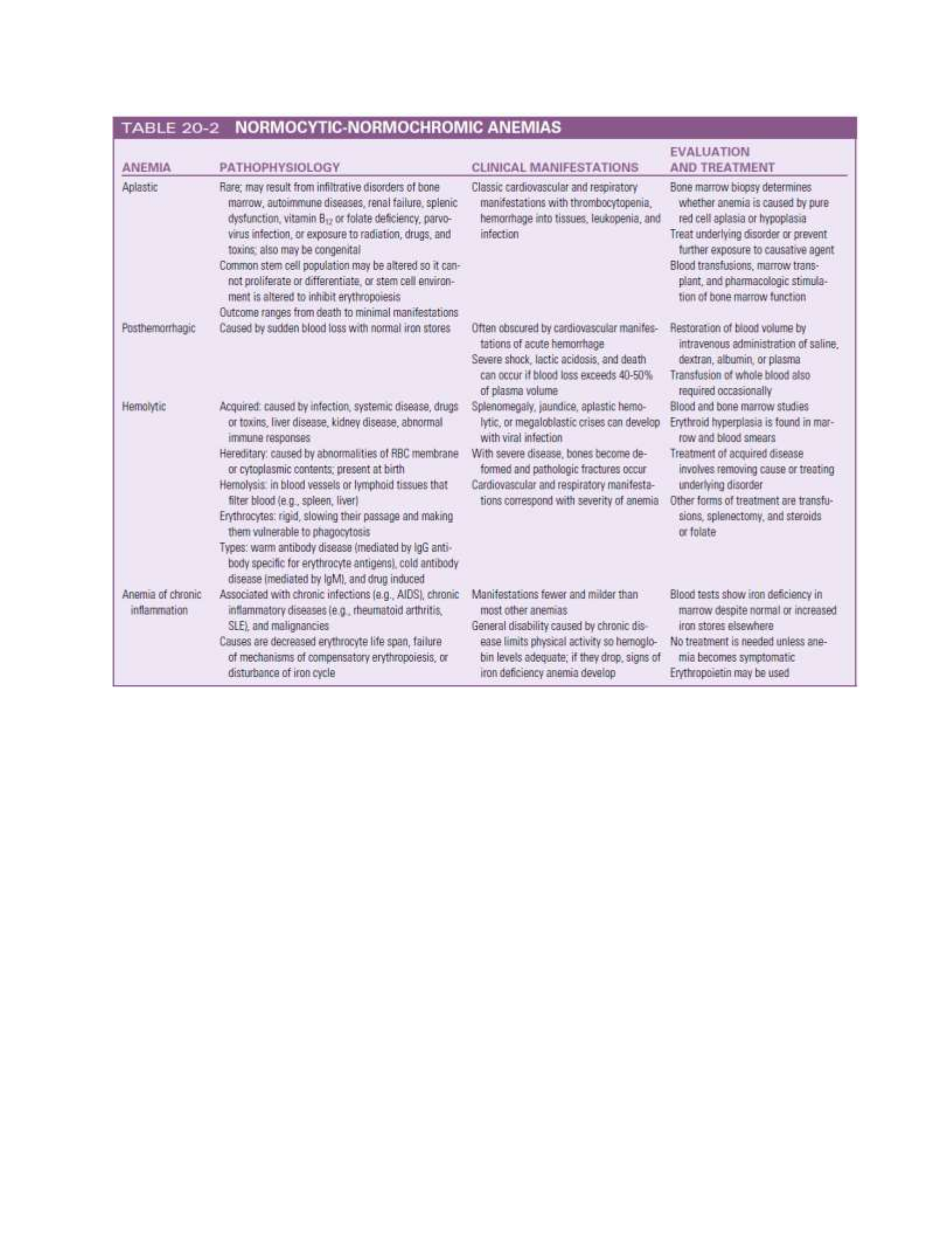
Normocytic-Normochromic Anemias
Normocytic-normochromic anemias (NNAs) are characterized by erythrocytes that are relatively
normal in size and hemoglobin content but insufficient in number. These anemias do not share any
common etiology, pathologic mechanism, or morphologic characteristics. They are less common
than the macrocytic-normochromic and the microcytic-hypochromic anemias. Five distinct
anemias—aplastic, posthemorrhagic, hemolytic, sickle cell, and anemia of chronic
inflammation—exemplify the diversity of the NNA characteristics.
BMET 221: PATHOPHYSIOLOGY
MYELOPROLIFERATIVE RED CELL DISORDERS
Hematologic dysfunction results from an overproduction of cells, as well as a deficiency. One or
more marrow elements may be produced in excess, responding to exogenous (e.g., exposure to
radiation, drugs) or endogenous (e.g., physiologic compensatory response, immune disorder)
signals. Excessive red cell production is classified as polycythemia. Polycythemia exists in two
forms: relative and absolute. Relative polycythemia results from hemo-concentration of the blood
associated with dehydration. It is of minor consequence and resolves with fluid administration or
treatment of underlying conditions. Absolute polycythemia consists of two forms: primary and
secondary. Secondary polycythemia, the most common of the two, is a physiologic response
resulting from erythropoietin secretion caused by hypoxia. This hypoxia is noted in individuals
living at higher altitudes (>10,000 ft), smokers with increased blood levels of CO, and individuals
with chronic obstructive pulmonary disease or coronary heart failure, or both. Abnormal types of
hemoglobin, which have a greater affinity for oxygen, also cause secondary polycythemia, as does
inappropriate secretion of erythropoietin by certain tumors (e.g., renal cell carcinoma, hepatoma,
and cerebellar hemangioblastomas). The absolute primary form of polycythemia is referred to as
polycythemia vera.

BMET 221: PATHOPHYSIOLOGY
Polycythemia Vera
Polycythemia vera (PV) is a chronic, clonal alteration characterized by overproduction of red
cells (frequently with increased white cells and platelets) accompanied by splenomegaly.
Hypercellularity of bone marrow, along with hyperplasia of myeloid, erythroid, and
megakaryocytes, is a distinguishing feature. PV is quite rare, occurring mostly in white males of
Eastern European Jewish origin from 55 to 80 years of age, with a median age of 55 to 60 years,
but it has been observed in females and individuals less than 40 years of age. It is rarely seen in
children or in multiple members of a single family; however, an autosomal dominant form exists
that causes increased secretion of erythropoietin.
Pathophysiology
PV is a neoplastic, nonmalignant condition characterized by an abnormal proliferation of bone
marrow stem cells with subsequent self-destructive expansion of red cells. This aberrant
proliferation occurs despite normal to below normal erythropoietin levels. The underlying cause
remains unknown, with the most likely etiology thought to be an acquired genetic stem cell
alteration of the erythropoietin receptor that causes the abnormal proliferation. Laboratory studies
have found red cell precursors that are capable of growth independent of erythropoietin. These red
blood cell precursors also demonstrate sensitivity to other growth factors, such as interleukin-3
(IL-3), granulocyte-macrophage colony-stimulating factor (GM-CSF), or insulin-like growth
factor.
Clinical Manifestations
Clinical manifestations of PV are due to increased blood volume, which increases blood viscosity,
creating a hypercoagulable state resulting in clogging and occlusion of blood vessels. Tissue injury
(ischemia) and death (infarction) is the outcome of blood vessel blockage, and this occurs about
40% of the time. These outcomes are directly correlated with hematocrit levels. Increases in
numbers of thrombocytes, as well as production of dysfunctional platelets, also contribute to this
hypercoagulable condition. Circulatory alterations caused by the thick, sticky blood give rise to
other manifestations, such as plethora (ruddy, red color of the face, hands, feet, ears, and mucous
membranes) and engorgement of retinal and cerebral veins. Other symptoms may include
headache, drowsiness, delirium, mania, psychotic depression, chorea, and visual disturbances.
Death from cerebral thrombosis is approximately five times greater in individuals with PV.
Cardiovascular function, despite the vascular alterations, remains relatively normal. Cardiac
workload and output remain constant; however, increased blood volume does increase blood
pressure. Coronary blood flow may be affected, precipitating angina, although cardiovascular
infarctions are uncommon. Other cardiovascular manifestations include Raynaud phenomenon and
thromboangiitis obliterans. A unique feature of PV, and helpful in diagnosis, is the development
of intense, painful itching that appears to be intensified by heat or exposure to water (aquagenic
pruritus) so that individuals avoid exposure to water, particularly warm water when bathing or
showering. The intensity of itching is related to the concentration of mast cells in the skin and is
generally not responsive to antihistamines or topical lotions.

BMET 221: PATHOPHYSIOLOGY
ALTERATIONS OF LEUKOCYTE FUNCTION
Leukocyte function is affected if too many or too few white cells are present in the blood or if the
cells that are present are structurally or functionally defective. Phagocytic cells (granulocytes,
monocytes, macrophages) may lose their ability to act as effective phagocytes, and the
lymphocytes may lose their ability to respond to antigens. Other leukocyte alterations include
infectious mononucleosis and cancers of the blood—leukemia and multiple myeloma.
Leukemias
Leukemia is a clonal malignant disorder of the blood and blood-forming organs. The common
pathologic feature of all forms of leukemia is an uncontrolled proliferation of malignant
leukocytes, causing an overcrowding of bone marrow and decreased production and function of
normal hematopoietic cells. The classification of leukemia is based on (1) the predominant cell of
origin (either myeloid or lymphoid) and (2) the degree of differentiation that took place before the
cell became malignant (acute, with a rapid growth of immature blood cells, or chronic, with a slow
growth of more differentiated cells). Thus there are four types of leukemia: acute lymphocytic
(ALL) or myelogenous (AML) and chronic lymphocytic (CLL) or myelogenous (CML). Further
classification of acute leukemias is based on characteristics that may provide significant
therapeutic prognostic information, such as structure, number of cells, genetics, identification of
surface markers, and histochemical staining. Acute leukemia is characterized by undifferentiated
or immature cells, usually a blast cell. The onset of disease is abrupt and rapid. Disease progression
results in a short survival time.
In chronic leukemia, the predominant cell is more mature but does not function normally. The
onset of the disease is gradual, and the prolonged clinical course results in a relatively longer
survival time. Leukemia occurs with varying frequencies at different ages and is more common in
adults than in children.
Pathophysiology
Although the exact cause of leukemia is unknown, several risk factors and related genetic
aberrations are associated with the onset of malignancy. There is a statistically significant tendency
for leukemia to reappear in families. There is also an increased incidence of leukemia in association
with other hereditary abnormalities such as Down syndrome, Fanconi aplastic anemia, Bloom
syndrome, trisomy, Patau syndrome, and some immune deficiencies(ataxia-telangiectasia,
Wiskott-Aldrich syndrome, congenital X-linked agammaglobulinemia).
Several genetic translocations (mitotic errors) are observed in leukemic cells. One of these
translocations, the Philadelphia chromosome, is observed in 95% of those with CML and 30% of
adults with ALL. The Philadelphia chromosome results from a reciprocal translocation between
the long arms of chromosomes 9 and 22. A unique protein (bcr-abl protein) is encoded from two
genes (BCR from chromosome 22 and ABL from chromosome 9) artificially linked at the junction
of translocation. The bcr-abl protein affects a variety of cell cycle control genes leading to an
increased rate of cellular division, inhibition of DNA repair, and other dysregulations of cell
growth.

BMET 221: PATHOPHYSIOLOGY
Risk factors for the onset of leukemia include environmental factors as well as other diseases.
Increased risk has been linked to cigarette smoke, exposure to benzene, and ionizing radiation.
Large doses of ionizing radiation particularly result in an increased incidence of myelogenous
leukemia. Infections with HIV or hepatitis C virus increase the risk for leukemia, and it is now
widely accepted that some types of leukemia are caused by infection with the human T cell
leukemia/lymphoma virus-1 (HTLV-1). Drugs that cause bone marrow depression (e.g.,
chloramphenicol, phenylbutazone, and certain alkylating agents, such as Cytoxan) also can
predispose an individual to leukemia. AML is the most frequently reported secondary cancer after
high doses of chemotherapy for Hodgkin lymphoma, non-Hodgkin lymphoma, multiple myeloma,
ovarian cancer, and breast cancer. Acute leukemia also may develop secondary to certain acquired
disorders, including CML, CLL, polycythemia vera, myelofibrosis, Hodgkin lymphoma, multiple
myeloma, ovarian cancer, and sideroblastic anemia.
The leukemia blasts literally “crowd out” the marrow and cause cellular proliferation of the other
cell lines to cease. Normal granulocyticmonocytic, lymphocytic, erythrocytic, and megakaryocytic
progenitor cells cease to function, resulting in pancytopenia (a reduction in all cellular components
of the blood).
Acute Leukemias:
About 85% of ALL arise from the B cell line, and about 15% arise from T cell lineage. A small
percentage of ALL cases have neither B nor T cell origination and are called null cell. Acute
leukemias are seen in both genders and in all ages, with the incidence increasing dramatically in
individuals older than 50 years.
Clinical Manifestations
The clinical manifestations of all varieties of acute leukemia are generally similar. Signs and
symptoms related to bone marrow depression include fatigue caused by anemia, bleeding resulting
from thrombocytopenia, and fever caused by infection. Bleeding may occur in the skin, gums,
mucous membranes, and GI tracts. Visible signs include petechiae and ecchymosis, as well as
discoloration of the skin, gingival bleeding, hematuria, and midcycle or heavy menstrual bleeding.
Infection sites include the mouth, throat, respiratory tract, lower colon, urinary tract, and skin and
may be caused by gram-negative bacilli (Escherichia coli), Pseudomonas, and Klebsiella. Fever
is an early sign often accompanied by chills. Anorexia is accompanied by weight loss, diminished
sensitivity to sour and sweet tastes, wasting of muscle, and difficulty swallowing. Liver, spleen,
and lymph node enlargement occurs more commonly in ALL than in CML. Liver and spleen
enlargement commonly occur together. The leukemic individual often experiences abdominal pain
and tenderness and also breast tenderness. Neurologic manifestations are common and may be
caused by either leukemic infiltration or cerebral bleeding. Headache, vomiting, papilledema,
facial palsy, blurred vision, auditory disturbances, and meningeal irritation can occur if leukemic
cells infiltrate the cerebral or spinal meninges. Because most chemotherapeutic agents do not
penetrate the blood-brain barrier, leukemia cells can grow easily in these locations.
Chronic Leukemias:

BMET 221: PATHOPHYSIOLOGY
The two main types of chronic leukemia are (1) myelogenous (CML) and (2) lymphocytic (CLL).
Several forms of CML can occur, depending on the lineage of the malignant cells (e.g., chronic
neutrophilic leukemia [CNL], chronic eosinophilic leukemia [CEL]). Unlike cells in acute
leukemia, chronic leukemic cells are well differentiated and can be readily identified. Individuals
with chronic leukemia have a longer life expectancy, usually extending several years from the time
of diagnosis. The chronic leukemias account for the majority of cases in adults. The incidences of
CLL and CML increase significantly in individuals over 40 years of age, with prevalence in the
sixth through eighth decades. CML is a group of diseases called myeloproliferative disorders,
which also include polycythemia vera, primary thrombocytosis, and idiopathic myelofibrosis
(invasion of bone marrow by fibrous tissue).
Pathophysiology and Clinical Manifestations
Chronic leukemia advances slowly and insidiously. Individuals are generally unaware of the
condition until symptoms appear. When symptoms do appear, they present as splenomegaly,
extreme fatigue, weight loss, night sweats, and low-grade fever. Individuals with CML may
progress through three phases of the disease: a chronic phase lasting 2 to 5 years during which
symptoms may not be apparent, an accelerated phase of 6 to 18 months during which the primary
symptoms develop, and a terminal blast phase with a survival of only 3 to 6 months. The
accelerated phase is characterized by excessive proliferation and accumulation of malignant cells.
Splenomegaly is prominent and becomes painful, but lymphadenopathy generally is not present.
Liver enlargement also occurs, but liver function is rarely altered. Hyperuricemia is commonand
produces gouty arthritis. Infections, fever, and weight loss also are seen often.
The terminal blast phase is characterized by rapid and progressive leukocytosis with an increase
in basophils. In the later stages of the terminal phase, which then resembles AML, blast cells or
promyelocytes predominate, and the individual experiences a “blast crisis.”
The Philadelphia chromosome is a useful diagnostic marker for CML and is observed in 95% of
individuals with CML. The median age for persons with Philadelphia chromosome-positive CML
is 40 to 45 years. The Philadelphia chromosome, although present in red cells, white cells, and
platelets, appears to affect only white cell function and production. Although it is difficult to
identify alterations within the cell’s structure, absent or low levels of the enzyme neutrophil
alkaline phosphatase, along with decreased phagocytic capabilities, indicate that cells fail to
differentiate normally. The only known cause of CML is exposure to ionizing radiation.
CLL involves predominantly malignant transformation of B cells; rarely (<5%) are T cells
involved. The malignant transformation is thought to be caused by failure of the normal
mechanisms of programmed cell destruction (apoptosis), allowing these cells to have an extended
life, thus the chronic nature of the disease. These cells fail to develop into antibody-producing cells
and fail to respond to stimulation by helper T cells. Suppression of normal antibody production is
the most significant effect in CLL. Individuals are thus at risk for recurrent bacterial and other
infections that are commonly sensitive to antibodies. Anemia, thrombocytopenia, and neutropenia
are typically present with overt CLL. Invasion of most organs by leukemic cells is uncommon, but
infiltration of lymph nodes, liver, spleen, and salivary glands is observed. Central nervous system

BMET 221: PATHOPHYSIOLOGY
involvement and elevated blood levels of calcium are rare, whereas elevated levels of lactic
dehydrogenase (LDH) and uric acid are common.
ALTERATIONS OF LYMPHOID FUNCTION
Lymphadenopathy
Lymphadenopathy is characterized by enlarged lymph nodes. Lymph node enlargement occurs
because of an increase in size and number of its germinal centers caused by proliferation of
lymphocytes and monocytes (immature phagocytes) or invasion by malignant cells. Normally,
lymph nodes are not palpable or are barely palpable.
Enlarged lymph nodes are characterized by being palpable and often also may be tender or painful
to touch, although not in all situations. Localized lymphadenopathy (reactive lymph nodes) usually
indicates drainage of an area associated with an inflammatory or infectious lesion. Generalized
lymphadenopathy, associated with infection, occurs less often and is generally seen in the presence
of malignant or nonmalignant disease. Lymphadenopathy is of more significance in adult disease
than in children. The location and size of the enlarged nodes are important factors in diagnosing
the cause of the lymphadenopathy, as are the individual’s age, gender, and geographic location.
Generalized lymphadenopathy occurs with non-Hodgkin lymphomas, chronic lymphocytic
leukemia, histiocytosis, and disorders that produce lymphocytosis. In general, lymphadenopathy
results from four types of conditions: (1) neoplastic disease, (2) immunologic or inflammatory
conditions, (3) endocrine disorders, or (4) lipid storage diseases. Diseases of unknown cause,
including autoimmune diseases and reactions to drugs, also may lead to generalized
lymphadenopathy.
Malignant Lymphomas
Lymphomas consist of a diverse group of neoplasms that develop from the proliferation of
malignant lymphocytes in the lymphatic system. The most recent classification of lymphomas was
published by the World Health Organization (WHO) and is derived from the Revised European-
American Lymphoma (REAL) Classification. This classification is based on the cell type from
which the lymphoma probably originated. The groups include Hodgkin lymphoma and two that
were previously classified as non-Hodgkin lymphoma (B cell neoplasms, T cell and NK cell
neoplasms). With the new classification, multiple myeloma, which was previously classified
independently, is included as a B cell lymphoma.
Hodgkin Lymphoma
Pathophysiology
Hodgkin lymphoma (HL) is characterized by its progression from one group of lymph nodes to
another, the development of systemic symptoms, and the presence of Reed-Sternberg (RS) cells.
It is widely accepted that the RS cell represents the malignant transformation of lymph cells. The
RS cells are often large and binucleate, with occasional mononuclear variants. The RS cellsare
necessary for the diagnosis of HL; however, they are not specific to HL. In rare instances, cells

BMET 221: PATHOPHYSIOLOGY
resembling RS cells can be found in benign illnesses, as well as in other forms of cancer, including
non-Hodgkin lymphomas and solid tissue cancers and in infectious mononucleosis.
Classical HL appears to be derived from a B cell in the germinal center that has not undergone
successful immunoglobulin gene rearrangement and would normally be induced to undergo
apoptosis. Survival of this cell may be linked to infection with Epstein-Barr virus (EBV).
Laboratory and epidemiologic studies have linked HL with EBV infections and EBV DNA, RNA,
and proteins are frequently observed in HL cells. The RS cells secrete and release cytokines (e.g.,
IL-10, transforming growth factor-beta [TGF-β]) that result in the accumulation of inflammatory
cells that produces the local and systemic effects. Classical HL is subclassified into four types
based on the morphology of RS cells, and the characteristics of the inflammatory cell infiltrate in
the tumor.
Clinical Manifestations
Many clinical features of HL can be explained by the complex action of cytokines and other growth
factors that are secreted and released by the malignant cells. These substances induce infiltration
and proliferation of inflammatory cells, resulting in an enlarged, painless lymph node in the neck
(often the first sign of HL). The discovery of an asymptomatic mediastinal mass on routine chest
x-ray is not uncommon. The cervical, axillary, inguinal, and retroperitoneal lymph nodes are
commonly affected in HL.
Local symptoms caused by pressure and obstruction of the lymph nodes are the result of the
lymphadenopathy. About a third of individuals will have some degree of systemic symptoms.
Intermittent fever, without other symptoms of infection, drenching night sweats, itchy skin
(pruritus), and fatigue are relatively common. These constitutional symptoms accompanied by
weight loss are associated with a poor prognosis. The Cotswold staging classification system used
for HL is able to establish a correlation between the anatomic extent of the disease and the
prognosis. This classification system is based on the individual’s medical history, examination
(presence of symptoms and palpable lymph nodes), and other radiologic and hematologic results.
Prognostic indicators include clinical stage, histologic type, tumor cell concentration and tumor
burden, constitutional symptoms, and age.
Although HL rarely arises in the lung, mediastinal and hilar node adenopathy can cause secondary
involvement of the trachea, bronchi, pleura, or lungs. Retroperitoneal nodes can involve vertebral
bodies and nerves and also can cause displacement of ureters. Spinal cord involvement is more
common in the dorsal and lumbar regions than in the cervical region. Skin lesions, although
uncommon, include psoriasis and eczematoid lesions, causing itching and scratching. As a result
of direct invasion from mediastinal lymph nodes, pericardial involvement can cause pericardial
friction rub, pericardial effusion, and engorgement of neck veins. The GI tract and urinary tract
are rarely involved. Anemia is often found in individuals with HL accompanied by a low serum
iron level and reduced iron-binding capacity. Other laboratory findings include elevated
sedimentation rate, leukocytosis, and eosinophilia. Leukopenia occurs in advanced stages of HL.
Splenic involvement in HL depends on histologic type. In mixed cellularity and lymphocytic

BMET 221: PATHOPHYSIOLOGY
deletion types of HL, the spleen is involved in 60% of cases. With lymphocyte and nodular
sclerosis types, 34% of cases involve the spleen.
Non-Hodgkin Lymphomas
The previously used generic classification of non-Hodgkin lymphoma has been reclassified in
the WHO/REAL scheme into (1) B cell neoplasms, a group that consists of a variety of
lymphomas including myelomas that originate from B cells at various stages of differentiation,
and (2) T cell and NK cell neoplasms, a group that includes lymphomas that originate from either
T or NK cells. These cancers are differentiated from HL by lack of RS cells and other cellular
changes not characteristic of HL. Because malignant changes can occur at various stages of B cell,
T cell, or NK cell development, these cancers present with a variety of clinical states. In the
following section, the types of tumors previously classified as non-Hodgkin lymphoma are
considered together, and myeloma is described separately.
Pathophysiology
As with all cancers, lymphomas most likely originate from mutations in cellular genes (many of
which are environmentally induced) in a single cell that lead to loss of control of proliferation and
other aspects of cell growth. The most common type of chromosomal alteration in non-Hodgkin
lymphoma (NHL) is translocation, which disrupts the genes encoded at the breakpoints. Risk
factors include a family history, exposure to a variety of mutagenic chemicals, irradiation,
infection with certain cancer-related viruses (e.g., Epstein-Barr virus, human herpesvirus, HIV,
HTLV-1, hepatitis C), and immune suppression related to organ transplantation. Gastric infection
with Helicobacter pylori increases the risk for gastric lymphomas. NHL is a disease of middle age,
usually found in persons over 50 years old.
Clinical Manifestations
Clinical manifestations of NHL usually begin as localized or generalized lymphadenopathy,
similar to HL. The cervical, axillary, inguinal, and femoral chains are the most commonly affected
sites. Generally, the swelling is painless and the nodes have enlarged and transformed over a period
of months or years. Other sites of involvement are the nasopharynx, GI tract, bone, thyroid, testes,
and soft tissue. Some individuals have retroperitoneal and abdominal masses with symptoms of
abdominal fullness, back pain, ascites (fluid in the peritoneal cavity), and leg swelling.
ALTERATIONS OF PLATELETS AND COAGULATION
Disorders of Platelet Function
Quantitative or qualitative abnormalities of platelets can interrupt normal blood coagulation and
prevent hemostasis. The quantitative abnormalities are thrombocytopenia, a decrease in the
number of circulating platelets, and thrombocythemia, an increase in the number of platelets.
Qualitative disorders affect the structure or function of individual platelets and can coexist with
the quantitative disorders. Qualitative disorders usually prevent platelet adherence and
aggregation, thereby preventing formation of a platelet plug.
Thrombocytopenia

BMET 221: PATHOPHYSIOLOGY
Thrombocytopenia is defined as a platelet count below 150,000/mm3 of blood, although most
individuals do not consider the decrease significant unless it falls below 100,000/mm3, and the
risk for hemorrhage associated with minor trauma does not appreciably increase until the count
falls below 50,000/mm3. Spontaneous bleeding without trauma can occur with counts ranging
from 10,000/mm3 to 15,000/mm3. When this happens, skin manifestations (i.e., petechiae,
ecchymoses, and larger purpuric spots) are observed or frank bleeding from mucous membranes
occurs. Severe bleeding results if the count falls below 10,000/mm3 and can be fatal if it occurs in
the gastrointestinal tract, respiratory tract, or central nervous system. Before thrombocytopenia is
diagnosed, the presence of a pseudo-thrombocytopenia must be ruled out. This phenomenon is
seen in approximately 1 in 1000 to 10,000 samples and results from an error in platelet counting
when a blood sample is analyzed by an automated cell counter. Platelets in the blood can become
nonspecifically agglutinated by immunoglobulins in the presence of ethylenediaminetetraacetic
acid (EDTA) and are not counted, thus giving an apparent, but false, thrombocytopenia.
Thrombocytopenia also may be falsely diagnosed because of a dilutional effect observed after
massive transfusion of platelet-poor packed cells to treat a hemorrhage. This is observed when
more than 10 units of blood have been transfused within a 24-hour period. The precipitating
hemorrhage also depletes platelets, contributing to the pseudothrombocytopenic state. Splenic
sequestering of platelets in hypersplenism also stimulates thrombocytopenia. Hypothermia
(<25°C) also predisposes to a thrombocytopenic state, which is reversed when temperatures return
to normal, suggesting sequestering and release.
Thrombocythemia
Thrombocythemia (also called thrombocytosis) is defined as a platelet count greater than
400,000/mm3 of blood.28 Thrombocythemia may be primary or secondary (reactive) and is
usually asymptomatic until the count exceeds 1 million/mm3. Then intravascular clot formation
(thrombosis), hemorrhage, or other abnormalities can occur.
Pathophysiology
Essential (primary) thrombocythemia (ET) is a myeloproliferative disorder in which platelet
production increases, resulting in platelet counts in excess of 600,000/mm3. It can occur in
individuals at most any age. Manifestations include increased numbers of bone marrow
megakaryocytes, splenomegaly, and periodic episodes of hemorrhage or thrombosis, or both. The
thrombocythemia is secondary to increased plasma thrombopoietin levels resulting from defects
in the thrombopoietin receptor. The defective receptor cannot adequately bind and remove
thrombopoietin from the blood; thus circulating levels remain high. Along with increased platelets,
there may be a concomitant increase in the number of red cells, indicating a myeloproliferative
disorder; however, the increase in red cells is not to the extent seen in polycythemia vera.
Secondary thrombocythemia may occur after splenectomy because platelets that normally would
be stored in the spleen remain in circulating blood. The increase in platelets may be gradual, with
thrombocythemia not occurring for up to 3 weeks after splenectomy. Reactive thrombocythemia
may occur during some inflammatory conditions, such as rheumatoid arthritis and cancers. In these
conditions, excessive production of some cytokines (e.g., IL-6, IL-11) may induce increased

BMET 221: PATHOPHYSIOLOGY
production of thrombopoietin in the liver, resulting in increased megakaryocyte proliferation.
Reactive thrombocythemia may also occur during a variety of physiologic conditions, such as after
exercise.
Clinical Manifestations
Clinical manifestations vary among individuals. Those with ET are at risk for large-vessel arterial
or venous thrombosis, and ischemia in the fingers, toes, or cerebrovascular regions is common.
Digital ischemia is characterized by warm, congested red extremities with a burning sensation,
particularly on the forefoot sole and toes. The lower extremities are affected more often, and only
one side may be involved. Standing, exercising, or applying heat precipitates the pain, which is
relieved by elevation and cooling of the affected extremity. In extreme situations, acrocyanosis
and gangrene may result. Thrombosis of arteries is more common than of veins, and myocardial
and renal arteries may be involved. The carotid, mesenteric, and subclavian arteries also may be
affected. Myocardial ischemia and infarction have occurred without clear evidence of coronary
artery disease. Involvement of the nervous system is manifested by headache and dizziness, with
paresthesias, transient ischemic attacks, strokes, visual disturbances, and seizures also being
reported. Major thrombotic events, not directly related to platelet count, occur in about 20% to
30% of individuals with ET. Other risk factors (prior thrombosis, age, and duration of ET) are
better predictors of future thrombosis. Although thrombosis is the more common symptom,
hemorrhage can also occur. Sites for bleeding include the GI tract, skin, urinary tract, gums, joints,
and brain. GI bleeding may be mistaken for a duodenal ulcer. Hemorrhage is not severe and
generally occurs in the presence of very high platelet counts; transfusions are required only
occasionally. Bleeding and clotting may occur simultaneously, and individuals are not necessarily
prone to one or the other.
Alterations of Platelet Function
Qualitative alterations in platelet function are characterized by an increased bleeding time in the
presence of a normal platelet count. Associated clinical manifestations include spontaneous
petechiae and purpura and bleeding from the GI tract, genitourinary tract, pulmonary mucosa, and
gums. Congenital alterations in platelet function (thrombocytopathies) are quite rare and may be
categorized into several types of disorders: (1) platelet–vessel wall adhesion (e.g., defect in GPIb
expression [Bernard-Soulier syndrome]), (2) platelet-platelet interactions (e.g., defect in
GPIIb/IIIa expression [Glanzmann thrombasthenia]), (3) platelet granules and secretion (e.g.,
receptor defects [ADP, collagen]), (4) arachidonic acid pathways (e.g., thromboxane synthase
deficiency), (5) cytoskeletal function (e.g., Wiskott- Aldrich syndrome, and (6) membrane
phospholipid regulation (coagulation protein-platelet interactions) (e.g., Scott syndrome).
Acquired disorders of platelet function are more common than the congenital disorders and may
be categorized into three principal causes: (1) drugs, (2) systemic conditions, and (3) hematologic
alterations. Multiple drugs are known to affect platelet function by interfering with platelet
function in three ways: (1) inhibition of platelet membrane receptors, (2) inhibition of
prostaglandin pathways, and (3) inhibition of phosphodiesterase activity. Aspirin is the most
commonly used drug that affects platelets. It irreversibly inhibits cyclooxygenase function for
several days after administration. Nonsteroidal anti-inflammatory drugs also affect

BMET 221: PATHOPHYSIOLOGY
cyclooxygenase, although in a reversible fashion. Diet can affect platelet function (Health Alert:
Dark Chocolate, Wine, and Platelet-Inhibitory Functions). Systemic disorders that affect platelet
function are chronic renal disease, liver disease, cardiopulmonary bypass surgery, and severe
deficiencies of iron or folate. Hematologic disorders associated with platelet dysfunction include
chronic myeloproliferative disorders, multiple myeloma, leukemias, and myelodysplastic
syndromes.
Disorders of Coagulation
Disorders of coagulation are usually caused by defects or deficiencies of one or more of the clotting
factors. Qualitative or quantitative abnormalities interfere with or prevent the enzymatic reactions
that transform clotting factors, circulating as plasma proteins, into a stable fibrin clot. Some
clotting factor defects are inherited and involve one single factor, such as the hemophilias and von
Willebrand disease, caused by deficiencies of clotting factors. Other coagulation defects are
acquired and tend to result from deficient synthesis of clotting factors by the liver. Causes include
liver disease and dietary deficiency of vitamin K. Other coagulation disorders are attributed to
pathologic conditions that trigger coagulation inappropriately, engaging the clotting factors and
causing detrimental clotting within blood vessels. For example, any cardiovascular abnormality
that alters normal blood flow by acceleration, deceleration, or obstruction can create conditions in
which coagulation proceeds within the vessels. An example of this is thromboembolic disease, in
which blood clots obstruct blood vessels. Coagulation is also stimulated by the presence of tissue
factor that is released by damaged or dead tissues. Vasculitis, or inflammation of the blood vessels,
along with vessel damage activates platelets, which in turn activates the coagulation cascade. In
extensive or prolonged vasculitis, blood clot formation can suppress mechanisms that normally
control clot formation and dissolution, leading to clogging of the vessels. In each of these acquired
conditions, normal hemostatic function proves detrimental to the body by consuming coagulation
factors excessively or by overwhelming normal control of clot formation and breakdown
(fibrinolysis).
Impaired Hemostasis
Impaired hemostasis, or the inability to promote coagulation and the development of a stable
fibrin clot, is commonly associated with liver dysfunction, which may be caused by either specific
liver disorders or lack of vitamin K.
Vitamin K deficiency. Vitamin K, a fat-soluble vitamin, is required for the synthesis of
prothrombin; the procoagulant factors II, VII, IX, and X; and the anticoagulant factors (proteins C
and S). Parenteral administration of vitamin K is the treatment of choice and usually results in
correction of the deficiency. Fresh frozen plasma also may be administered but is usually reserved
for individuals with life-threatening hemorrhages or those who require emergency surgery.
Liver disease. Individuals who have liver disease present with a broad range of hemostatic
derangements that may be characterized by defects in the clotting or fibrinolytic system and by
platelet dysfunction. The usual sequence of events is an initial reduction in clotting factors, which
parallels the degree of liver cell damage or destruction. Factor VII is the first to decline because of
its rapid turnover, followed by a decrease in the levels of factors II and X. Factor IX levels are less

BMET 221: PATHOPHYSIOLOGY
affected and do not decline until the liver destruction is well advanced. Protein C (an antithrombin)
levels decline early, similar to levels of factor VII, and protein S (also an antithrombin) levels
decline in the later stages of liver disease. Declines of factor V levels are of special importance
because factor V plasma levels appear to be a direct reflection of liver cell damage. Other
alterations of hemostasis in liver disease include an increase in fibrinolytic activity that either is
primary in origin or is a manifestation secondary to disseminated intravascular coagulation (DIC).
This increased fibrinolysis results from excessive fibrinolytic activators and decreased levels of
inhibitors, such as α2-antiplasmin.
Thrombocytopenia and thrombocytopathies are manifestations of liver disease. Thrombocytopenia
is caused by splenomegaly, which often accompanies liver disease. Splenic pooling of platelets is
the major cause of thrombocytopenia. Thrombocytopathies are associated with elevated levels of
fibrin split products, ethanol, or drugs. Treatment of hemostasis alterations in liver disease must
be comprehensive to cover all aspects of dysfunctions. Fresh frozen plasma (FFP) administration
is the treatment of choice; however, not all individuals tolerate the volume needed to adequately
replace all deficient factors. Alternative modalities include the addition of exchange transfusions
and platelet concentration to FFP administration.
Consumptive Thrombohemorrhagic Disorders
Consumptive thrombohemorrhagic disorders are a heterogeneous group of conditions that
demonstrate the entire spectrum of hemorrhagic and thrombotic pathologic findings. The
symptoms of these disorders also range from the subtle to the devastating and are generally
considered to be intermediary disease processes that complicate a vast number of primary disease
states. These disorders are also characterized by confusion and controversy related to their
diagnosis, treatment, and management. No one term is capable of covering all the possible varieties
of these disorders; however, DIC is most commonly used in the clinical setting to describe a
pathologic condition that is associated with hemorrhage and thrombosis.
Disseminated intravascular coagulation. Disseminated intravascular coagulation (DIC) is an
acquired clinical syndrome characterized by widespread activation of coagulation, resulting in
formation of fibrin clots in medium and small vessels throughout the body. Widespread clotting
may lead to blockage of blood flow to organs, resulting in multiple organ failure. The magnitude
of clotting may cause consumption of platelets and clotting factors, leading to severe bleeding.
The clinical course of DIC is largely determined by the intensity of the stimulus, the response of
the host, and the comorbidities, ranging from an acute, severe, life-threatening process that is
characterized by massive hemorrhage and thrombosis to a chronic, low-grade condition. The
chronic condition is characterized by subacute hemorrhage and diffuse microcirculatory
thrombosis. DIC may be localized to one specific organ or generalized, involving multiple organs.
Pathophysiology
Pathophysiology of Disseminated Intravascular Coagulation (DIC). Tissue factor initiates clot
formation and this effect is increased by a decrease in natural anticoagulants (tissue factor
inhibitor, antithrombin-III, and protein C). There also is a reduction in clot breakdown or
fibrinolysis by plasmin. The combined effect is to cause thrombosis. The thrombotic activity

BMET 221: PATHOPHYSIOLOGY
consumes (uses up) coagulation factors and platelets, which can increase bleeding. Slow
degradation of the fibrin clot produces fibrin degradation products (FDPs). FDPs have inhibitory
effects on thrombin and platelets. The inhibition of coagulation, combined with the depletion of
factors and platelets, then creates a bleeding tendency. Uncontrolled DIC will eventually lead to
multiple end-organ failure.
CELLULAR PROLIFERATION: CANCER
Cancer is a leading cause of suffering and death in the developed world and recently in the
developing world. The incidence of cancer increases markedly with advancing age and is strongly
affected by gender, life-style, ethnicity, infection, inflammation, and genetics. Because of intensive
research, we now understand that cancer is a collection of more than 100 different diseases, each
caused by a specific and often unique accumulation of genetic and epigenetic alterations.
Environment, heredity, and behavior interact to modify the risk of developing cancer and the
response to treatment. Improvements in treatment strategies and supportive care, coupled with
new, often individualized therapies based on advances in our fundamental understanding of the
basic pathophysiology of malignancy, have contributed to an increasing number of effective
options for these diverse, often lethal, disorders collectively called cancer.
The term cancer derives from the Greek word for crab, karkinoma, which the physician
Hippocrates used to describe the appendage-like projections from tumors. The word tumor
originally referred to any swelling that is caused by inflammation, but is now generally reserved
for describing a new growth, or neoplasm. Not all tumors or neoplasms, however, are cancer. The
term cancer refers to a malignant tumor and is not used to refer to benign growths such as lipomas
or hypertrophy of an organ. Yet it is important to recognize that benign neoplasms also can be life-
threatening if they enlarge in critical locations. For example, a benign meningioma at the base of
the skull may cause symptoms by compressing adjacent normal brain tissue.
Tumor Classification and Nomenclature
The careful evaluation of each cancer is important for many reasons. Different cancers will have
different causes, different rates and patterns of progression, and different responses to treatment.
The classification starts with knowing the tissue and organ of origin, the extent of distribution to
other sites, and the microscopic and immuno-histochemical appearance of the lesion. Increasingly,
it also includes a detailed description of the critical genetic changes in the cancer.
Benign and Malignant
Benign tumors, which are not referred to as cancers, are made of fairly well-differentiated cells
and well-organized stroma, the surrounding capsule of connective tissue. They retain recognizable
tissue structure and do not invade beyond their capsule, nor do they spread to regional lymph nodes
or distant locations. Mitotic cells are very rarely present during microscopic analysis. Benign
tumors are generally named according to the tissues from which they arise, and include the suffix
“-oma.” For example, a benign tumor of the smooth muscle of the uterus is a leiomyoma, and a
benign tumor of fat cells is a lipoma. Benign tumors will usually have a subset of the genetic
lesions found in advanced cancers.

BMET 221: PATHOPHYSIOLOGY
Malignant tumors are distinguished from benign tumors by more rapid growth rates and specific
microscopic alterations, including loss of differentiation and absence of normal tissue
organization. One of the microscopic hallmarks of cancer cells is anaplasia, the loss of cellular
differentiation. Malignant cells are also pleomorphic, with marked variability of size and shape.
They often have large darkly stained nuclei and mitotic cells are common. Malignant tumors may
have a substantial amount of stroma, but it is disorganized, with loss of normal tissue structure.
Malignant tumors lack a capsule and grow to invade nearby blood vessels, lymphatics, and
surrounding structures. The most important and most deadly characteristic of malignant tumors is
their ability to spread far beyond the tissue of origin, a process known as metastasis.
In general, cancers are named according to the cell type from which they originate. Cancers arising
in epithelial tissue are called carcinomas, and if they arise from or form ductal or glandular
structures are named adenocarcinomas. Hence, a malignant tumor arising from breast glandular
tissue is a mammary adenocarcinoma. Cancers arising from mesenchymal tissue (including
connective tissue, muscle, and bone) usually have the suffix sarcoma. For example, malignant
cancers of skeletal muscle are known as rhabdomyosarcomas. Cancers of lymphatic tissue are
called lymphomas, whereas cancers of blood-forming cells are called leukemias. However, many
cancers, such as Hodgkin disease and Ewing sarcoma, are named for historical reasons that do not
follow this nomenclature convention.
Carcinoma in Situ
Cancers develop incrementally, as they accumulate specific genetic lesions. Careful surveillance
for cancer often detects abnormal growths in epithelial tissues that have atypical cells and increased
proliferation rate compared with normal surrounding tissues. These early stage growths are
localized to the epithelium but have not penetrated the local basement membrane or invaded the
surrounding stroma. Based on these characteristics, they are not malignant but are often called
carcinoma in situ (CIS). CIS is commonly found in a number of sites, including the cervix, skin,
oral cavity, esophagus, and bronchus. In glandular epithelium, in situ lesions occur in the stomach,
endometrium, breast, and large bowel. In the breast, ductal carcinoma in situ (DCIS) can fill the
mammary ducts but has not progressed to local tissue invasion. CIS lesions can have one of the
following three fates: they can remain stable for a long time, they can progress to invasive and
metastatic cancers, or they can regress and disappear. CIS can vary from low-grade to high-grade
dysplasia, with the high-grade lesions having the highest likelihood of becoming invasive cancers.
Knowing how to best treat low-grade CIS lesions is challenging, because the proportion that
progress to cancer versus the proportion that will never cause clinical problems is usually not
known. Although most persons prefer removal of any CIS as opposed to “watchful waiting,” this
topic continues to be a source of great debate.
Classification of Tumors—Classic Histology and Modern Genetics
Because our knowledge about the molecular alterations in cancer can influence the choices of
therapy, it becomes increasingly important for clinicians to accurately molecularly classify each
cancer. The classification, and hence the treatment decisions, of cancers was originally based on
gross and light microscopic appearance, and is now commonly accompanied by immuno-

BMET 221: PATHOPHYSIOLOGY
histochemical analysis of protein expression. Increasingly, this is supplemented by a more
extensive molecular analysis of the tumors. Sometimes a single gene is examined (for example, to
determine if there is a characteristic chromosomal translocation diagnostic of chronic myelogenous
leukemia [CML]), and sometimes a panel of genes and proteins are examined (e.g., in breast
cancer) to determine if the tumor expresses estrogen receptor, progesterone receptor, and the
epidermal growth factor (EGF) receptor HER2, or if there are mutations in specific genes that
include response to therapy. In a research setting, and increasingly in clinical settings, gene
expression and mutation analysis can be measured using polymerase chain reaction (PCR),
microarray, or advanced DNA sequencing technology, so that the status of a large number of genes
can be assessed. These analyses can be used to classify tumors more precisely and may predict the
most effective therapy. This detailed analysis of each tumor is a form of personalized medicine
that offers therapy based on a very detailed knowledge of each individual’s characteristics and
their specific cancer. This enhanced molecular characterization subdivides cancers into
therapeutically and prognostically relevant smaller groups. As an example, breast cancers can now
be subclassified into over four types (luminal A, luminal B, basal-like, and others) based on their
expression of specific markers, such as estrogen receptor, HER2/Neu, and other specific genes and
proteins.
Tumor Markers
During surveillance or diagnosis of cancer as well as following therapy, specific biochemical
markers of tumors have proven to be helpful. These tumor markers are substances produced by
both benign and malignant cells that are either present in or on tumor cells or found in blood, spinal
fluid, or urine. Some tumor markers have been known for many decades. For diseases associated
with a tumor marker, there is indeed a “blood test for cancer.”
Tumor markers include hormones, enzymes, genes, antigens, and antibodies. Liver and germ cell
tumors secrete a protein known as alpha fetoprotein (AFP) into the blood, and prostate tumors
secrete prostate specific antigen (PSA) into the blood. If the tumor marker itself has biologic
activity, then it can cause symptoms, a phenomenon known as a paraneoplastic syndrome. For
example, the adrenal medulla normally secretes the catecholamine epinephrine (adrenaline).
Benign tumors of the adrenal medulla (pheochromocytoma) can produce catecholamines (e.g.,
adrenaline) in vast excess, leading to rapid pulse rate, high blood pressure, diaphoresis (i.e.,
sweating), and tremors. Detection of elevated blood or urine levels of catecholamines helps to
confirm the diagnosis, and treatment of the disease relieves the symptoms.
Tumor markers can be used in three ways: (1) to screen and identify individuals at high risk for
cancer; (2) to help diagnose the specific type of tumor in individuals with clinical manifestations
relating to their tumor, as in adrenal tumors or enlarged liver or prostate; and (3) to follow the
clinical course of a tumor. For example, a falling PSA level after radiation or surgical therapy for
prostate cancer indicates successful treatment, and a later rise in the PSA level may indicate a
recurrence. There are several significant problems in using tumor marker assays to screen
populations of healthy individuals for cancer. Testing large populations will always detect a few
normal individuals with test results at the high end of the normal distribution (the “false positives”),
which can lead to expensive and invasive additional tests, and unnecessary concern. Similarly,

BMET 221: PATHOPHYSIOLOGY
some individuals with disease will have test results in the normal range (“false negatives”).
Furthermore, some non-malignant conditions also can produce tumor markers. The presence of an
elevated tumor marker therefore may suggest a specific diagnosis, but it is not used alone as a
definitive diagnostic test. Identification of ideal sensitive and specific tumor markers that are
elevated early in the course of common cancers remains a high priority because the early detection
of cancer often improves the treatment outcome.
Cancer Cells in the Laboratory
Cancer cells behave differently than normal cells in several important ways including microscopic
differences and the genetic differences. There are also differences in cancer cell behavior that can
be analyzed in the laboratory. Cancer cells are sometimes described as transformed cells, because
they can be created from normal cells. Once transformed, cancer cells display distinct growth
properties in the laboratory. They often have markedly decreased requirements for external growth
factors. Transformed cells, unlike normal cells, lack contact inhibition and continue to crowd,
eventually piling up on each other. Normal cells usually will not grow unless they are attached to
a firm surface (such as a petri dish). However, cancer cells are often anchorage independent; that
is, they continue to divide even when suspended in a soft agar gel. Normal cells have a limited life
span in the laboratory; they may divide in a petri dish 10 or 50 times, but then they cease growing.
Cancer cells usually are immortal in that they seem to have an unlimited life span and will
continue to divide for years under appropriate laboratory conditions. One of the most commonly
used laboratory cell lines, HeLa cells, was derived from a cervical cancer specimen obtained in
1951 that continues to grow and divide in laboratories around the world. Cancerous cells can be
assayed in mice as well; normal human cells injected into a special type of mouse (genetically
engineered to lack an immune system to prevent rejection of human cells) will not grow. However,
transformed cells from humans can continue to grow, invade normal tissues, and even metastasize
in these mice.
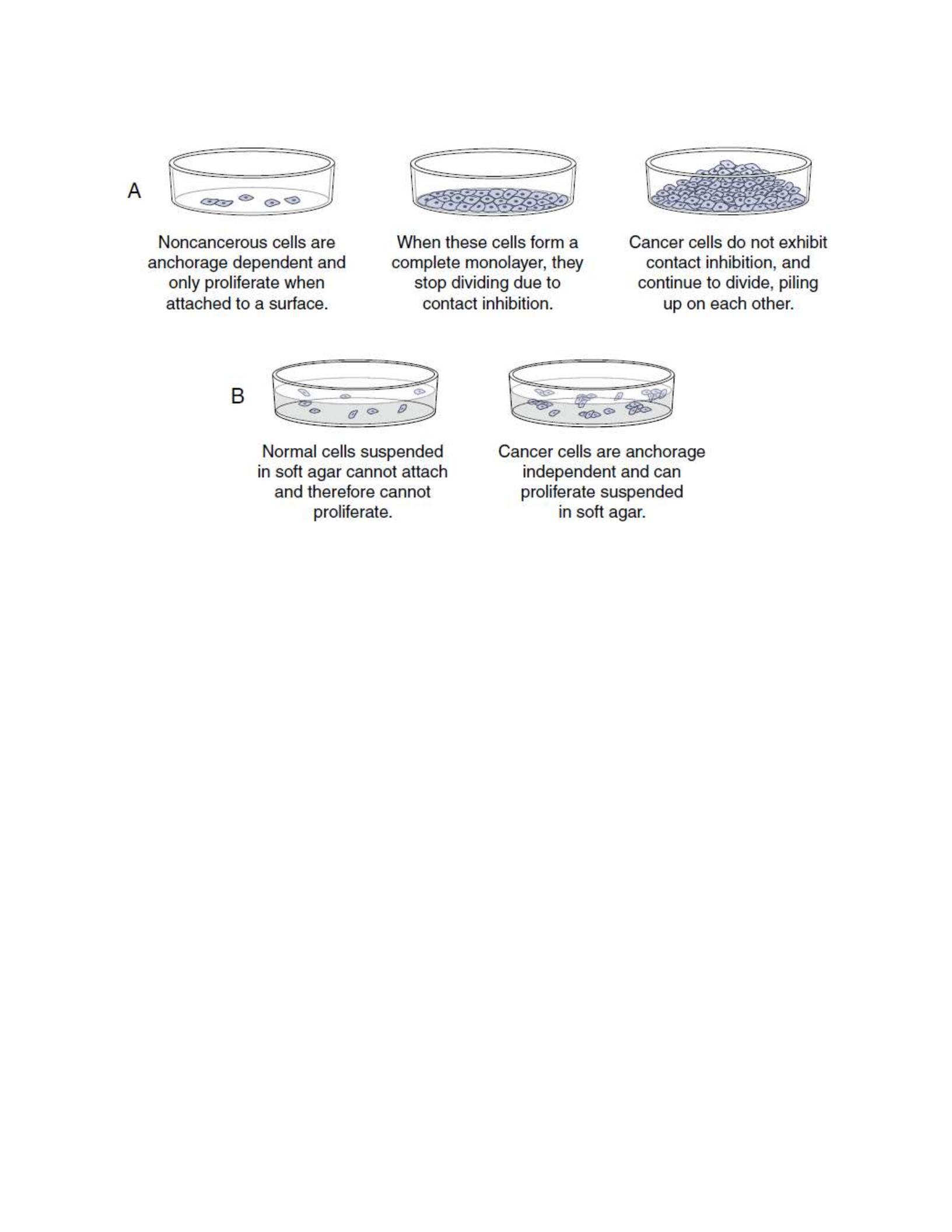
BMET 221: PATHOPHYSIOLOGY
Cancer Metabolism
Cancer cells live in a distinct milieu from normal cells and have different nutritional requirements
from non-proliferating cells. The successful cancer cell divides rapidly, with the consequent
requirement for the building blocks of new cells. Cancers often must grow in a hypoxic and acidic
environment. Cancers also are parasites, able to selectively extract nutrients from the bloodstream
without any evolutionary pressure for balanced metabolism. Nonmalignant cells in the presence
of adequate oxygen normally generate adenosine triphosphate (ATP) by mitochondrial oxidative
phosphorylation (OXPHOS), generating 36 ATP molecules from each glucose molecule that is
broken down to water and carbon dioxide. Only in the absence of sufficient oxygen do normal
cells perform anaerobic glycolysis, generating only two ATP molecules per molecule of glucose,
with lactic acid as a byproduct. However, even in the presence of oxygen, cancer cells perform
glycolysis, not OXPHOS.
Although this aerobic glycolysis was originally postulated to be caused by some form of cancer-
specific mitochondrial dysfunction, it is now apparent that this is instead a highly regulated and
beneficial adaptation for cancer cells. This shift from OXPHOS to glycolysis allows lactate and its
metabolites to be used for the more efficient production of lipids and other molecular building
blocks needed for rapid cell growth. Furthermore, many cancer genes promote this switch to
aerobic glycolysis. Alterations in a number of cancer genes, including receptor tyrosine kinases,
AKT, PTEN, TP53, and MYC, inhibit OXPHOS and promote the activity of glycolytic and related
metabolic pathways that support the rapid growth of cancers. Clinically the high glucose utilization
of a cancer can be exploited for its detection. 18F-Fluorodeoxyglucose (FDG) is incorporated into
cells in the same way as glucose, with two key differences. Because it is missing a key hydroxyl
group it cannot be broken down by glycolysis and, thus, FDG accumulates in cells. Because it is
tagged with 18F, it can be imaged by positron emission tomography (a PET scan). Small metastatic
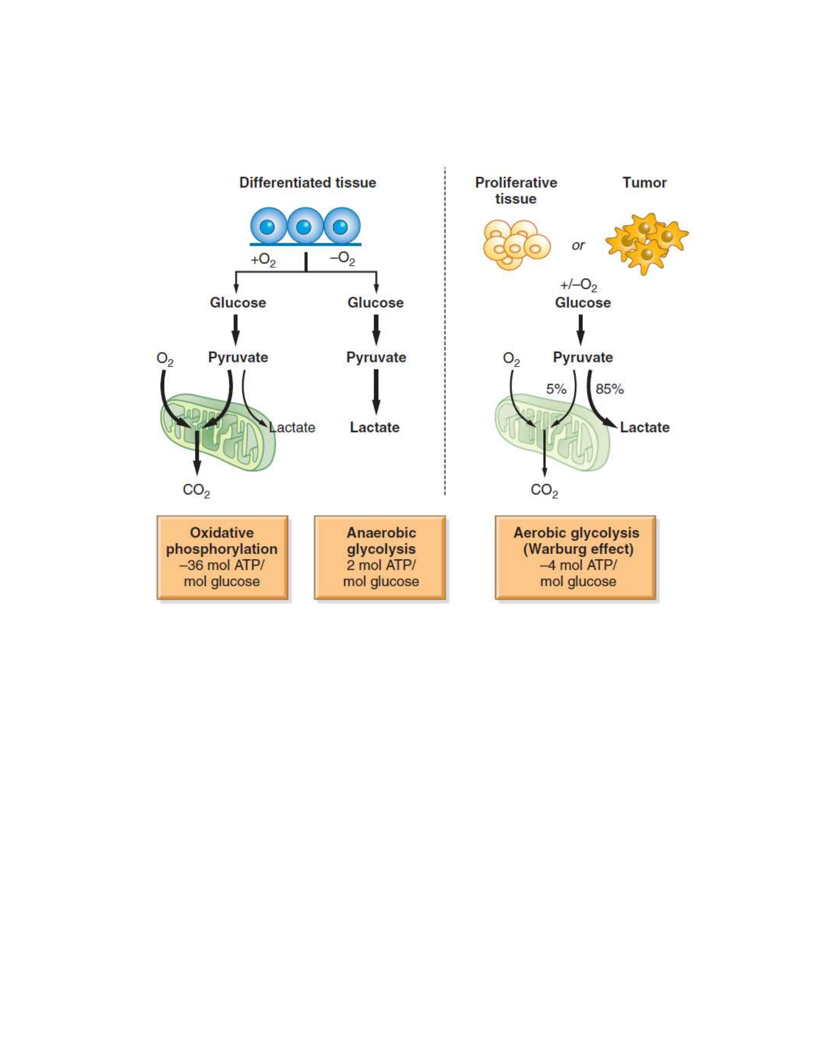
BMET 221: PATHOPHYSIOLOGY
tumor masses that are consuming huge amounts of glucose can readily be detected with this
imaging method
Cancer Invasion and Metastasis
Metastasis is the spread of cancer cells from the site of the original tumor to distant tissues and
organs through the body. Metastasis is a defining characteristic of cancer, contributes significantly
to the pain and suffering from cancer, and is the major cause of death from cancer. Cancer that has
not metastasized can often be cured by a combination of surgery, chemotherapy, and radiation.
These same therapies are frequently ineffective against cancer that has metastasized. For example,
in appropriately treated women with low-stage breast cancer, the 5-year survival rate is often
greater than 90%. Tragically, less than 30% of women with metastatic breast cancer are alive 5
years after diagnosis. A growing body of basic and clinical research is defining the biologic
principles of metastasis, with the hope that this improved understanding will lead to novel
diagnostic approaches and better therapies to prevent and treat metastatic cancers.
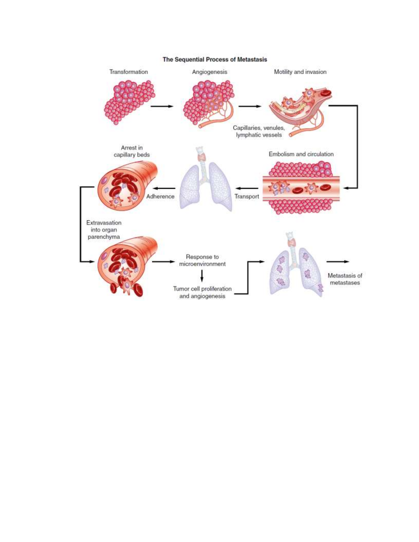
BMET 221: PATHOPHYSIOLOGY
Invasion, or local spread, is a prerequisite for metastasis and is the first step in the metastatic
process. In its earliest stages local invasion may occur by direct tumor extension. Eventually,
however, cells migrate away from the primary tumor and invade the surrounding tissues.
Mechanisms important in local invasion include recruitment of macrophages and other cell types
to the primary tumor, where they promote digestion of connective tissue capsules and other
structural barriers by secreted proteases; changes in cell-to-cell adhesion, often by changes in the
expression of cell adhesion molecules such as cadherins and integrins, making the cancer cells
more slippery and mobile; and increased motility of individual tumor cells5. To transition from
local to distant metastasis, the cancer cells must also be able to invade local blood and lymphatic
vessels, a task facilitated by stimulation of neoangiogenesis and lymphangiogenesis by factors
such as VEGF. Finally, a successful metastatic cell must be able to survive in the circulation, attach
in an appropriate new microenvironment, and multiply to produce an entire new tumor, similar to
the characteristics of a cancer stem cell. Different cancers have different patterns of spread,
determined by a combination of factors. Cancers often spread first to regional lymph nodes through
the lymphatics and then to distant organs through the bloodstream. A cancer’s ability to establish
a metastatic lesion in a new location requires that the cancer both attach to specific receptors and

BMET 221: PATHOPHYSIOLOGY
survive in the specific environment. Because metastasis requires successful completion of each
and every step, there may be many opportunities to interrupt this potentially lethal pathway.
Clinical Manifestations of Cancer
Diagnosis and Staging
Cancer can be discovered in many ways: after screening tests, from routine exams, and after
investigation of symptoms. The symptoms a cancer produces are as diverse as the types of cancer.
The location of the cancer can determine symptoms by physical pressure, obstruction, and loss of
normal function, or a cancer can cause problems far away from its source by pressing on nerves or
secreting bioactive compounds. Whatever the initial complaint, once the diagnosis is suspected
and a tumor has been identified, it is essential that tumor tissue be obtained to establish a definitive
diagnosis and correctly classify the disease.
Once tissue is obtained, it is examined microscopically by the pathologist for the histologic
hallmarks of cancer detailed in the beginning of this chapter. The classification of the cancer can
be further facilitated by a variety of clinically available tests, including immuno-histochemical
stains, flow cytometry, electron microscopy, chromosome analysis, and nucleic acid–based
molecular studies. If the diagnosis of cancer is established, it is critical to determine if the cancer
has spread, known as the stage of the cancer. Staging initially involves determining the size of
the tumor, the degree to which it has locally invaded, and the extent to which it has spread
(metastasized)
Specific molecular tests are increasingly used in staging as well. Diverse schemes are used for
staging different tumors. In general, a four-stage system is used, with carcinoma in situ regarded
as a special case. Cancer confined to the organ of origin is stage 1; cancer that is locally invasive
is stage 2; cancer that has spread to regional structures, such as lymph nodes, is stage 3; and cancer
that has spread to distant sites, such as a liver cancer spreading to lung or a prostate cancer
spreading to bone, is stage 4. One common scheme for standardizing staging is the World Health
Organization’s TNM system: T indicates tumor spread, N indicates node involvement, and M
indicates the presence of distant metastasis. The prognosis generally worsens with increasing
tumor size, lymph node involvement, and metastasis. Staging also may alter the choice of therapy,
with more aggressive therapy being delivered to more invasive disease.
Paraneoplastic Syndromes
Paraneoplastic syndromes are symptom complexes that are triggered by a cancer but are not
caused by direct local effects of the tumor mass. They are most commonly caused by biologic
substances released from the tumor (e.g., hormones) or by an immune response triggered by the
tumor. For example, a small fraction of carcinoid tumors releases hormones, including serotonin,
into the bloodstream that cause flushing, diarrhea, wheezing, and rapid heartbeat. A number of
cancers trigger an antibody response that attacks the nervous system, causing a variety of
neurologic disorders that can precede other symptoms of cancer by months. Although infrequent,
paraneoplastic syndromes are significant because they may be the earliest symptom of an unknown

BMET 221: PATHOPHYSIOLOGY
cancer and, in affected individuals, can be serious, often irreversible, and sometimes life-
threatening.
Pain
Pain is one of the most feared complications of advanced cancer. Although pain can be one of the
presenting symptoms of cancer, most commonly there is little or no pain during the early stages of
malignant disease. Significant pain, however, occurs in a large fraction of those individuals who
are terminally ill with cancer. Pain is strongly influenced by fear, anxiety, sleep loss, fatigue, and
overall physical deterioration. It occurs through an interaction among physiologic, cultural, and
psychologic components.
Cancer-associated pain can arise from a variety of direct and indirect mechanisms. Direct pressure,
obstruction, invasion of a sensitive structure, stretching of visceral surfaces, tissue destruction,
infection, and inflammation all can cause pain. Pain can occur at the site of the primary tumor or
can result from a distant metastatic lesion. Furthermore, pain may be referred away from the
involved site and manifest, for example, as back pain. Specific sites are more prone to cancer-
associated pain. Bone metastases, common in advanced breast and prostate cancer, can cause
significant pain because of periosteal irritation, medullary pressure, vertebral collapse, and
pathologic fractures. Brain tumors (primary or metastatic) can, depending on the location, cause
headache, seizures, or neurologic deficits. Pain in the abdomen may be caused by bowel
obstruction, or inflammation and infection. Hepatic malignancies can stretch the liver, resulting in
a dull pain or a feeling of fullness over the right upper abdominal quadrant. Mucosal surfaces can
develop painful ulcerative lesions from the cancer, chemotherapy, and radiation or leukopenia (or
both). The diagnosis and treatment of pain is one of the primary responsibilities of the medical
team. The individual’s perception and, hence, reporting of pain can vary widely and be affected
by such factors as age and cultural background. The first priority of treatment is to control pain
rapidly and completely as judged by the individual. The second priority is to prevent recurrence of
pain. Objective measurements of pain are increasingly being included along with the reporting of
more traditional vital signs. Many institutions are using specialized pain management teams that
are trained to recognize different types of acute and chronic pain, as well as the individual’s
response to that pain. Many modalities are available to treat pain, ranging from combinations of
NSAIDs and narcotics to palliative surgery and radiation therapy. Individual controlled analgesia
provides many benefits, not the least of which is regaining some control over one’s own body.
Although cancer pain is a complex problem arising from multiple sources, individuals should be
assured that suffering is not inevitable and that relief is attainable
Fatigue
Fatigue is the most frequently reported symptom of cancer and cancer treatment. The exact
mechanisms that produce fatigue are poorly understood. Suggested causes include sleep
disturbances, various biochemical changes secondary to disease and treatment, numerous
psychosocial factors, level of activity, nutritional status, and other environmental and physical
factors. The physiologic understanding of fatigue probably includes mechanisms for decreased
muscle contractility. Overall, studies of muscle function suggest that some individuals with cancer

BMET 221: PATHOPHYSIOLOGY
may lose portions of muscle function needed to perform normal physical activities. Other areas of
research include muscle function consequences from metabolic products of cancer treatment and
associated muscle loss from circulating cytokines (e.g., tumor necrosis factor [TNF] and
interleukin-1 [IL-1]). Similar to pain, fatigue is a subjective clinical manifestation. Fatigue is
described by individuals with cancer as tiredness, weakness, lack of energy, exhaustion, lethargy,
inability to concentrate, depression, sleepiness, boredom, lack of motivation, and decreased mental
status. Some of these symptoms have been termed “chemo brain,” or mild cognitive impairment.
The changes in cognitive function can be caused by the cancer itself or by the stress associated
with the diagnosis of cancer, however, because symptoms similar to “chemo brain” also occur in
individuals who have not received chemotherapy.
Cachexia
The syndrome of cachexia includes a constellation of symptoms including anorexia, early satiety
(filling), weight loss, anemia, asthenia (marked weakness), taste alterations, and altered protein,
lipid, and carbohydrate metabolism. Cachexia is the most severe form of malnutrition associated
with cancer and results in wasting, emaciation, and decreased quality of life. Cachexia occurs even
with seemingly adequate caloric intake because metabolic disturbances, including insulin
resistance, hypertriglyceridemia, muscle wasting, and general increased derangement of
metabolism, lead to significant inefficiency of energy usage. Anorexia, or loss of appetite,
frequently worsens the weight loss associated with this abnormal metabolic state. Anorexia itself
can be caused by pain, depression, chemotherapy, and/ or radiotherapy. Alterations in taste,
making foods seem bland or distasteful, by these same causes also can account for the anorexia
present in individuals with cancer. Altered carbohydrate metabolism causes a syndrome
resembling diabetes mellitus. Individuals show hyperinsulinemia, insulin resistance,
hyperglycemia, and abnormal glucose tolerance test results. These disturbances cause increased
gluconeogenesis, which produces glucose from amino acids. In starvation, protein usually is spared
to protect vital structures; however, in cancer, protein and fatty acids are used to meet energy
needs. An unusual and frustrating component of cancer care is the person’s early satiety, or a sense
of being full after only a few mouthfuls of food. Cytokines, including TNF-α, IL-6, and interferon-
γ, appear to cause the metabolic alterations associated with tissue loss in cancer wasting. Tumor
metabolites may also contribute to a cachectic state. For example, a factor found in the urine of
some individuals with cancer induces the catabolism of muscle. This factor, originally called
proteolysis-inducing factor, is a partial fragment of an antimicrobial peptide, dermcidin, normally
expressed in the skin.
Anemia
Anemia is commonly associated with malignancy, with 20% of persons diagnosed with cancer
having hemoglobin concentrations less than 9 g/dl (normal value = 15 g/dl). Mechanisms that
cause anemia in persons with cancer include chronic bleeding (resulting in iron deficiency), severe
malnutrition, cytotoxic chemotherapy, and malignancy in blood-forming organs. Chronic bleeding
and iron deficiency can accompany colorectal or genitourinary malignancy. Iron also is
malabsorbed in persons with gastric, pancreatic, or upper intestinal cancer. Often there is a defect
in the reutilization of iron because of lack of transfer of iron from the storage pool to blood cell

BMET 221: PATHOPHYSIOLOGY
precursors. This defect may be caused by increased secretion of IL-6 and hepcidin. Defects in
erythropoietin production and shortened duration of red cell survival have also been documented.
In addition, anorexia can cause both iron and folate deficiency. Megaloblastic (large red cell)
anemias also may develop after methotrexate treatment. Administration of erythropoietin, which
stimulates production of erythrocytes, has been effective in correcting anemia in persons with
cancer; fewer red blood cell transfusions were required in most of the studied subjects. In addition,
anemias occurring after chemotherapy or radiotherapy have been treated successfully with
erythropoietin. However, recent studies have shown that aggressive use of erythropoietin increases
the risk of blood clots and can decrease cancer survival.
Leukopenia and Thrombocytopenia
Direct tumor invasion of the bone marrow causes both leukopenia (a decreased total white blood
cell count) and thrombocytopenia (a decreased number of platelets). More commonly, many
chemotherapeutic drugs are toxic to the bone marrow, often causing granulocytopenia and
thrombocytopenia. Granulocytopenia also can result from radiation therapy if it encompasses
significant areas of the bone marrow. The duration of granulocytopenia and hence the riskof
serious infection can be lessened by treatment with recombinant human granulocyte colony-
stimulating factor (rhG-CSF, filgrastim). rhG-CSF stimulates white blood cell precursors in the
marrow to proliferate and differentiate rapidly. Thrombocytopenia is a major cause of hemorrhage
in persons with cancer and is often treated with platelet transfusions. Thrombocytopenia also is an
accompanying disorder of disseminated intravascular coagulation that occurs in persons with acute
promyelocytic leukemia and severe infections.
Infection
Infection is the most significant cause of complications and death in persons with malignant
disease. When the absolute granulocyte count falls below 500 cells per microliter, the risk of
serious microbial (bacterial and fungal) infection increases. Persons with cancer also have debility
with advanced disease, and immunosuppression from the underlying cancer and the radiotherapy
and chemotherapy used to treat it. Surgery also can lower resistance to infection because removal
of large quantities of tissue, together with hemorrhage, dead spaces, and poor tissue perfusion, can
create favorable sites for infection. Hospital-related (nosocomial) infections increase because of
indwelling medical devices, inadequate wound care, and the introduction of microorganisms from
visitors and other individuals.
Gastrointestinal Tract
The entire gastrointestinal (GI) tract relies on rapidly growing cells to produce an effective barrier
to trauma and infection and to provide an absorptive surface for nutrients. Both chemotherapy and
radiation therapy may cause a decreased cell turnover, thereby leading to oral ulcers (stomatitis),
malabsorption, and diarrhea. The disruption of barrier defenses also increases the risk for infection,
especially invasion by a person’s own GI flora. Therapy-induced nausea, thought to be caused by
an agent’s direct action upon the central nervous system’s vomiting centers, historically has been
a major obstacle in therapy. Aggressive antinausea (antiemetic) therapy, including the centrally
acting serotonin 5-hydroxytryptamine (5-HT3) antagonists (such as ondansetron or dolasetron),

BMET 221: PATHOPHYSIOLOGY
has allowed better tolerance of highly emetogenic protocols. Other popular antiemetics include
steroids and phenothiazines. Synthetic cannabinoids, the active ingredients in marijuana, increase
appetite in addition to having antinausea properties. Analgesia often includes opiate agents, vital
in treating severe cases of mucosal lesions. Supplemental nutrition through enteral or parenteral
routes may be needed to combat malnutrition. Good oral hygiene may help prevent complications
arising from mucosal membrane breakdown.
Hair and Skin
Alopecia (hair loss) results from chemotherapy effects on hair follicles. Alopecia is usually
temporary, although hair may regrow with a different texture initially. Not all chemotherapeutic
agents cause alopecia. Decreased renewal rates of the epidermal layers in the skin may lead to skin
breakdown and dryness, altering the normal barrier protection against infection. Radiation therapy
may cause skin erythema (redness) and contribute to breakdown.
Treatment of Cancer
The diagnosis of cancer has a profound effect on individuals and their families. Responses range
from depression to resigned fatalism to an aggressive no-holds-barred pursuit of therapy. The
choice of therapy should be based on full consideration by the individual, the family, and the
medical team of the individual’s diagnosis, prognosis, and therapeutic options. Many types of
cancer can be effectively treated with chemotherapy, radiotherapy, surgery, and combinations of
these modalities. Caregivers must recognize that many individuals seek additional nonscience-
based explanations and therapies and often use alternative therapies, either concurrently or
sequentially. Alternative therapies can be biologically harmless or harmful; rarely is there any
evidence these approaches are medically effective and in the worst cases they can be expensive,
delay the use of effective therapies, and produce toxic side effects. A challenge for the medical
team is to provide the same level of psychosocial comfort and support that alternative therapies
can provide, while also providing scientifically rational evidence-based therapies.
Chemotherapy
The era of modern chemotherapy began with the observation in World War II that mustard gas
exposure caused suppression of the bone marrow. Related compounds, such as nitrogen mustard
and cyclophosphamide, were then tested and produced clinical responses in hematologic
malignancies, including lymphomas. Also in the late 1940s, based on the remarkable clinical
observation that the vitamin folic acid could increase leukemia growth, antifolate drugs were
developed (leading ultimately to methotrexate) that produced remissions in previously untreatable
leukemias. All chemotherapeutic agents take advantage of specific vulnerabilities in target cancer
cells. Antimetabolites, such as methotrexate and l-asparaginase, block normal growth pathways in
all cells, but leukemia and other cancer cells are exquisitely sensitive to folic acid and asparagine
deprivation, whereas nonmalignant cells are far less sensitive. Similarly, some cancer cells are
highly sensitive to DNA-damaging agents, such as cyclophosphamide and anthracyclines, because
of the oncogenic mutations that accelerate the cell cycle and DNA synthesis. Cellular checkpoints
prevent normal cells treated with microtubule-directed drugs, such as vincristine and the taxanes,
from undergoing mitosis, whereas cancer cells treated with these agents lack normal checkpoints,

BMET 221: PATHOPHYSIOLOGY
continue through mitosis, and undergo mitotic catastrophe. Single chemotherapeutic agents often
shrink cancers, but these drugs given alone rarely, if ever, provide a cure. Hence, chemotherapy
drugs are usually given in combinations designed to attack a cancer from many different
weaknesses at the same time and to limit the dose and therefore the toxicity of any single agent.
Cancers contain a very large number of cells, and commonly a small fraction of those cells may
be resistant to a particular drug. However, those cells are likely to be sensitive to the second or
third drug in a chemotherapy cocktail. Scheduling of drug administration is also very important,
with many studies showing cancers are more likely to develop drug resistance if there are
significant delays between planned courses of chemotherapy. The newest highly targeted agents
used to treat cancer exploit specific vulnerabilities uncovered by molecular analysis in specific
diseases. These new drugs are still used in combination with conventional chemotherapy and to be
effective they must be used in diseases in which the molecular target is present. For example,
imatinib is highly effect in treating CML and gastrointestinal stromal tumor (GIST) but ineffective
in virtually all other cancers. Fortunately, because these drugs are so tightly targeted they have
much less toxicity than conventional chemotherapies that have targets in virtually all cells.
Chemotherapy can be used for several distinct purposes. Induction chemotherapy seeks to cause
shrinkage or disappearance of tumors. In Hodgkin disease, for example, chemotherapy alone can
be used in some cases to cure the disease. In other settings, chemotherapy may shrink the tumor
and improve symptoms without ultimately providing a cure. Adjuvant chemotherapy is given
after surgical excision of a cancer with the goal of eliminating micrometastases. Neoadjuvant
chemotherapy is given before localized (surgical or radiation) treatment of a cancer. As with
induction chemotherapy, the effectiveness, or lack thereof, of neoadjuvant therapy can be
measured (for example, with follow-up scans). Neoadjuvant therapy can shrink a cancer so that
surgery may spare more normal tissue. For example, in the bone cancer osteogenic sarcoma,
neoadjuvant therapy often converts a large tumor mass into a much smaller mass, allowing the
surgeon to perform a limb-sparing excision rather than an amputation.
Radiation Therapy
Radiation therapy is used to kill cancer cells while minimizing damage to normal structures.
Ionizing radiation damages cells by imparting enough energy to cause molecular damage,
especially to DNA. The damage may be lethal, in which the cell is killed by radiation; potentially
lethal, in which the cell is so severely affected by radiation that modifications in its environment
will cause it to die; or sublethal, in which the cell can subsequently repair itself. Cellular
compartments with rapidly renewing cells are, in general, more radiosensitive. Effective cell
killing by radiation also requires good local delivery of oxygen, something not always present in
large cancers. Radiation produces slow changes in most cancers and irreversible changes in normal
tissues as well. Because of these irreversible changes, each tissue has a maximum lifetime dose of
radiation it can tolerate. Radiation is well suited to treat localized disease in areas that are hard to
reach surgically, for example, in the brain and pelvis. A number of radiation delivery methods are
available, with external beam being the most common. Radiation sources, such as small 125I-
labeled capsules (also called seeds), can also be temporarily placed into body cavities, a delivery
method termed brachytherapy. Brachytherapy is useful in the treatment of cervical, prostate, and
head and neck cancers.

BMET 221: PATHOPHYSIOLOGY
Surgery
Surgery plays many roles in the care of individuals with cancer. The multiple approaches to
obtaining tissue for diagnosis have been discussed. Surgery is often the definitive treatment of
cancers that do not spread beyond the limits of surgical excision. It is also indicated for the relief
of symptoms, for instance, those caused by tumor mass obstruction. In selected high-risk diseases,
surgery plays a role in the prevention of cancer. For example, individuals with familial
adenomatous polyposis because of germline mutations of the APC gene have close to a 100%
lifetime risk of colon cancer, so a prophylactic colectomy is indicated. Similarly, women with
BRCA1/2 mutations have a markedly increased risk of breast and ovarian cancer, and often choose
prophylactic mastectomy or bilateral salpingo-oophorectomy (removal of ovaries and fallopian
tubes), or both. Key principles apply specifically to cancer surgery, including obtaining adequate
surgical margins during a resection to prevent local recurrences, placing needle tracks and biopsy
incision scars (that may be contaminated with cancer cells) carefully so they can be removed in
subsequent incisions, avoiding the spread of cancer cells during surgical procedures through
careful technique, and paying attention to obtaining adequate tissue specimens during biopsies so
that the pathologist can be confident of the diagnosis. Additionally, the surgeon provides critical
staging information by inspection, sampling, and removal of local and region lymph nodes during
procedures.

