


PUHE 112:
HUMAN PHYSIOLOGY
Internal Environment
•
•
•
•
•
•
Physiology : The study of the functions of the human body.
Looks at mechanisms by which the various organs and tissues
carry out their specific activities.
Emphasis is often placed on the processes that control and
regulate these functions.
In order for the body to function optimally, conditions within the
body, referred to as the
internal environment
, must be very
carefully regulated
The maintenance of relatively constant or steady-state internal
conditions is referred to as
homeostasis
.
Cells and tissues of the body will survive and function efficiently
only when these internal conditions are properly maintained.
Internal Environment
•
•
•
•
•
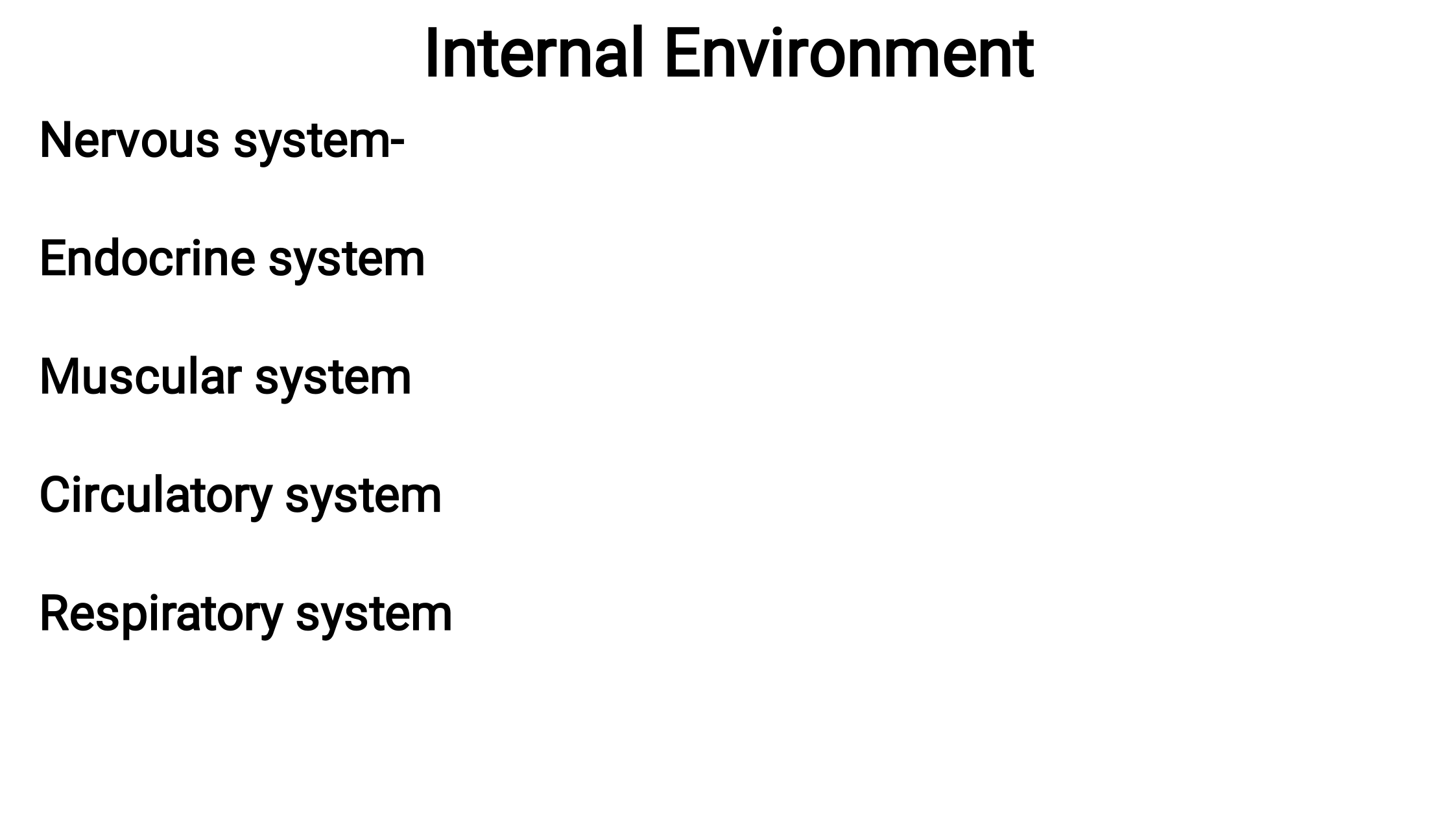
Nervous system- Regulates muscular activity and glandular
secretion; responsible for all activities associated with the mind
Endocrine system -Regulates metabolic processes through
secretion of hormones
Muscular system- Allows for body movement; contributes to
thermoregulation
Circulatory system -Transports nutrients, O2, waste, CO2,
electrolytes, and hormones throughout the body
Respiratory system- Obtains oxygen and eliminates carbon
dioxide; regulates acid-base balance (pH)
Internal Environment
•
•
•
•
•
•
Gastrointestinal system -Digests food to provide nutrients to the
body,
Renal system- Eliminates waste products from the body;
regulates blood volume and blood pressure; regulates acid-base
balance (pH)
For example,
Most metabolic reactions within cells consume oxygen and
glucose. These substances must then be replaced.
In addition, these reactions produce metabolic wastes including
carbon dioxide and urea, which must then be eliminated.
Therefore, it is more accurate to say that the internal
environment is in a
dynamic steady state
Internal Environment
•
•
•
Physiology includes not only of how each of these systems
carries out its functions, but also of the mechanisms involved
that regulate these activities in order to maintain homeostasis
under a variety of conditions.
In order to maintain homeostasis, the body must be able to
monitor and sense changes in the internal environment.
It must also be able to compensate, or make adjustments, for
these changes.

Negative feedback
•
•
•
•
•
Most of the body’s
compensatory homeostatic mechanisms
function by way of
negative feedback
.
This is a response that causes the level of a variable to change
in a direction opposite to that of the initial change.
If for example blood pressure increases, the arterial
baroreceptors are stimulated and an increased number of nerve
impulses are transmitted to the CNS through afferent pathways.
The region of the brain regulating the cardiovascular system
responds to this sensory input by altering efferent nerve activity
to the heart.
The result is a decrease in heart rate and therefore a decrease in
blood pressure back to its baseline value.
Negative feedback mechanism
•
•
•
When a physiological variable becomes too high or too low, a
control system elicits a negative feedback response consisting
of one or a series of changes that returns the variable to within
its normal physiological range.
Negative feedback mechanism—a control system in which a
stimulus initiates a response that reverses or reduces the
stimulus, thereby stopping the response until the stimulus
occurs again and there is a need for the response.
Positive feedback mechanism—a control system that requires an
external interruption or brake. Has the potential to become a self-
perpetuating and harmful cycle, therefore is rare in the body
Metabolism and Homeostasis
•
•
•
Metabolism is the sum of all of the chemical and physical
changes that take place in the body.
Metabolic rate is the amount of energy and heat production per
unit of time.
The body constantly responds to internal and external changes,
yet remains stable; its many aspects of metabolism are kept
within normal limits (usually a range of values, not a single value).
Main regulatory systems
•
i.
ii.
i.
ii.
iii.
•
Two regulatory systems in the body influence the activity of all
the other organ systems so that Homeostasis is maintained.
Nervous system
Endocrine system
The
nervous system
has three functional components
Sensory division of the peripheral nervous system
Central nervous system
Motor division of the peripheral nervous system
The
endocrine system
, which carries out its effects by secreting
hormones
. These hormones are transported in the blood to the
specific tissues upon which they exert their effects.
Main regulatory systems
•
•
In general, the nervous system primarily regulates muscular
activity and glandular secretion and the endocrine system
primarily regulates metabolic activity in the body’s cells.
However, these two systems may work together in the regulation
of many organs, as well as influence each other’s activity
CELLULAR COMMUNICATION
•
•
i.
ii.
iii.
Each cell is surrounded by a plasma membrane that separates the
cytoplasmic contents of the cell, or the intracellular fluid, from the fluid
outside the cell, the extracellular fluid.
Homeostatic functions of the plasma membrane include
serves as a
permeability barrier
that insulates or protects the
cytoplasm from immediate changes in the surrounding environment.
allows the cell to maintain a cytoplasmic composition very different
from that of the extracellular fluid; the functions of neurons and
muscle cells depend on this difference.
contains many enzymes and other components such as antigens and
receptors that allow cells to interact with other cells, neurotransmitters,
blood-borne substances such as hormones, and various other
chemical substances, such as drugs.
Plasma membrane
•
i.
ii.
iii.
iv.
•
•
The major components of the plasma membrane include:
Phospholipids
Cholesterol
Proteins
Carbohydrates
The basic structure of the plasma membrane is formed by
phospholipids
, which are one of the more abundant of the
membrane components.
Phospholipids are
amphipathic
molecules that have polar (water-
soluble) and nonpolar (water-insoluble) regions.
Plasma membrane
•
•
•
They are composed of a phosphorylated glycerol backbone,
which forms a hydrophilic polar head group and a nonpolar
region containing two hydrophobic fatty acid chains.
In an aqueous environment such as the body, these molecules
are arranged in a formation referred to as the
lipid bilayer
consisting of two layers of phospholipids.
The polar region of the molecule is oriented toward the outer
surface of the membrane where it can interact with water; the
nonpolar, hydrophobic fatty acids are in the center of the
membrane away from the water.
Membrane properties
•
•
•
•
The functional significance of the lipid bilayer is that it creates a
semipermeable barrier
.
Lipophilic, or non-water-soluble, substances can readily cross the
membrane by simply passing through its lipid core.
Important examples of these substances include gases, such as
oxygen and carbon dioxide, and fatty acid molecules, which are
used to form energy within muscle cells.
Most hydrophilic, or water-soluble, substances are repelled by
this hydrophobic interior and cannot simply diffuse through the
membrane.
Fluidity of plasma membrane
•
•
•
•
Instead, these substances must cross the membrane using
specialized transport mechanisms.
Examples of lipid-insoluble substances that require such
mechanisms include nutrient molecules, such as glucose and
amino acids, and all species of ions (Na+, Ca++, H+, Cl–, and
HCO3–).
Therefore, the plasma membrane plays a very important role in
determining the composition of the intracellular fluid by selectively
permitting substances to move in and out of the cell.
The surrounding pH can determine whether a molecule is in a
protonated form (positively charged, lipid insoluble) or in an
unprotonated form (uncharged, lipid soluble). Charged substances
do not readily cross the membrane, as do uncharged substances.
Fluidity of plasma membrane
•
•
•
•
Another important property of the lipid bilayer is that the
phospholipids are not held together by chemical bonds.
This enables molecules to move about freely within the
membrane, resulting in a structure that is not rigid in nature, but
instead, very fluid and pliable.
Also contributing to membrane fluidity is the presence of
cholesterol
. Cholesterol has a steroid nucleus that is lipid soluble.
Membrane fluidity
is very important in terms of function in many
cell types. For example, skeletal muscle activity involves
shortening and lengthening of muscle fibbers.
Fluidity of plasma membrane
•
•
Furthermore, as white blood cells leave the blood vessels and
enter the tissue spaces to fight infection, they must squeeze
through tiny pores in the wall of the capillary requiring significant
deformation of the cell and its membrane.
Finally, in all cells, many processes that transport substances
across the plasma membrane require the embedded proteins to
change their conformation and move about within the bilayer.
Functions of proteins on plasma membrane
•
i.
ii.
iii.
iv.
v.
•
•
These proteins provide a variety of important cellular functions by
forming the following structures:
Channels
Carrier molecules
Enzymes
Chemical receptors
Antigens
Some proteins may form
channels
through the cell membrane that allow
small, water-soluble substances such as ions to enter or leave the cell.
Other proteins may serve as
carrier molecules
that selectively transport
larger water-soluble molecules, such as glucose or cellular products,
across the membrane.

Functions of proteins on plasma membrane
•
•
•
Regulators of specific chemical reactions,
enzymes
are extrinsic
proteins found on the internal (e.g., adenylate cyclase) or external
(e.g., acetylcholinesterase) surfaces of the membrane.
Chemical receptors
are found on the outer surface of the cell
membrane and selectively bind with various endogenous
molecules as well as with drugs.
Other proteins found on the external surface of the plasma
membrane are
antigens
. These molecules serve as cell “markers”
that allow the body’s immune system to distinguish between its
own cells and foreign cells or organisms such as bacteria and
viruses.
Carbohydrates in the plasma membrane
•
•
•
The plasma membrane contains a small amount of carbohydrate
(2 to 10% of the mass of the membrane) on the outer surface.
This carbohydrate is found attached to most of the protein
molecules, forming glycoproteins, and to some of the
phospholipid molecules (<10%), forming glycolipids.
Consequently, the external surface of the cell has a carbohydrate
coat, or glycocalyx.

Functions of proteins on plasma membrane
i.
ii.
iii.
iv.
Repelling negatively charged substances: many of the
carbohydrates are negatively charged, creating an overall
negative charge on the surface of the cell that repels negatively
charged extracellular molecules.
Cell-to-cell attachment: the glycocalyx of one cell may attach to
the glycocalyx of another cell, which causes the cells to
become attached.
Receptors: carbohydrates may also serve as specific
membrane receptors for extracellular substances such as
hormones.
Immune reactions: carbohydrates play a role in the ability of
cells to distinguish between “self” cells and foreign cells.
Membrane transport
•
•
•
•
The lipid bilayer arrangement of the plasma membrane renders it
selectively permeable.
Uncharged or nonpolar molecules, such as oxygen, carbon
dioxide, and fatty acids, are lipid soluble and may permeate
through the membrane quite readily.
Charged or polar molecules, such as glucose, proteins and ions,
are water soluble and impermeable, unable to cross the
membrane unassisted.
These substances require protein channels or carrier molecules
to enter or leave the cell.
Passive diffusion through the membrane
•
•
•
•
Molecules and ions are in constant motion and the velocity of their
motion is proportional to their temperature.
This passive movement of molecules and ions from one place to
another is referred to as
diffusion
.
When a molecule is unevenly distributed across a permeable
membrane with a higher concentration on one side and a lower
concentration on the opposite side, there is said to be a
concentration
gradient
or a concentration difference.
In a
dynamic equilibrium
, number of molecules moving in one
direction across the membrane is equal to the number of molecules
moving in the opposite direction. At this point, although the diffusion
of molecules continues, no further
net
diffusion takes place.
Passive diffusion through the membrane
•
•
•
•
•
•
The rate of diffusion of a substance is influenced by several factors.
It is proportional to the concentration gradient; the permeability of the
membrane; and the surface area of the membrane.
For example, as the permeability of the membrane increases, the rate
of diffusion increases.
It is inversely proportional to the molecular weight of the substance
and the thickness of the membrane.
Movement of ions, in particular, depends not only on a concentration
gradient but also on an
electrical gradient
.
Positively charged ions (cations) are attracted to a negatively charged
area and negatively charged ions (anions) are attracted to a positively
charged area. Ions of a similar charge tend to repel each other and
oppose diffusion.
Factors Influencing Rate of Diffusion of a Substance
i.
ii.
iii.
iv.
v.
Concentration gradient
Permeability of membrane
Surface area of membrane
Molecular weight of substance
Thickness of membrane
Osmosis
•
•
•
•
•
Water is a small polar molecule that can easily diffuse across plasma
membranes through small intermolecular spaces.
Osmosis
is the net movement of water through a semipermeable
membrane down its own concentration gradient from an area of high
water concentration to an area of low water concentration.
Water moves toward an area of higher
solute
concentration. The
solute particles may be thought of as ‘drawing’ the water toward them.
The
osmotic pressure
of a solution is the pressure or force by which
water is drawn into the solution through a semipermeable membrane.
The magnitude of this pressure depends on the number of solute
particles present.
An increase in the number of particles in the solution results in an
increase in the osmotic pressure and, therefore, an increase in the
movement of water toward it.

Osmosis
•
•
•
•
The plasma membrane is
semipermeable
because it is not
permeable to all solute particles present.
As a result, it maintains a concentration difference for many ions
and molecules across itself, although water crosses the
membrane freely in either direction.
The movement of water in and out of the cell will occur
whenever there is a difference in osmotic pressure between the
intracelluar fluid and the extracellular fluid.
For example, an increase in the osmotic pressure of the
extracellular fluid (more solute, lower water concentration) will
cause water to leave the cell by osmosis. On the other hand, a
decrease in the osmotic pressure in the extracellular fluid (less
solute, higher water concentration) will cause water to enter the
Mediated transport
•
•
•
i.
ii.
iii.
In
mediated transport
, carrier proteins embedded within the
plasma membrane assist in the transport of larger polar
molecules into or out of the cell.
When a given substance attaches to a specific binding site on
the carrier protein, the protein undergoes a conformational
change such that this site with the bound substance moves from
one side of the plasma membrane to the other.
The substance is then released. Mediated transport displays
three important characteristics influencing its function:
Specificity
Competition
Saturation
Characteristics of Mediated transport
•
•
•
Specificity
-Each of these proteins may bind only with select
substances that “fit” into its binding site.
Competition-
different substances with similar chemical
structures may be able to bind to the same carrier protein and
therefore compete for transport across the membrane.
Saturation
. The greater the number of carrier proteins utilized at
any given time, the greater the rate of transport is. Initially, as the
concentration of a substance increases, the rate of transport
increases; however, a finite number of carrier proteins exist in a
given cell membrane. Once all these proteins are utilized in the
transport process,
Two forms of Mediated transport
i.
ii.
•
•
Facilitated diffusion
Active transport
With
facilitated diffusion
, carrier proteins move across the
membrane in either direction and will transport a substance
down its concentration gradient. Substances are moved from an
area of high concentration to an area of low concentration — a
passive process that requires no energy. Eg glucose, which is a
large polar molecule. A concentration gradient is always
available for diffusion into the cell.
With
active transport
, energy is expended to move a substance
against its concentration gradient from an area of low
concentration to an area of high concentration.
ELECTRICAL COMMUNICATION (NERVOUS SYSTEM)
•
•
•
•
Function of neurons is to convey information to other cells in the
form of electrical signals.
These signals occur due to ion influx (movement) across the
plasma membrane.
A given stimulus will cause its effect by altering the permeability
to one or more ions. The involved ions will then diffuse into or
out of the cell according to their concentration and electrical
gradients, causing a change in the membrane potential.
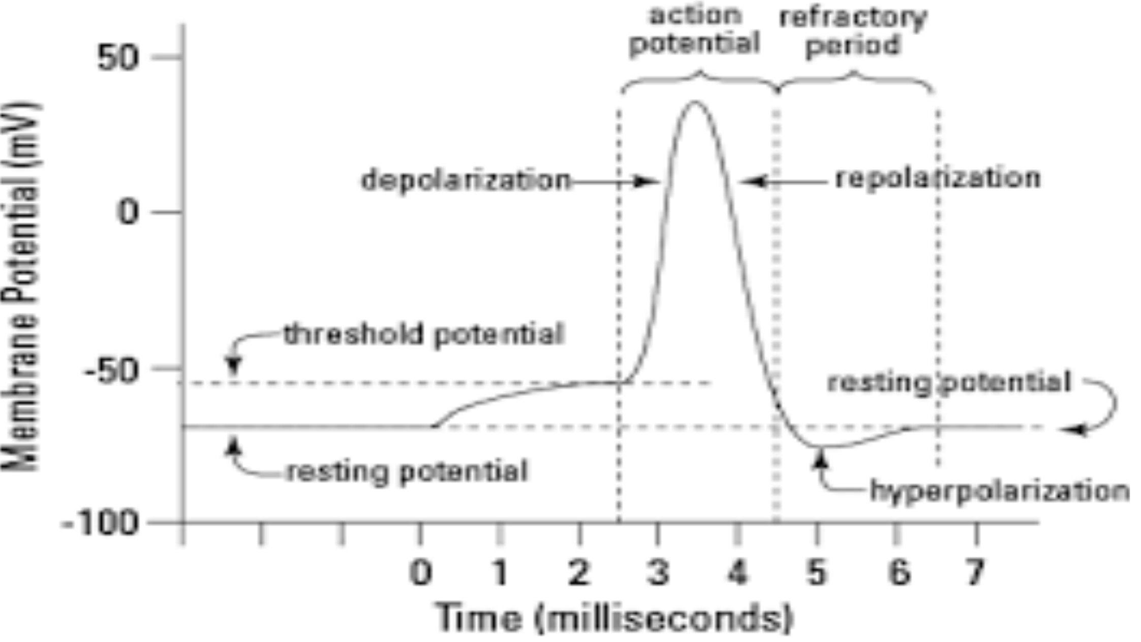
The resting membrane potential in a typical neuron is –70 mV.
Movement of the membrane potential toward zero (less negative)
is referred to as depolarization.
ELECTRICAL COMMUNICATION (NERVOUS SYSTEM)
•
•
•
•
A neuron can receive input from other neurons via a chemical
called a neurotransmitter.
If this input is strong enough, the neuron will pass the signal to
downstream neurons.
Transmission of a signal within a neuron (in one direction only,
from dendrite to axon terminal) is carried out by the opening and
closing of voltage-gated ion channels, which cause a brief reversal
of the resting membrane potential to create an action potential.
As an action potential travels down the axon, the polarity changes
across the membrane. Once the signal reaches the axon terminal, it
stimulates other neurons.
ELECTRICAL COMMUNICATION (NERVOUS SYSTEM)
•
•
•
•
•
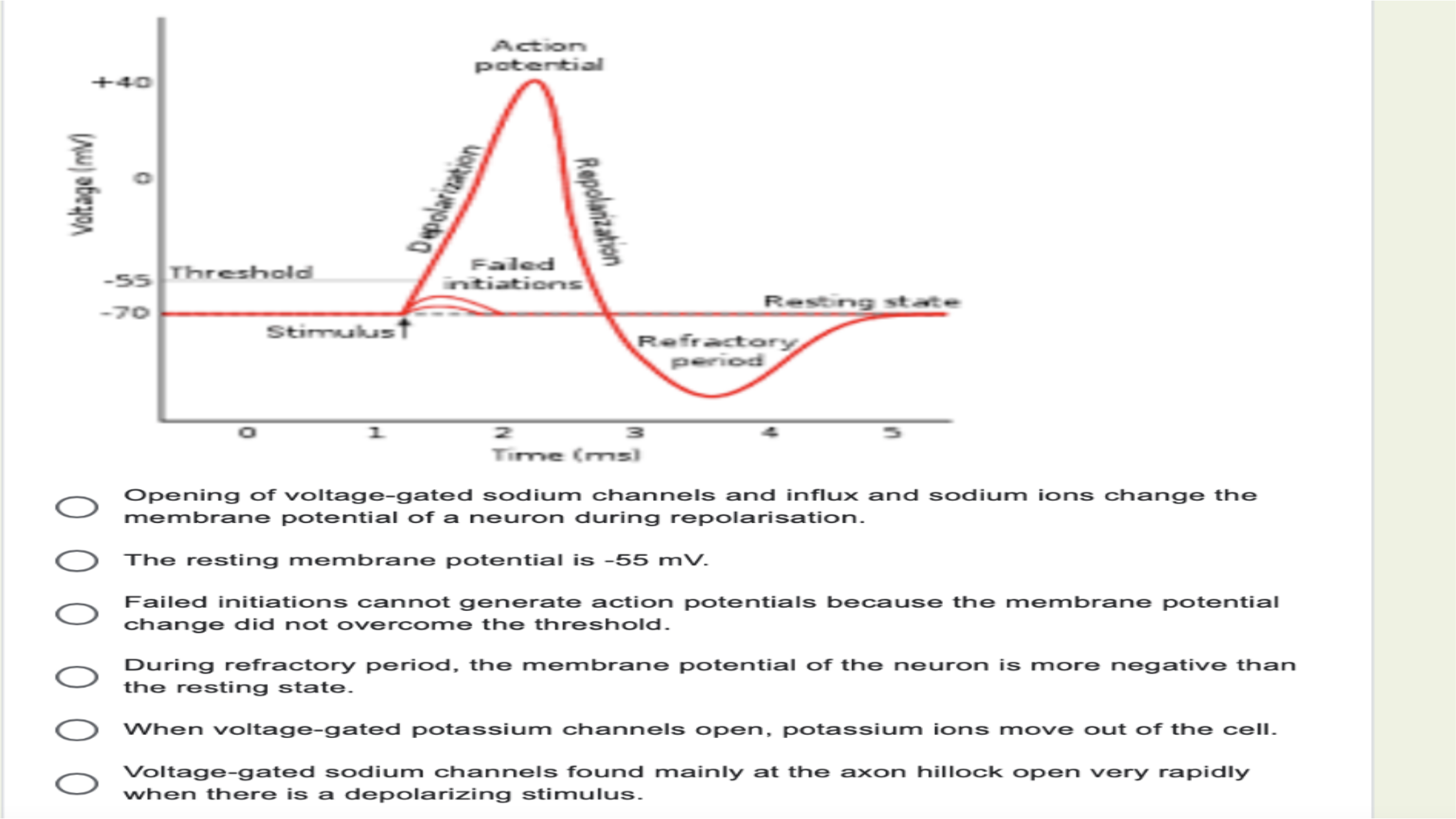
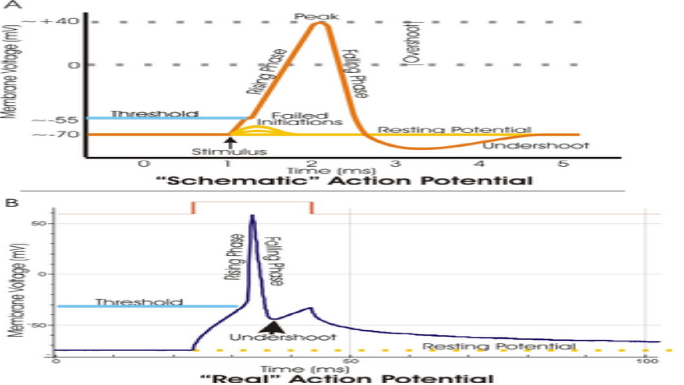
(1) A stimulus from a sensory cell or another neuron causes the
target cell to depolarize toward the threshold potential.
(2) If the threshold of excitation is reached, all Na+ channels
open and the membrane depolarizes.
(3) At the peak action potential, K+ channels open and K+ begins
to leave the cell. At the same time, Na+ channels close.
(4) The membrane becomes hyperpolarized as K+ ions continue
to leave the cell. The hyperpolarized membrane is in a refractory
period and cannot fire. (5) The K+ channels close and the Na+/
K+ transporter restores the resting potential.
Depolarization and the Action Potential

•
•
•
•
When neurotransmitter molecules bind to receptors located on
a neuron’s dendrites, voltage-gated ion channels open.
At excitatory synapses, positive ions flood the interior of the
neuron and depolarize the membrane, decreasing the difference
in voltage between the inside and outside of the neuron.
A stimulus from a sensory cell or another neuron depolarizes
the target neuron to its threshold potential (-55 mV), and Na+
channels in the axon hillock open, starting an action potential.
Once the sodium channels open, the neuron completely
depolarizes to a membrane potential of about +40 mV. The
action potential travels down the neuron as Na+ channels open.

•
•
•
•
•
The membrane potential is the basis for the conduction of nerve
impulses along the cell membranes of neurons.
Ions that are important in the formation of a nerve impulse include
sodium (Na+) and potassium (K+).
The sodium-potassium pump maintains the resting potential of a
neuron
Two mechanisms have evolved to transmit nerve signals. First, within
cells, electrical signals are conveyed along the cell membrane.
Second, for communication between cells, the electrical signals
generally are converted into chemical signals conveyed by small
messenger molecules called neurotransmitters.

•
•
•
•
•
The return of the membrane potential to its resting value is
referred to as repolarization.
Movement of the membrane potential further away from zero
(more negative) is referred to as hyperpolarization.
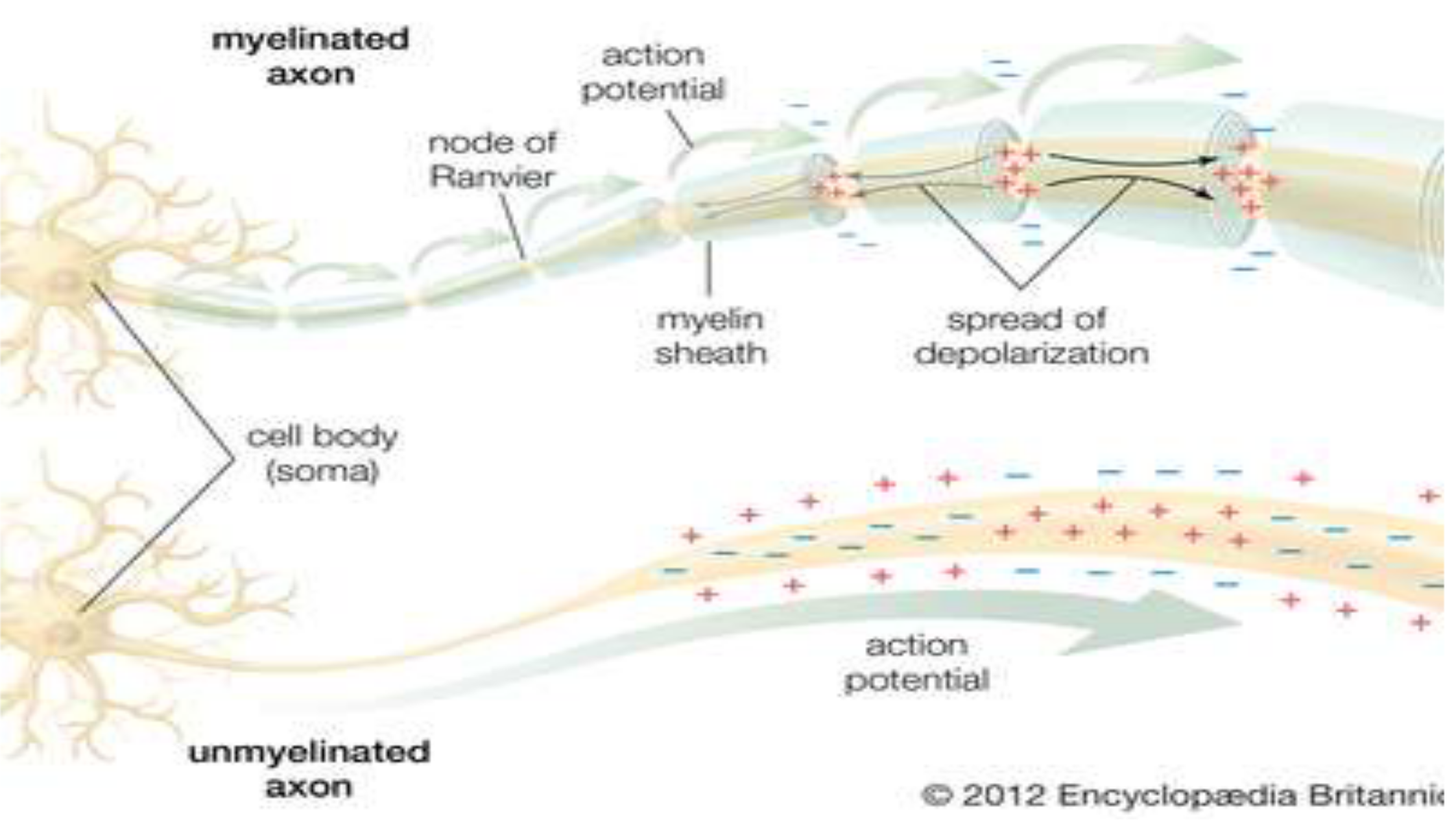
The mechanism by which the signal is transmitted along the cell
membrane is referred to as local current flow or the movement of
positively charged ions.
In the area of a stimulus causing a depolarization, the inside of
the cell becomes positive (less negative) relative to the outside
of the cell.
Because opposite charges attract, the (+) charges in this area are
attracted to and move toward the negative charges on the
adjacent areas of the internal surface of the cell membrane

•
•
•
•
As a result, these adjacent areas become depolarized due to the presence
of these (+) charges.
his process continues and the electrical signal travels along the cell
membrane away from the initial site of the stimulus; however, these graded
or local potentials travel only short distances.
The cell membrane is not well insulated and the current (positive charges)
tends to drift away from the internal surface of the cell membrane.
Consequently, as the signal travels along the membrane, the number of (+)
charges causing the depolarization of the next region of membrane
continually decreases and the magnitude of the depolarization therefore
decreases.
Actereorm
potent
al
—-
40
‘oe
=
—
-g5
Threshold
Fo
Reso
mg
wt
«t=
Stirvastust
Li
a
=
St
os
s
000000
Torre
(rms!
Opening
of
voltage-gated
sodium
channels
and
influx
and
sodium
ions
change
the
membrane
potential
of
a
neuron
during
repolarisation.
The
resting
membrane
potential
is
-55
mW.
Failed
initiations
cannot
generate
action
potentials
because
the
membrane
potential
change
did
not
overcome
the
threshold.
During
refractory
period,
the
membrane
potential
of
the
neuron
is
more
negative
than
the
resting
state.
When
voltage-gated
potassium
channels
open,
potassium
ions
move
out
of
the
cell.
Woltage-gated
sodium
channels
found
mainly
at
the
axon
hillock
open
wery
rapidly
when
there
is
a
depolarizing
stimulus.
o
L
>
|
a
|
action
peafracbhors
potenhal
porod
OO
'
depolarization
—-s
|
7
wembrane
Potential
(mV
|
3
|
threshold
potenbal
?
\
:
,
7,
:
pei
eee
4
——AEOC
area
ee
eee
eae
a
resting
potential
hyparpolarization
rT
0
a
Tr
T
C1
o
1
2
3
4&
& &
F
Time
(milliseconds)
axon
©
2012
Encyclopsedia
Britannix
>
—
+
a4)
=
=
o
=
5
2.
—
=
FO
menial
ca
ch
1
= =
|
=
Tir
+
(rris)
eS
I
Sot
Oo
tio
mm:
Reo
te
ra
til
-
Resting
Poteniial
_
= =
i
Titre
grrs])
Ree
OF
fiom
PS
ra
Tico

•
•
•
The activation gates of the voltage-gated Na+ channels open,
permitting the influx of Na+ ions and further depolarization
toward threshold.
At the threshold potential, all voltage-gated Na+ channels are
open, resulting in the “spike” of the action potential.
Approximately 1 msec after the activation gates open, the
inactivation gates of the Na+ channels close; in addition, the
activation gates of the K+ channels open, resulting in the
repolarization of the neuron.

The mechanism of action of a chemical synapse
•
•
•
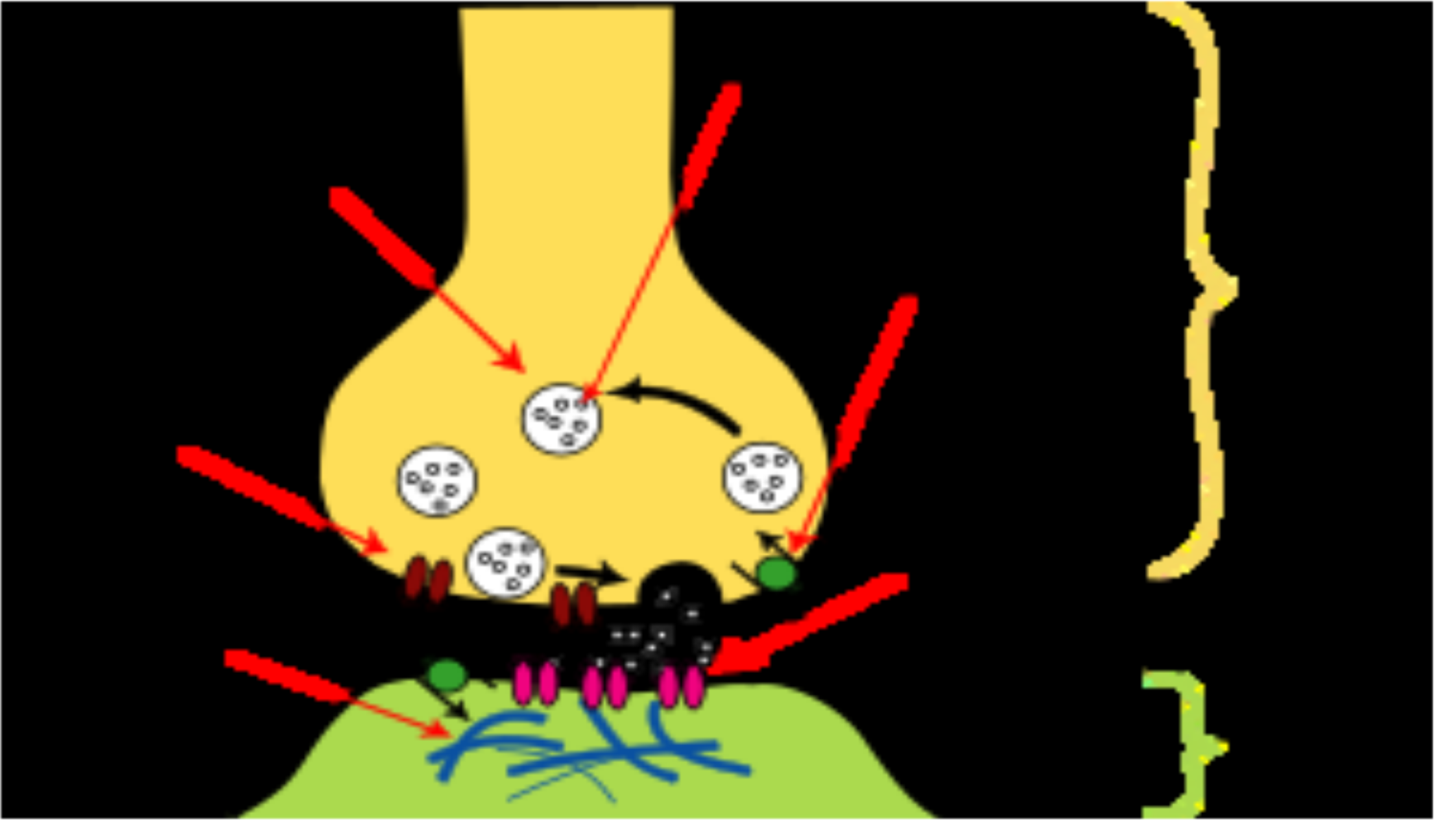
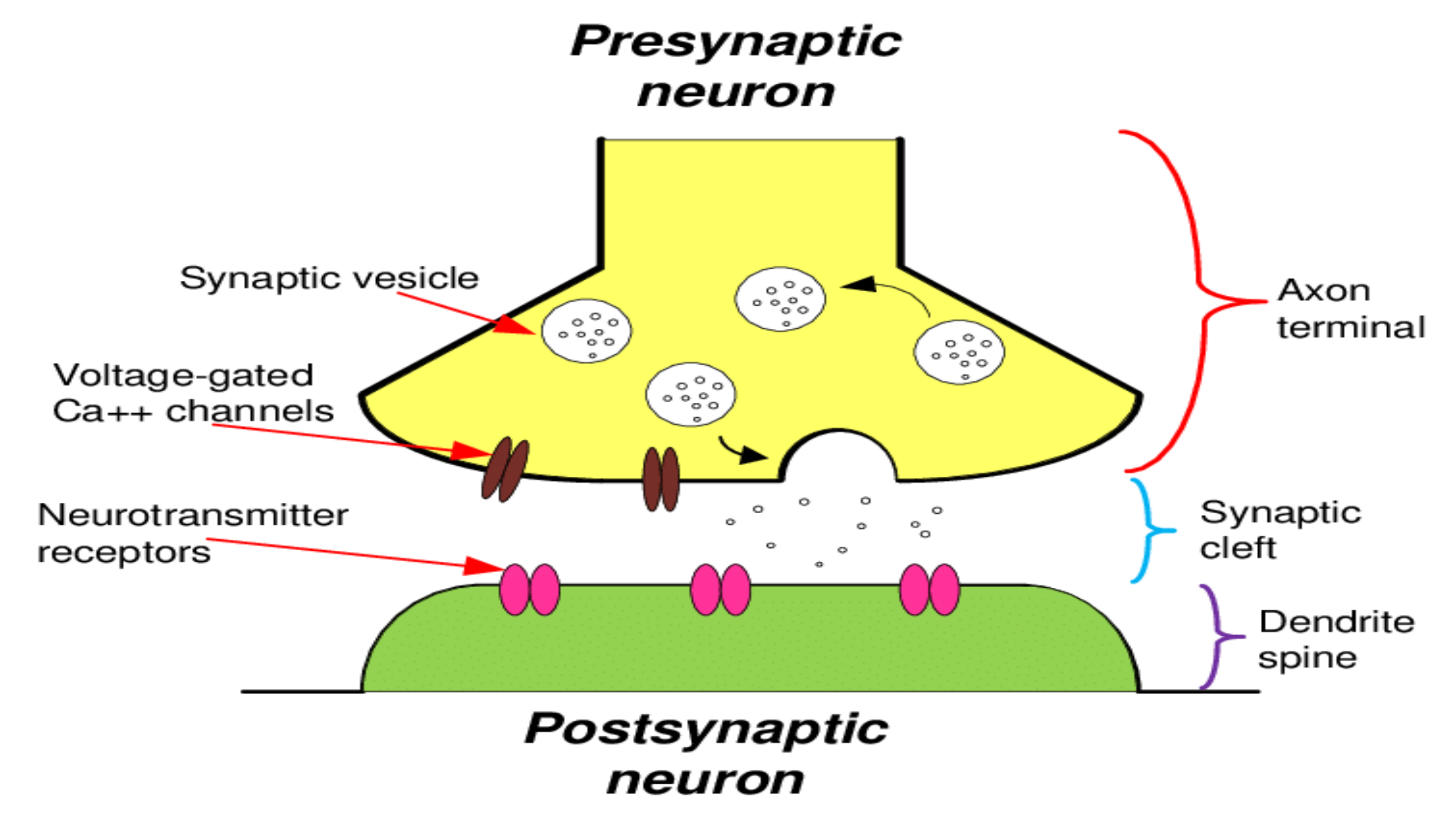
The axon terminal broadens to form a swelling referred to as the
synaptic knob. Within the synaptic knob are many synaptic
vesicles that store the preformed neurotransmitter.
Also found in the membrane of the synaptic knob are voltage-
gated Ca++ channels. When the electrical impulse, or action
potential, has been transmitted along the length of the axon and
reaches the axon terminal, the accompanying change in voltage
causes the voltage-gated Ca++ channels to open.
Because calcium is in greater concentration in the extracellular
flfluid compared to the intracellular flfluid, Ca++ ions enter the
cell down their concentration gradient.

•
•
•
•
The Ca++ ions then induce the release of the neurotransmitter
from synaptic vesicles into the synaptic cleft by causing the
vesicles to fuse with the presynaptic membrane, thereby facilitating
the process of exocytosis.
This change at the synapse is in the form of a graded potential only.
At any given synapse, the change in membrane potential is not
great enough to reach threshold and generate an action potential.
Instead, many graded potentials generated at one or more
synapses are conducted over the cell membrane toward the axon
hillock.
If the depolarization caused by multiple graded potentials added
together is sufficient for the axon hillock to reach threshold, then
an action potential is generated .
_
Neurotransmitters
Synaptie
vesicle
Neurotransmitter
Voltage-
e-uptake
pump
Axon
gated
Cat
terminal
channel
Neuro-
transmitter
receptors
Sunantic
[
Post-synaptic
tt
P
density
4
'
_
Dendritic
|
spine
Presynaptic
neuron
Synaptic
vesicle
Axon
terminal
Voltage-gated
Ca++
channels
Neurotransmitter
°
-
oynaptic
receptors
°
°
cleft
Dendrite
spine
Postsynaptic
neuron
common neurological disorders
i.
ii.
iii.
iv.
v.
vi.
Headaches. Headaches are one of the most common
neurological disorders and can affect anyone at any age. ...
Epilepsy and Seizures. ...
Stroke. ...
ALS: Amyotrophic Lateral Sclerosis. ...
Alzheimer's Disease and Dementia. ...
Parkinson's Disease.
CHEMICAL COMMUNICATION
•
•
This process is mediated by Hormones. Hormones are chemical
messengers that exert a regulatory effect on the cells of the body.
Hormones are classifified into three biochemical categories
i.Steroids
ii.Proteins/peptides
iii.Amines
1. Steroids
•
•
•
•
•
Steroid hormones are produced by the adrenal cortex, testes,
ovaries, and placenta.
Synthesized from cholesterol, these hormones are lipid soluble
therefore, they cross cell membranes readily and bind to
receptors found intracellularly.
However, because their lipid solubility renders them insoluble in
blood, these hormones are transported in the blood bound to
proteins.
Furthermore, steroid hormones are not typically preformed and
stored for future use within the endocrine gland.

Steroids
•
•
Because they are lipid soluble, they could diffuse out of the cells and
physiological regulation of their release would not be possible.
Finally, steroid hormones are absorbed easily by the gastrointestinal tract
and therefore may be administered orally.
2. Protein/peptide hormones
•
•
•
•
•
Are derived from amino acids.
These hormones are preformed and stored for future use in
membrane-bound secretory granules.
When needed, they are released by exocytosis.
Protein/peptide hormones are water soluble, circulate in the
blood predominantly in an unbound form, and thus tend to have
short half-lives.
Because these hormones are unable to cross the cell
membranes of their target tissues, they bind to receptors on the
membrane surface.
Protein/peptide hormones
•
•
•
•
Protein/peptide hormones cannot be administered orally
because they would be digested in the gastrointestinal tract.
Instead, they are usually administered by injection (e.g., insulin).
Because small peptides are able to cross through mucus
membranes, they may be given sublingually or intranasally.
For example, Miacalcin®, the synthetic form of the hormone
calcitonin, is prepared in the form of a nasal spray.
3.Amine hormones
•
•
•
•
•
Amine hormones include the thyroid hormones and the
catecholamines.
The thyroid hormones tend to be biologically similar to the
steroid hormones.
They are mainly insoluble in the blood and are transported
predominantly (>99%) bound to proteins.
As such, these hormones have longer half-lives (triiodothyronine,
T3, = 24 h; thyroxine, T4, = 7 days).
Furthermore, thyroid hormones cross cell membranes to bind
with intracellular receptors and may be administered orally (e.g.,
synthryoid). In contrast to steroid hormones, however, thyroid
hormones have the unique property of being stored
extracellularly in the thyroid gland as part of the thyroglobulin

•
•
•
•
•
The catecholamines are biologically similar to protein/peptide
hormones.
These hormones are soluble in the blood and are transported in
an unbound form.
Therefore, the catecholamines have a relatively short half-life.
Because these hormones do not cross cell membranes, they
bind to receptors on the membrane surface.
Finally, the catecholamines are stored intracellularly in secretory
ranules for future use.
Functional classifification of hormones
•Hormones are classifified into two functional categories:
1.Trophic hormones
2.Nontrophic hormones
Trophic hormones
•
•
•
•
A trophic hormone acts on another endocrine gland to stimulate
secretion of its hormone.
For example, thyrotropin, or thyroid-stimulating hormone (TSH),
stimulates the secretion of thyroid hormones.
Adrenocorticotropin, or adrenocorticotropic hormone (ACTH),
stimulates the adrenal cortex to secrete the hormone cortisol.
Both trophic hormones are produced by the pituitary gland; in
fact, many trophic hormones are secreted by the pituitary.
The pituitary gland is sometimes referred to as the “master
gland” because its hormones regulate the activity of other
endocrine glands.
Nontrophic hormones
•
•
•
A nontrophic hormone acts on nonendocrine target tissues.
For example, parathormone released from the parathyroid glands
acts on bone tissue to stimulate the release of calcium into the
blood.
Aldosterone released from the cortical region of the adrenal
glands acts on the kidney to stimulate the reabsorption of
sodium into the blood.
Hormone interactions
•
•
•
Multiple hormones may affect a single target tissue
simultaneously.
Therefore, the response of the target tissue depends not only on
the effects of each hormone individually, but also on the nature
of the interaction of the hormones at the tissue.
The three types of hormone interactions include:
1)Synergism
2)Permissiveness
3)Antagonism

•
•
•
•
•
Synergism occurs when two hormones interact at the target
tissue such that the combination of their effects is more than
additive.
In other words, their combined effect is greater than the sum of
their separate effects.
For example, epinephrine, cortisol, and glucagon are three
hormones that each increase the level of blood glucose.
The magnitude of their individual effects on glucose levels tends
to be low to moderate.
However, the simultaneous activity of all three hormones results
in an increase in blood glucose that is several times greater than
the sum of their individual effects.
Permissiveness
•
•
•
•
In permissiveness, one hormone enhances the responsiveness
of the target tissue to a second hormone; in other words, the first
hormone increases the activity of the second.
For example, the normal maturation of the reproductive system
requires reproductive hormones from the hypothalamus, pituitary,
and gonads as well as the presence of thyroid hormone.
Although thyroid hormone by itself has no effect on the
reproductive system, if it is absent the development of this
system is delayed.
Therefore, thyroid hormone is considered to have a permissive
effect on the reproductive hormones, facilitating their actions
causing sexual maturation.

Antagonism
•
•
•
•
When the actions of one hormone oppose the effects of another,
the result is antagonism.
For example, insulin decreases blood glucose and promotes the
formation of fat.
Glucagon, on the other hand, increases blood glucose and
promotes the degradation of fat.
Therefore, the effects of insulin and glucagon are antagonistic.
Mechanisms of hormone action
•
•
•
•
The binding of a hormone to its receptor initiates intracellular
events that direct the hormone’s action.
Ultimately, all hormones produce their effects by altering
intracellular protein activity.
However, the mechanism by which this occurs depends on the
location of the hormone receptor.
Receptors are typically located on the cell surface or in the cell
nucleus. As a result, most hormones carry out their effects by
means of two general mechanisms:
i.Signal transduction and second messenger systems
ii.Gene activation
Signal transduction and second messenger systems
•
•
•
•
Protein/peptide hormones and the catecholamines are water-
soluble substances and, accordingly, are unable to cross the
plasma membrane to enter the cell.
Therefore, these hormones must bind to their specifific receptors
on the cell surface.
This receptor binding causes a response within the cell by way
of signal transduction or by the production of intracellular
second messenger molecules.
The original, extracellular hormone is considered the first
messenger because it carried the signal to the target tissue.
Signal transduction and second messenger systems
•
•
•
•
•
•
The most common second messenger activated by protein/
peptide hormones and catecholamines is cyclic adenosine
monophosphate (cAMP).
The pathway by which cAMP is formed and alters cellular
function.
The process begins when the hormone binds to its receptor.
These receptors are quite large and span the plasma membrane.
On the cytoplasmic surface of the membrane, the receptor is
associated with a G protein that serves as the transducer
molecule.
In other words, the G protein acts as an intermediary between the
receptor and the second messengers that will alter cellular
i i Th i f d G i b
Gene action
•
•
•
•
•
Steroid hormones and thyroid hormone carry out their effects by
way of gene activation.
In contrast to the protein/peptide hormones, which alter existing
enzyme activity, these hormones induce the synthesis of new
enzymes that then inflfluence cellular metabolism.
Hormones in this category are lipophilic and easily enter the cells
of the target tissue by diffusing through the plasma membrane.
The hormone continues into the cell nucleus where it binds to its
receptor forming a hormone–receptor complex.
Hormone receptors are also capable of binding to DNA at
specifific attachment sites referred to as hormone response
elements (HRE).

•
•
Each of the steroid hormones binds with its receptor and
attaches to a different HRE.
Binding of the hormone–receptor complex to the DNA activates
specifific genes within the target cell, resulting in the formation
of mRNA molecules.
CIRCULATORY SYSTEM
•
•
•
•
Blood consists of cellular elements (red blood cells, white blood
cells, and platelets) as well as plasma, the fluid in which the blood
cells are suspended.
Normally, total circulating blood volume is about 8% of body weight
(about 5 l in women and 5.5 l in men). Adipose tissue is relatively
avascular and therefore contains little blood compared to other
tissues.
The cellular elements of the blood have a short life span and must
be continuously replaced.
The formation of red blood cells, white blood cells, and platelets,
collectively, is referred to as hematopoiesis. This process takes
place in the red bone marrow. In adults, red bone marrow is found
in the pelvis, ribs, and sternum
Plasma
•
•
•
The fluid portion of the blood, the plasma, accounts for 55 to 60%
of total blood volume and is about 90% water.
The remaining 10% contains proteins (8%) and other substances
(2%) including hormones, enzymes, nutrient molecules, gases,
electrolytes, and excretory products.
All of these substances are dissolved in the plasma (e.g., oxygen)
or are colloidal materials (dispersed solute materials that do not
precipitate out, e.g., proteins).

•The three major plasma proteins include:
i.Albumin
ii.Globulins
iii.Fibrinogen

Blood
cells
and
its
types
with
functions
a
er
#
Lymphocytes
Neutrophil
White
Blood
Cells
Erythrocytes
•
•
•
•
•
•
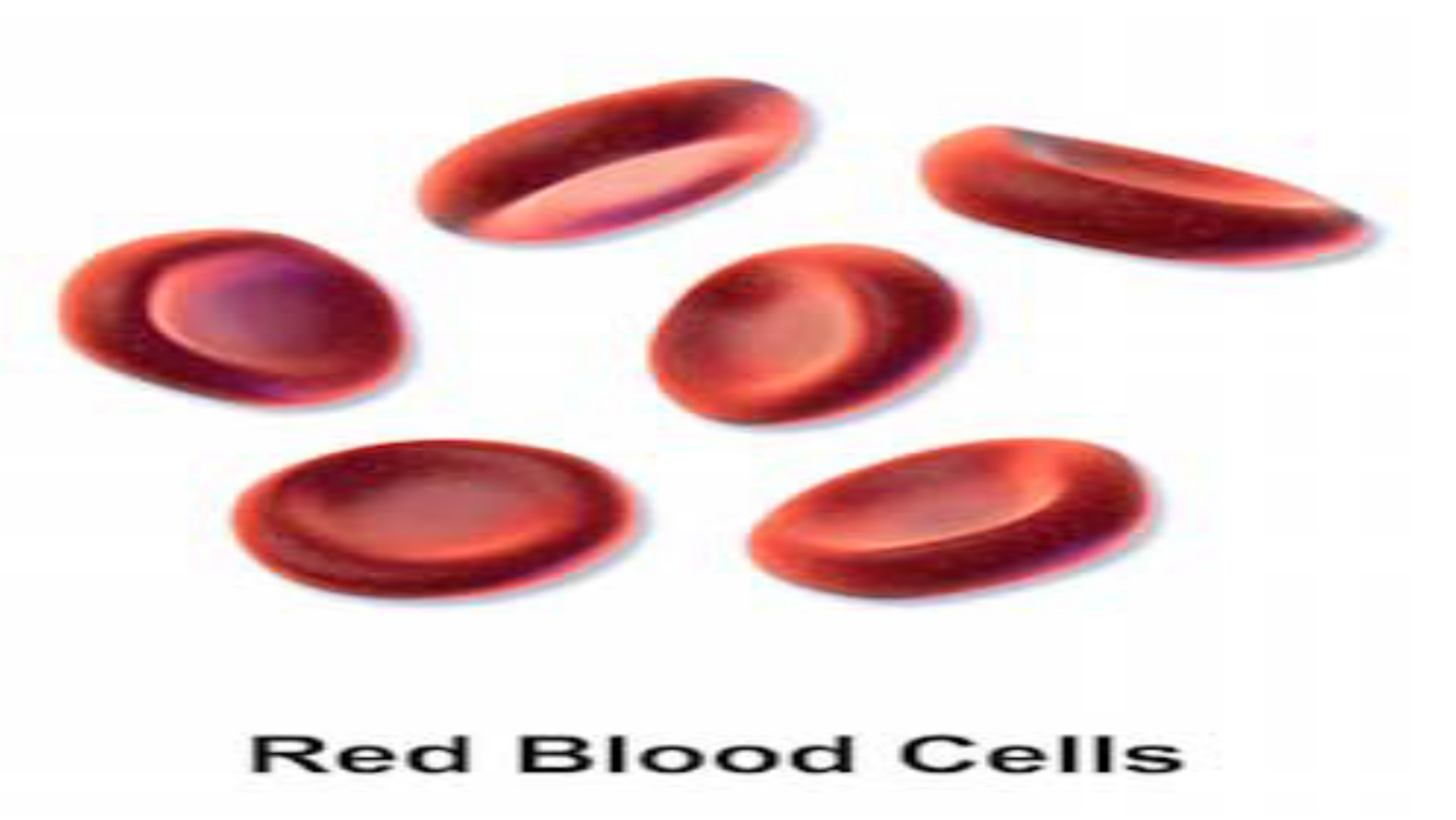
The most numerous of the cellular elements in the blood are the
erythrocytes (red blood cells).
On average, there are 5 million red blood cells per microliter (ml) of
blood, or a total of about 25 to 30 trillion red blood cells in the adult
human body.
The percentage of the blood made up of red blood cells is referred to
as hematocrit.
An average hematocrit is about 45% (42% females, 47% males). As
such, the viscosity of the blood is determined primarily by these
elements.
Transport oxygen from the lungs to the cells of the body.
Pick up carbon dioxide from other tissues and unload it in the lungs.
2
oo
Rredi
Biood
Cells.
Erythrocytes
•
•
•
•
•
•
•
•
•
•
Biconcave disc which is round and flat, sort of like a shallow bowl.
Disk diameter of approximately 6.2-8.2 µm.
They have a thick rim and a thin sunken center.
Nucleus Absent.
Can change shape without breaking.
Production of RBCs is controlled by erythropoietin.
RBC contains hemoglobin (33%).
The iron found in hemoglobin gives the blood its red color.
RBCs cannot repair themselves.
Life span of 120 days.
White Blood Cells (Leukocytes)
•
•
•
•
•
•
Account for only about 1% of the blood.
4500-11,000/mm3
They are the cells that make up the majority of the immune
system.
It is the part of the body that protects itself against foreign
substances and various types of infections.
They are made in the bone marrow from multi-potent cells called
hematopoietic stem cells.
They exist in all parts of the body, including the connective tissue,
lymph system, and the bloodstream.
White Blood Cells (Leukocytes)
•
•
Leukopenia is a low white blood cell count that can be caused by
damage to the bone marrow from things like medications,
radiation, or chemotherapy.
Leukocytosis is a high white blood cell count that can be caused
by a number of conditions, including various types of infections,
inflammatory disease in the body.
White Blood Cells (Leukocytes)
•
•
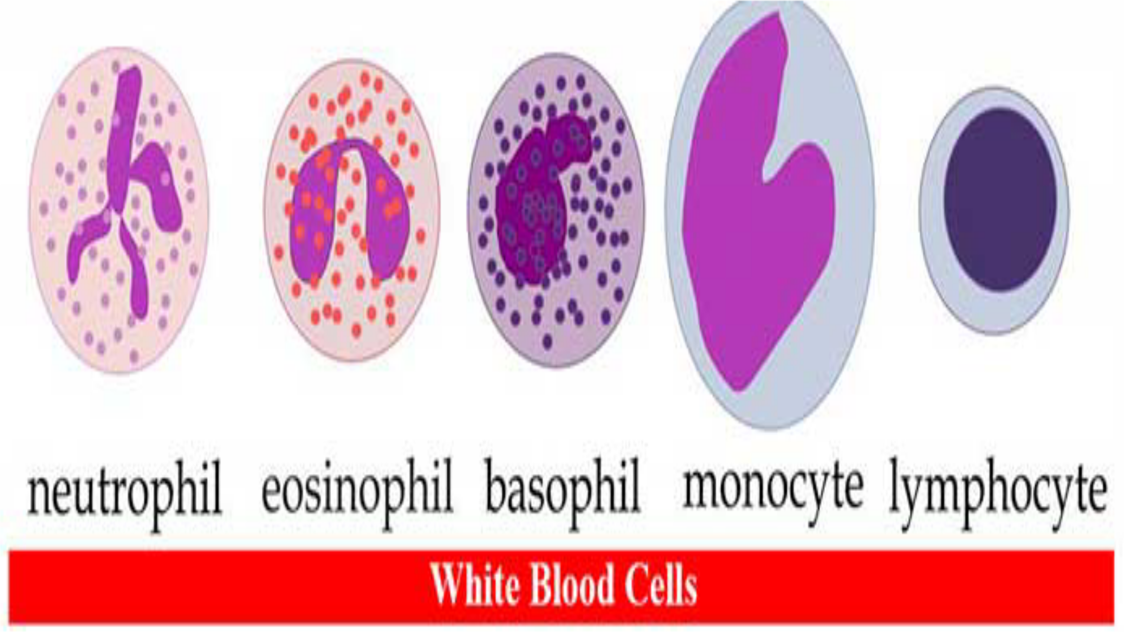
They are divided into Granulocytes (having visible granules or
grains inside the cells) and Agranulocytes (free of visible grains
under the microscope).
There are five main types of WBCs.: Neutrophils (granulocytes),
Eosinophils (granulocytes), Basophils (granulocytes),
Lymphocytes (non-granulocytes) and Monocytes (non-
granulocytes).
White
Blood
Cells

•
•
They are divided into Granulocytes (having visible granules or
grains inside the cells) and Agranulocytes (free of visible grains
under the microscope).
There are five main types of WBCs.: Neutrophils (granulocytes),
Eosinophils (granulocytes), Basophils (granulocytes),
Lymphocytes (non-granulocytes) and Monocytes (non-
granulocytes).
Neutrophils (granulocytes)
•
•
•
•
•
•
•
•
•
Most common type of white blood cell.
Accounts for 62% of Leukocytes
Multi-lobed Nucleus present.
Contain very fine cytoplasmic granules.
2000 to 7500 cells per mm3
Medium-sized white blood cells.
Also called polymorphonuclear (PMN) because they have a
variety of nuclear shapes.
Diameter of 10–12 μm.
Life span of 6 hours to few days.
Functions of neutrophils
Kills bacteria through the process of phagocytosis.
They also release a burst of super oxides that have the ability to
kill many bacteria at the same time
Eosinophils (granulocytes)
•
•
•
•
•
•
•
i.
ii.
40-400 cells per mm3
Have large granules
Nucleus is divided into two lobes (bi-lobed nucleus)
Diameter of 10–12 μm.
Accounts for 2.3%
Life span of 8–12 day
Functions
Kills parasites and have a role in allergic reactions.
Releases toxins from their granules to kill pathogens.
Basophils (granulocytes)
•
•
•
•
•
•
•
0-100 cells per mm3
Colorful when stained and looked at under the microscope
They have a pale nucleus that is usually hidden by granules.
Bi-lobed or Tri-lobed nucleus present.
Diameter of 12–15 μm.
Accounts for 0.4%
Life span of few hours to few days.
Functions of Basophils
i.
ii.
iii.
iv.
Functions in allergic reactions.
Secrete anticoagulants and antibodies that have function
against hypersensitivity reactions in the bloodstream.
Basophils contain histamine, which dilates the vessels to bring
more immune cells to the area of injury.
Secrete heparin which is an anticoagulant that promotes
mobility of other WBCs by preventing clotting.
Lymphocytes (Agranulocytes)
•
•
•
•
•
•
Small rounded cells
Nucleus Present
1300 to 4000 per mm3
Diameter of 7-8 μm (Small) and 12-15 μm (Large)
Accounts for 30%
Life span of years for memory cells and weeks for all else.
Functions of Lymphocytes (Agranulocytes)
i.
ii.
iii.
iv.
v.
T lymphocytes (T cells) are responsible for cell-mediated
immunity.
B lymphocytes are responsible for humoral immunity or
antibody production.
They can recognize and have a memory of invading bacteria
and viruses.
Function in destroying cancer cells.
They present antigens to activate other cells of the immune
system.

Monocytes (Agranulocytes)
•
•
•
•
•
•
•
•
i.
ii.
Largest of the types of white blood cells
Kidney shaped nucleus present.
200 to 800 monocytes per mm3
Turn into macrophages when they exit the bloodstream.
Diameter of 15-30 μm.
Accounts for 5.3%
Life span of few hours to few days.
Functions
Enters the tissue, where they become larger and turn into
macrophages.
Destroy old, damaged and dead cells in the body.
Platelets (Thrombocytes)
•
•
•
•
Nucleus Absent.Platelets (Thrombocytes)
Do not reproduce.
Small fragments of bone marrow cells.
150,000–400,000 platelets in each microliter of human blood.
Functions of Platelets
•
•
•
•
•
•
•
Platelets are the parts of cells that the body uses for clotting.
Helps to promote other blood clotting mechanisms. Example:
Secrete procoagulants (clotting factors) to promote blood clotting.
They secrete vasoconstrictors which constrict blood vessels,
causing vascular spasms in broken blood vessels.
They secrete chemicals that attract neutrophils and monocytes to
sites of inflammation.
Dissolve blood clots when they are no longer needed.
Digest and destroy bacteria.
They secrete growth factors to maintain the linings of blood
vessels.
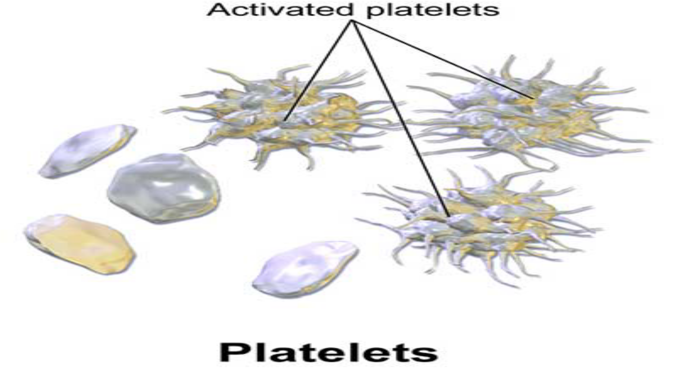
Activated
platelets
Piatelets
Platelets
•
•
•
•
Platelets are essential for many aspects of hemostasis, or the
cessation of blood loss. Several substances are found within the
cytoplasm of platelets that contribute to the arrest of bleeding as
well as vessel repair:
i.Actin and myosin molecules, and thrombosthenin, are
contractile proteins that enable platelets to contract.
ii.Fragments of the endoplasmic reticulum and the golgi
apparatus produce enzymes and store calcium.
iii.Mitochondria and enzyme systems form ATP and ADP.
Platelets
•
•
•
Enzyme systems produce prostaglandins; these are substances
involved with formation of platelet plugs as well as limitation of
clot growth.
Fibrin-stabilizing factor is a protein involved with blood
coagulation.
Growth factor facilitates vascular endothelial cell, vascular
smooth muscle cell, and fifibroblast multiplication and growth,
leading to repair of damaged blood vessels.
Hemostasis of blood
•
The prevention of blood loss from a damaged blood vessel is
referred to as hemostasis. Three inherent mechanisms
contribute to hemostasis:
a.Vascular constriction
b.Formation of platelet plug
c.Blood coagulation
Vascular constriction
•
•
•
. The fifirst mechanism to occur is vascular constriction.
Immediately after a blood vessel is cut or severed, the vascular
smoothmuscle automatically constricts.
This results in a decrease in the flow of blood through the vessel
that helps to limit blood loss.
Formation of a platelet plug
•
•
•
•
•
Formation of a platelet plug physically blocks small holes in
blood vessels.
Normally, platelets are unable to adhere to the endothelial lining
of the blood vessels.
The surface of the platelets contains a coat of glycoproteins that
repels the normal endothelium.
When platelets come into contact with a damaged vascular
surface, in particular collagen fibers in the vessel wall or
damaged endothelial cells, the platelets become activated.
These platelets become “sticky” and adhere to the damaged
tissue.
Coagulation
•
•
The third major step in hemostasis is coagulation, or the
formation of a blood clot.
This complex process involves a series of reactions that result in
formation of a protein fifiber meshwork that stabilizes the
platelet plug. Three essential steps lead to clotting
1.Activation of factor X
2.Conversion of prothrombin into thrombin
3.Conversion of fibrinogen into fibrin
All together, 12 clotting factors are in the plasma. These factors,
which are proteins synthesized in the liver, are normally found
circulating in plasma
THE RESPIRATORY SYSTEM
•
•
•
•
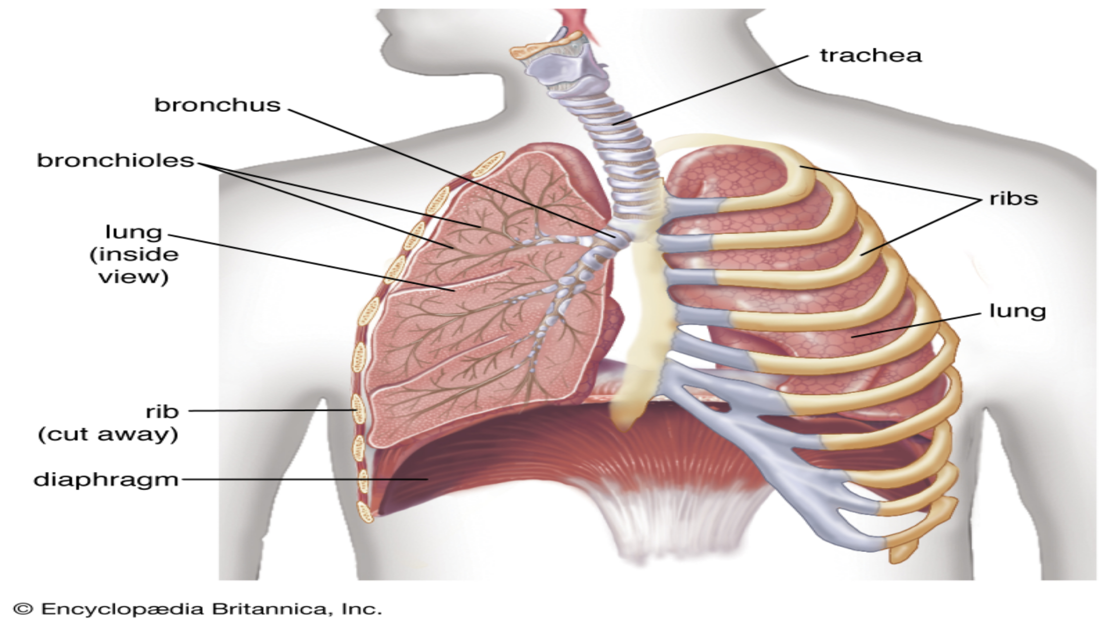
The respiratory system is the network of organs and tissues that
help human beings breathe. It includes the airways, lungs and
blood vessels.
The muscles that power the lungs are also part of the respiratory
system.
These parts work together to move oxygen throughout the body
and clean out waste gases like carbon dioxide.
Each group of parts has many separate components.
trachea
bronchus
bronchioles
lung
(inside
view)
rib
(cut
away)
diaphragm
©
Encyclopzedia
Britannica,
Inc.

NASAL
CAVITY
LARYNX
oe
SEA
LUAA
AN
TRACHEA
SO
NCn
Ee
7
=
Pic
BRONCHIOLES
De
lara
ends
SYSTEM
i
www.visiblebody.com
i
en
:
‘
LUNGS—4
12
y
c
¥
THE RESPIRATORY SYSTEM
•
•
•
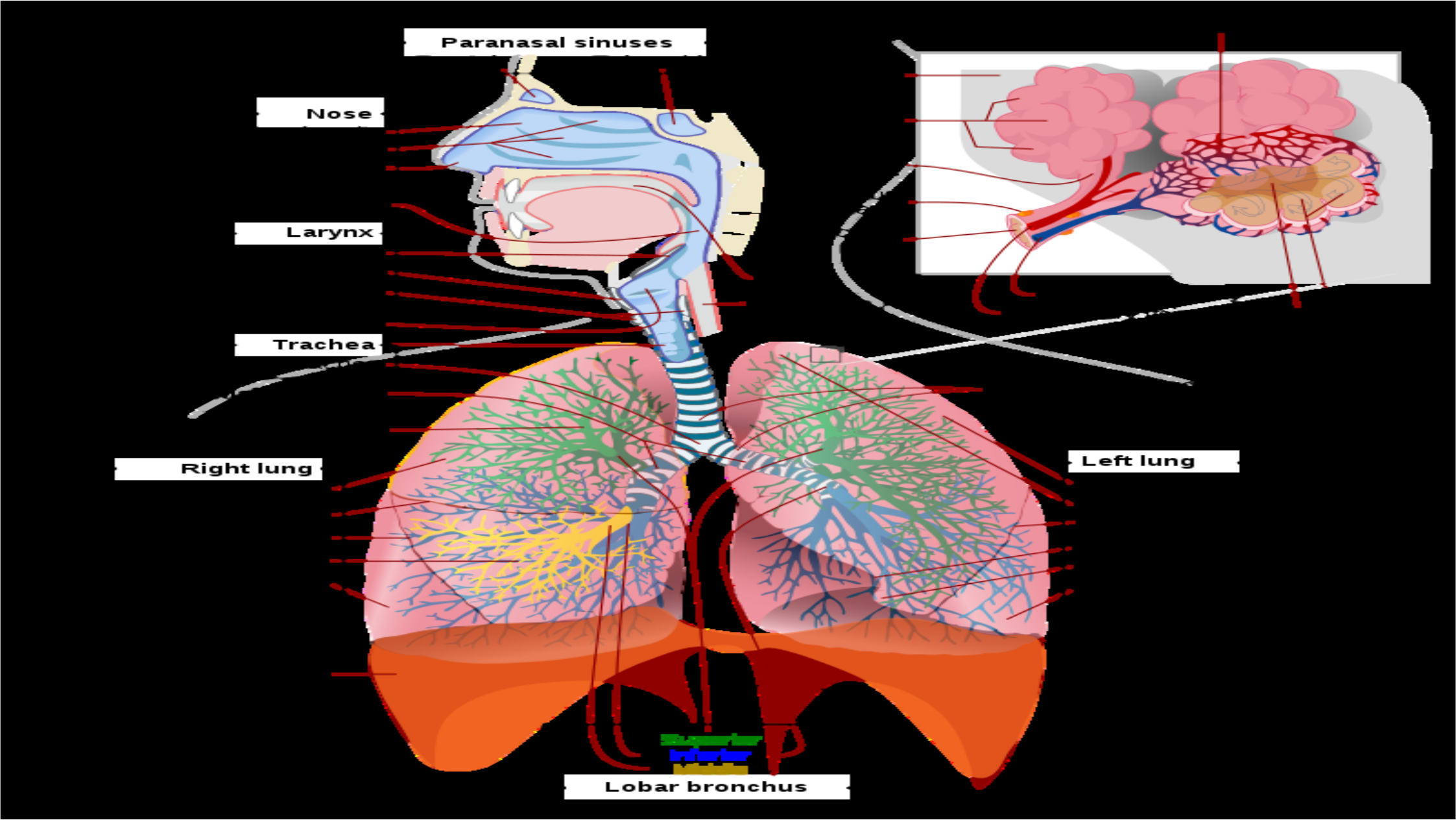
The airways deliver air to the lungs. the airways are a system
that includes :
Mouth and nose:Openings that pull air from outside the body
into the respiratory system.
Sinuses:Hollow areas between the bones in the head that help
regulate the temperature and humidity of the air Human beings
inhale.
THE RESPIRATORY SYSTEM
•
•
•
•
•
•
Pharynx (throat):Tube that delivers air from the mouth and nose
to the trachea (windpipe).
Trachea:Passage connecting the throat and lungs.
Bronchial tubes:Tubes at the bottom of the windpipe that
connect into each lung.
Lungs:Two organs that remove oxygen from the air and pass it
into the blood.
From the lungs, the bloodstream delivers oxygen to all organs
and other tissues.
Muscles and bones help move the air Human beings inhale into
and out of the lungs. Some of the bones and muscles in the
respiratory system include the:
THE RESPIRATORY SYSTEM
•
•
•
Diaphragm:Muscle that helps the lungs pull in air and push it out.
Ribs:Bones that surround and protect the lungs and heart.
When one breathes out, the blood carries carbon dioxide and
other waste out of the body.
THE RESPIRATORY SYSTEM
•
•
•
•
•
•
Other components that work with the lungs and blood vessels
include:
Alveoli:Tiny air sacs in the lungs where the exchange of oxygen
and carbon dioxide takes place.
Bronchioles:Small branches of the bronchial tubes that lead to
the alveoli.
Capillaries:Blood vessels in the alveoli walls that move oxygen
and carbon dioxide.
Lung lobes:Sections of the lungs — three lobes in the right lung
and two in the left lung.
Pleura:Thin sacs that surround each lung lobe and separate the
lungs from the chest wall.
THE RESPIRATORY SYSTEM
•
•
•
•
Some of the other components of the respiratory system include:
Cilia:Tiny hairs that move in a wave-like motion to filter dust
and other irritants out of the airways.
Epiglottis:Tissue flap at the entrance to the trachea that closes
when Human beings swallow to keep food and liquids out of the
airway.
Larynx (voice box):Hollow organ that allows Human beings to
talk and make sounds when air moves in and out.
Paranasal
sinuses
Capilllarny
beds
yf
i
Sy
Frontal
|
Sphenoid
Connective
_
a
tissue
Mose
—
Masal
cawity
-
Masal
conchae
-
Masal
westibule
-
a,
4S
weolhar
_
Secs
Le
A“
bve
olar
Pharyns
Larynx
—
Epiqglottis
Thyroid
cartilage
-+
Cricoid
cartilage
-
Pulmonary
artery
Pulmonary
weinm
Tracheal
and
bronchi
rings
Wocal
folds
——
—_—
Trachea
Carina
of
trachea
_Didin
bronchi
Lingular
division
bronchi
a
Right
lung
—
Left
lung
Superior
lobe
=~
Superior
lobe
.
.
Aes
of
beth
lun
Horizontal
fissure
-
P g
Oblique
fissure
Oblique
fissure
Middle
lobe
Inferior
lobe
.
4
Cardiac
notch
Lingula
of
lung
Inferior
lobe
Ab
we
oll
AS
CrIUMn
Diaphragm
a
\
SUPperiar
S
Inferior
Middle
Llobar
bronchus
—
CONDITIONS AND DISORDERS OF THE THE
RESPIRATORY SYSTEM
•
•
•
Many conditions can affect the organs and tissues that make up
the respiratory system.
Some develop due to irritants Human beings breathe in from the
air, including viruses or bacteria that cause infection. Others
occur as a result of disease or getting older.
Conditions that can cause inflammation (swelling, irritation and
pain) or otherwise affect the respiratory system include:
CONDITIONS AND DISORDERS OF THE THE
RESPIRATORY SYSTEM
a.Allergies:Inhaling proteins, such as dust, mold, and pollen, can
cause respiratoryallergiesin some people. These proteins can
cause inflammation in the airways.
b.Asthma:A chronic (long-term) disorder,asthmacauses
inflammation in the airways that can make breathing difficult.
c.Infection:Infections can lead topneumonia(inflammation of the
lungs) orbronchitis(inflammation of the bronchial tubes).
Common respiratory infections include the flu (influenza) or a cold.
CONDITIONS AND DISORDERS OF THE THE
RESPIRATORY SYSTEM
d.Disease:Respiratory disorders includelung cancerandchronic
obstructive pulmonary diseases (COPD). These illnesses can
harm the respiratory system’s ability to deliver oxygen throughout
the body and filter out waste gases.
e.Aging:Lung capacity decreases as Human beings get older.
f.Damage:Damage to the respiratory system can cause breathing
problems.
Transport of Carbon Dioxide
•
•
•
Carbon dioxide is transported in the blood from the tissue to the
lungs in three ways:
(i) dissolved in solution
(ii) buffered withwater as carbonic acid
(iii) bound to proteins,particularly haemoglobin.
Approximately 75% of carbon dioxide is transport in the red blood
cell and 25% in the plasma. The relatively small amount in
plasma is attributable to a lack of carbonic anhydrase in plasma
so association with water is slow;
Plasma plays little role in buffering and combination with plasma
proteins is poor.
2. Transport of Carbon Dioxide
•
•
•
•
•
•
75% of all CO2is transported through production of HCO3–ions in
the red blood cell. This is explained in the diagram below .
CO2diffuses into the red blood cells and is converted to H+and
HCO3–by an enzyme calledcarbonicanhydrase.
This HCO3–is transported back into the blood via a chloride-
bicarbonate exchanger (aka anion exchanger/AE).
bicarbonate ion diffuses out to the plasma to be exchanged for
chloride ions.
This is known as the chloride shift (Gibbs–Donnan equilibrium or
Hamburger effect). An ion exchange transporter protein in the cell
membrane called Band 3 for Cl– HCO3 – facilitates chloride shift.
The HCO3–can now act as a buffer against any hydrogen in the
blood plasma.
Transport of Carbon Dioxide
•
•
•
A build up of hydrogen ion in the red blood cell would also
prevent further conversion and production of bicarbonate ion.
However, hydrogen ions bind easily to reduced haemoglobin,
which is made available when oxygen is released; therefore,
freehydrogen ions are removed from solution. Reduced
haemoglobinis less acidic than oxygenated haemoglobin.
This is another way of stating the Haldane effect, which explains
that, at any given PCO2, the carbon dioxide content of
deoxygenated blood is greater than that of oxygenated blood.

•
•
As a result of the shift of chloride ions into the red cell and the
buffering of hydrogen ions onto reduced haemoglobin, the
intercellular osmolarity increases slightly and water enters
causing the cell to swell.
This can be measured as an increase in mean corpuscular
volume (MCV). The reverse process occurs as the red blood cell
passes through the lung.
3. Bound to haemoglobin and other proteins
•
•
•
•
Carbon dioxide combines rapidly to the terminal uncharged
amino groups (R-NH2) to form carbamino compounds.
In most proteins, it is only the terminal amino acid group that
combines with carbon dioxide.
Haemoglobin is different when forming carbaminohaemoglobin.
Reduced haemoglobin is the only effective protein buffer of
hydrogen ion at physiological pH because of its high content of
the amino acid histidine.
About 30% of exhaled carbon dioxide is combined with
haemoglobin protein.
Bound to haemoglobin and other proteins
•
•
•
The amount of carbon dioxide held in blood in the carbamino
form is small but it accounts a third of the difference between
venous and arterial carbon dioxide content.
The Haldane effect reflects the difference in carbon dioxide
content between oxygenated and reduced haemoglobin at the
same PCO2.
This effect is partly attributable to the ability of haemoglobin to
buffer hydrogen ions and partly due to the fact that reduced
haemoglobin is 3.5 times more effective in combining with
carbon dioxide than oxyhaemoglobin.
Transport of Oxygen
•
•
•
•
•
Once oxygen has entered the blood from the lungs, it is taken up by
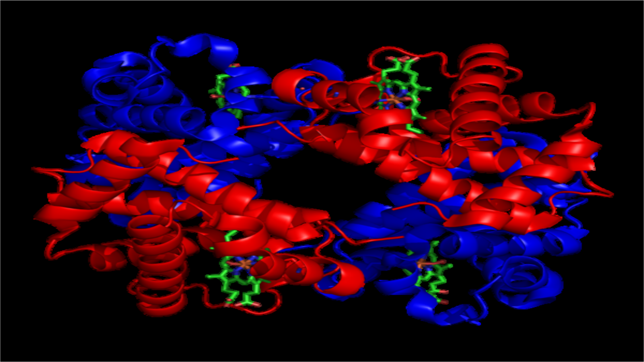
haemoglobin (Hb) in the red blood cells.
Haemoglobin is a protein found in red blood cells that is comprised
of four subunits: two alpha subunits and two beta subunits.
Each subunit has a heme group in the centre that contains iron
and binds one oxygen molecule.
This means each haemoglobin molecule can bind four oxygen
molecules, forming oxyhaemoglobin.
Haemoglobin molecules with a greater number of oxygen
molecules bound are brighter red, hence why oxygenated arterial
blood is brighter red and deoxygenated venous blood is darker red.
Transport of Oxygen
•
•
•
•
•
Haemoglobin changes shape based on how many oxygen
molecules are bound to it.
The change in shape also causes a change in affinity to oxygen.
As the number of oxygen molecules bound to haemoglobin
increases, the affinity of haemoglobin for oxygen increases. This
is known as cooperativity.
When no oxygen is bound, the haemoglobin is said to be in the
Tense State (T-state), with a low affinity for oxygen.
At the point where oxygen first binds, the haemoglobin alters its
shape into the Relaxed State (R-state), which has a higher affinity
for oxygen.

CO POISONING
•
•
•
•
Carbon Monoxide (CO) is a colourless, odourless gas that can be
released from faulty boilers or combustion engines.
Carbon Monoxide poisoning occurs when CO reacts with
haemoglobin at the site of oxygen binding.
Haemoglobin has an affinity for CO that is 210x greater than its
affinity for oxygen.
This means that once carbon monoxide binds to haemoglobin, it
is irreversible.
CO POISONING
•
•
Symptoms of CO poisoning are headache, nausea and tiredness, but
interestingly, respiration rate is usually spared as the partial pressure of
oxygen dissolved in the blood is maintained at normal levels.
Haemoglobin bound to CO has a cherry-red colour and this may be visible
in nails beds and mucous membranes of patients with CO poisoning.
The lymphatic system
•
•
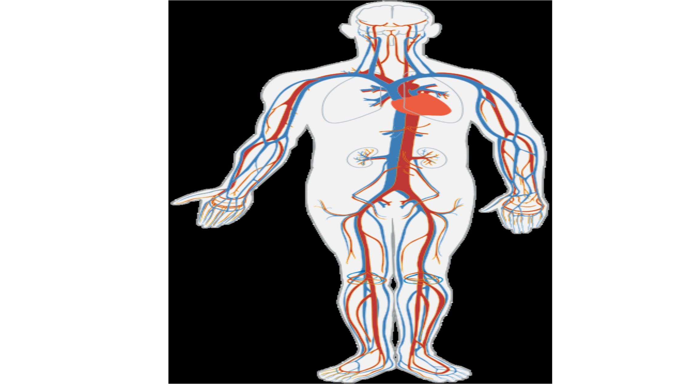
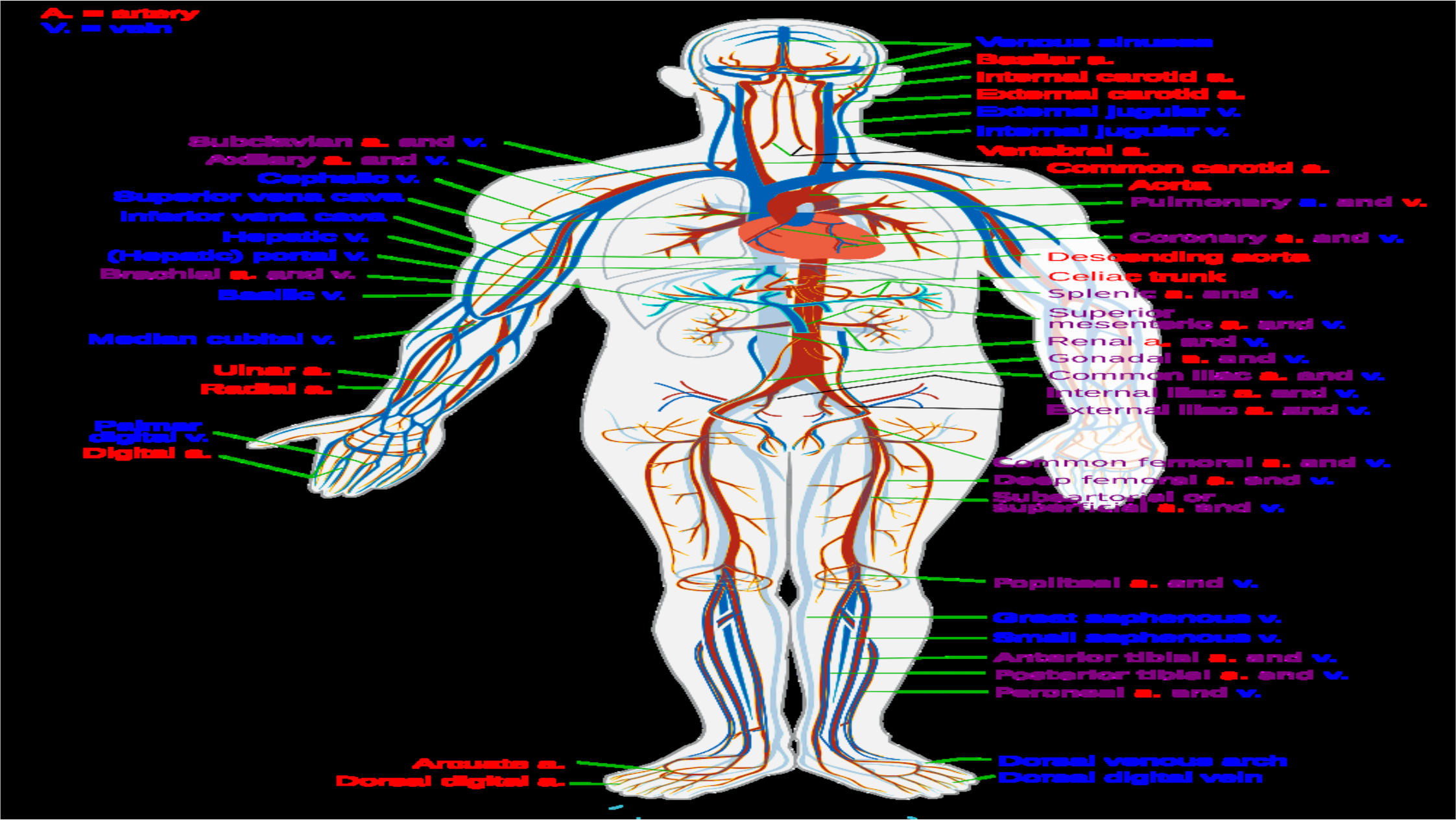
The cardiovascular system consists of the heart, blood vessels,
and blood.
Its primary function is to transport nutrients and oxygen-rich
blood to all parts of the body and to carry deoxygenated blood
back to the lungs.
artery
_
Subclavian
a.
and
wv.
24
=
aes
a.
ama
wv.
WSUS
Sinise:
sS
BasSsilar
za.
Internal
Swe
terna|l|
eSxternal
jugular
wv.
Internal
jerqguilar
w.
WW
rte
bral
=.
Carotid
=.
carotid
aA.
cephalic
~w.
ae
Superior
vena
cava
,
Inferior
vena
cava
:
Hepatic
wv.
‘
CHepatic)
portal
Brachial
a.
amd
~w.
i
>
SOM
mMmeMn
CAretid
aA.
~~.
Orta
>.
Pulmonary
a.
4
Heart
Clorawnary
rmescending
aorta.
and
~w.
ama
«~w_
Celiac
trunk
eSaAaSilic
wv.
Median
cubital
+.
LInar
A.
Radial
a.
Splenic
a.
ama
w.
Superior
—
mesenteric
A.
Ama
w.
_RPeenal
a.
amd
ww.
eS
onadal
a.
and
wv.
:
TS
oemnniaen
thac
Aa
ama
~~.
|
Internal
iliac
Aa.
And
wv.
acoA
SS
ieorsal
digital
ty
Se
=
Me
1
External
iliac
A.
And
w.
|
|
{eo
mi
non
femoral
a.
amd
wv.
reeelp
Femoral
Aa.
amd
wv.
Subsartortal
or
Super
Ficral
a.
ame
+.
Poepliteeal
&a.
anc
w.
CSreat
saphenous
wv.
Small
saphenous
w.
44
mbericr
tibial
a.
and
w.
Posterigr
tibial
a.
and
w.
Peraneal
a.
ama
~w.
SS
Doorsal
venous
arch
Dooarsal
Aigital
veim
Functions of the cardiovascular system
i.
ii.
iii.
iv.
v.
Circulates OXYGEN and removes Carbon Dioxide.
Provides cells with NUTRIENTS.
Removes the waste products of metabolism to the excretory
organs for disposal.
Protects the body against disease and infection.
Clotting stops bleeding after injury.
Parts of the cardiovascular system
i.
ii.
iii.
iv.
v.
vi.
vii.
viii.
Blood.
The heart.
The right side of the heart.
The left side of the heart.
Blood vessels.
Arteries.
Capillaries.
Veins.
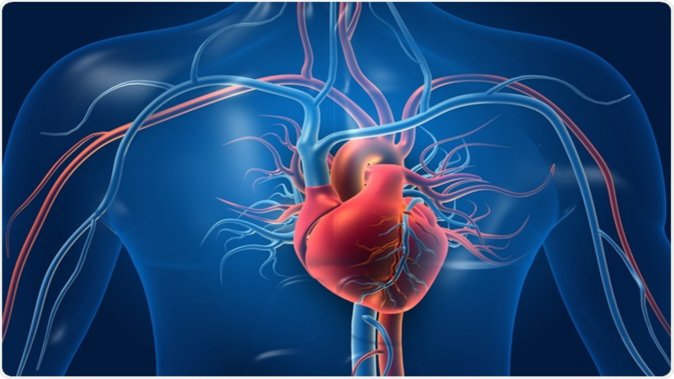
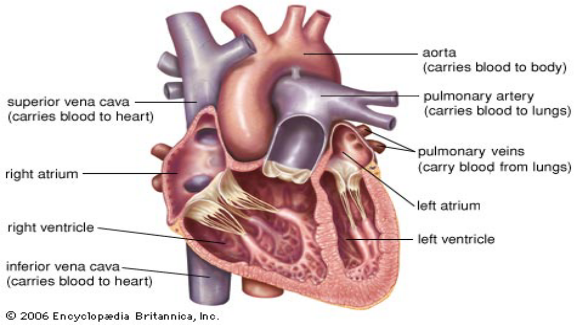
The heart
•
•
•
•
•
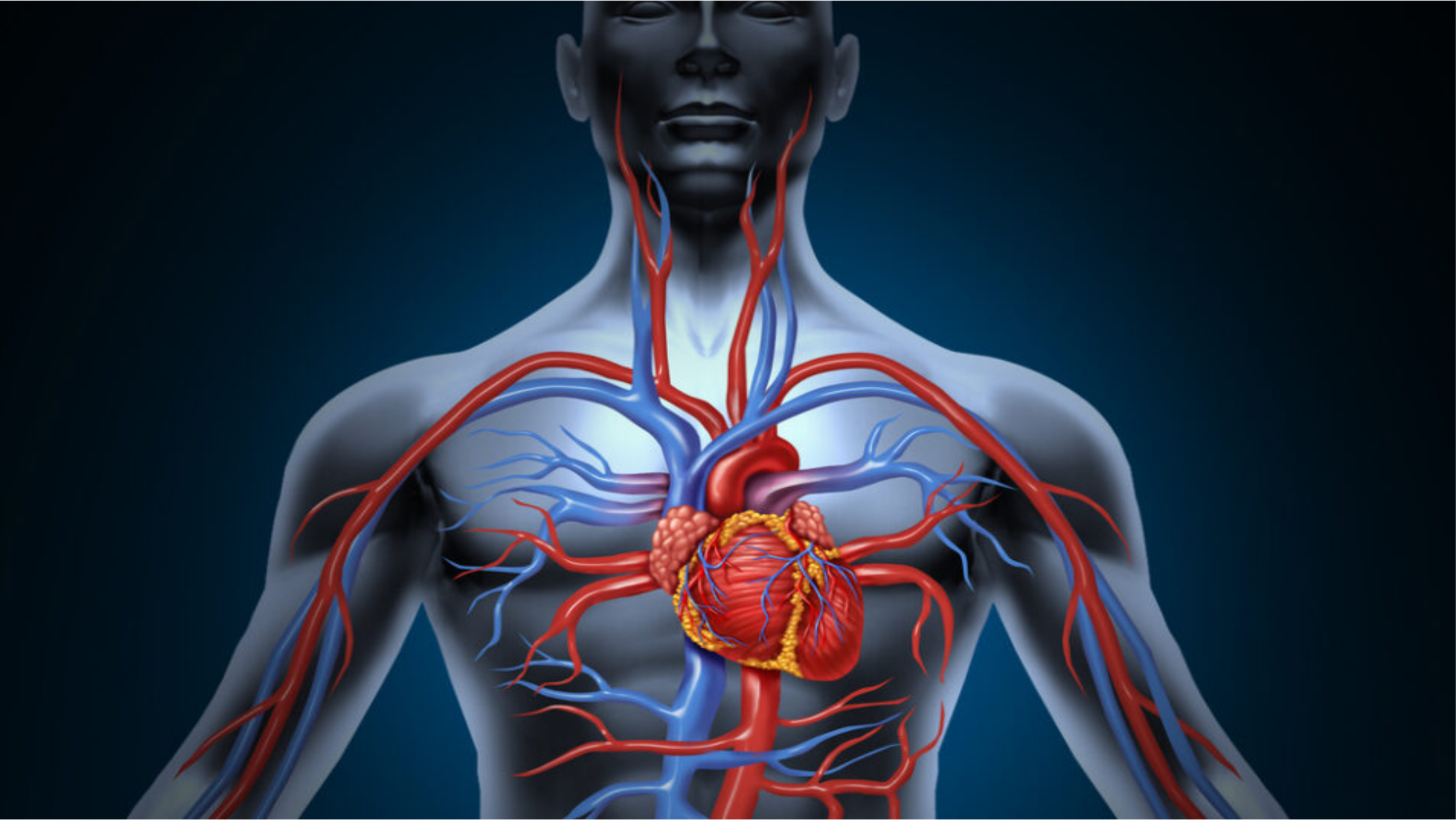
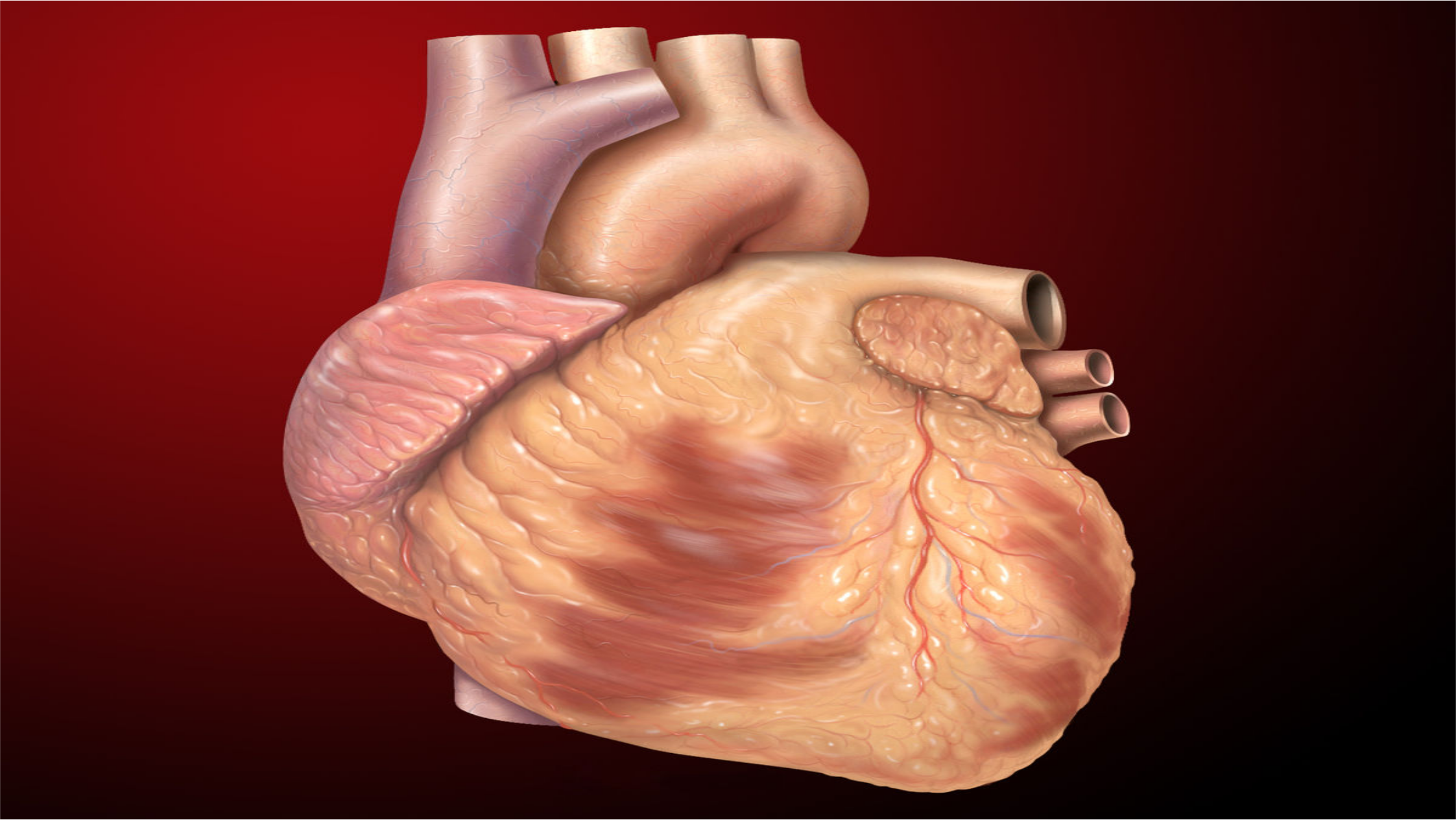
a muscular pumping organ located medial to the lungs along the
body’s midline in the thoracic region.
The bottom tip of the heart, known as its apex, is turned to the
left, so that about 2/3 of the heart is located on the body’s left
side with the other 1/3 on right.
The top of the heart, known as the heart’s base, connects to the
great blood vessels of the body: theaorta, vena cava, pulmonary
trunk, and pulmonary veins.
The heart controls two types of circulation
There are 2 primary circulatory loops in the human body:
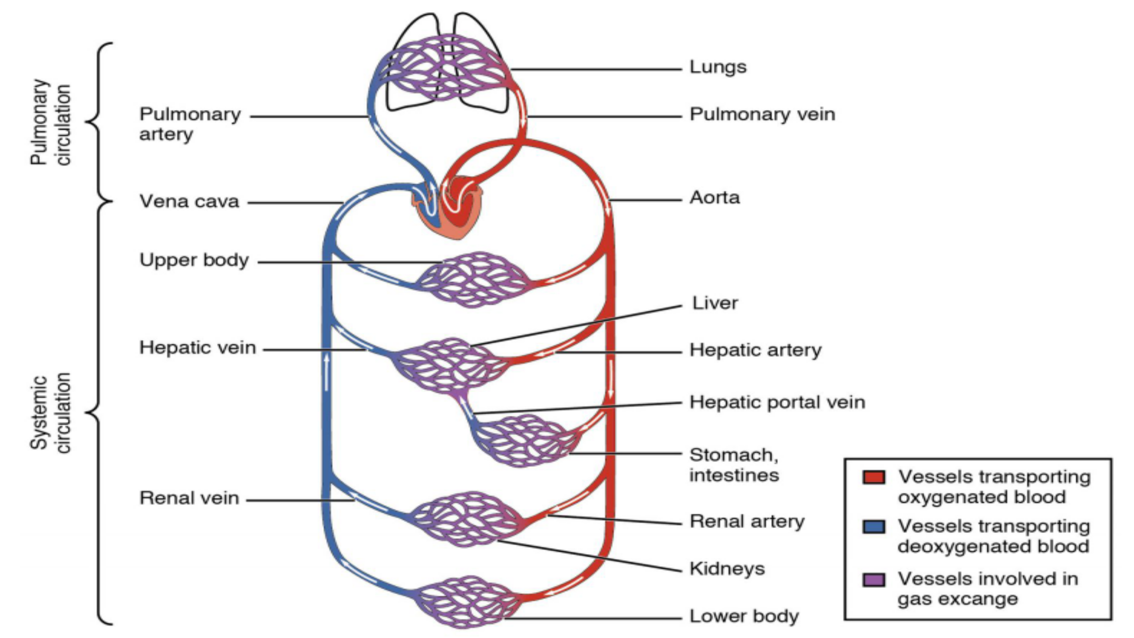
thepulmonary circulation loopand thesystemic circulation loop.

Pulmonary and systemic Circulation
1.Pulmonary circulation transports deoxygenated blood from the
right side of the heart to thelungs, where the blood picks up
oxygen and returns to the left side of the heart.
The pumping chambers of the heart that support the pulmonary
circulation loop are the right atrium and right ventricle.
2.Systemic circulation carries highly oxygenated blood from the
left side of the heart to all of the tissues of the body (with the
exception of the heart and lungs).
Systemic circulation removes wastes from body tissues and
returns deoxygenated blood to the right side of the heart. The left
atrium and left ventricle of the heart are the pumping chambers
for the systemic circulation loop.
Pulmonary
circulation
circulation
systemic
-
Lungs
Pulmonary
artery
Vena
cava
Aorta
Upper
body
Liver
Hepatic
vein
stomach,
intestines
Renal
vein
Renal
artery
Kidneys
Lower
body
Pulmonary
vein
Hepatic
artery
Hepatic
portal
vein
Gi
Vessels
transporting
oxygenated
blood
@
Vessels
transporting
deoxygenated
blood
Ge
Vessels
involved
in
gas
excange
aorta
(carries
blood
to
body)
pulmonary
artery
Su
ior
Vena cava
aie
.
(carnes
blood
to
lungs)
(carries
blood
to
heart)
™~nulmonary
veins
{carry
blood
from
lungs)
right
atrium
lett
atrium
right
ventricle
lett
ventricle
inferior
vena
cava—
(cames
blood
to
heart)
2006
Encyclopedia
Britannica,
Inc.
Blood vessels
•
•
•
•
•
Blood vessels are the body’s highways that allow blood to flow
quickly and efficiently from the heart to every region of the body
and back again.
The size of blood vessels corresponds with the amount of blood
that passes through the vessel.
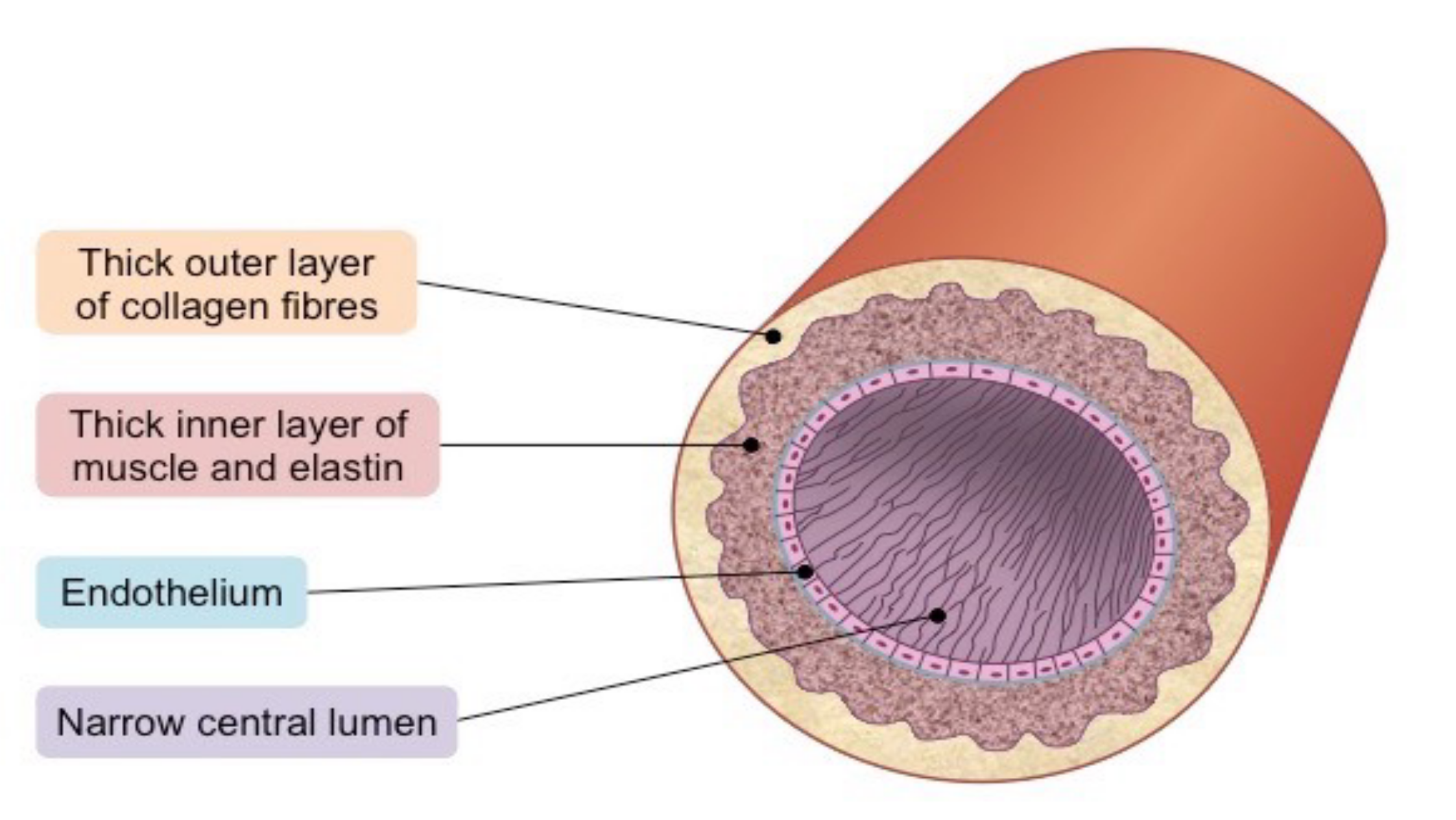
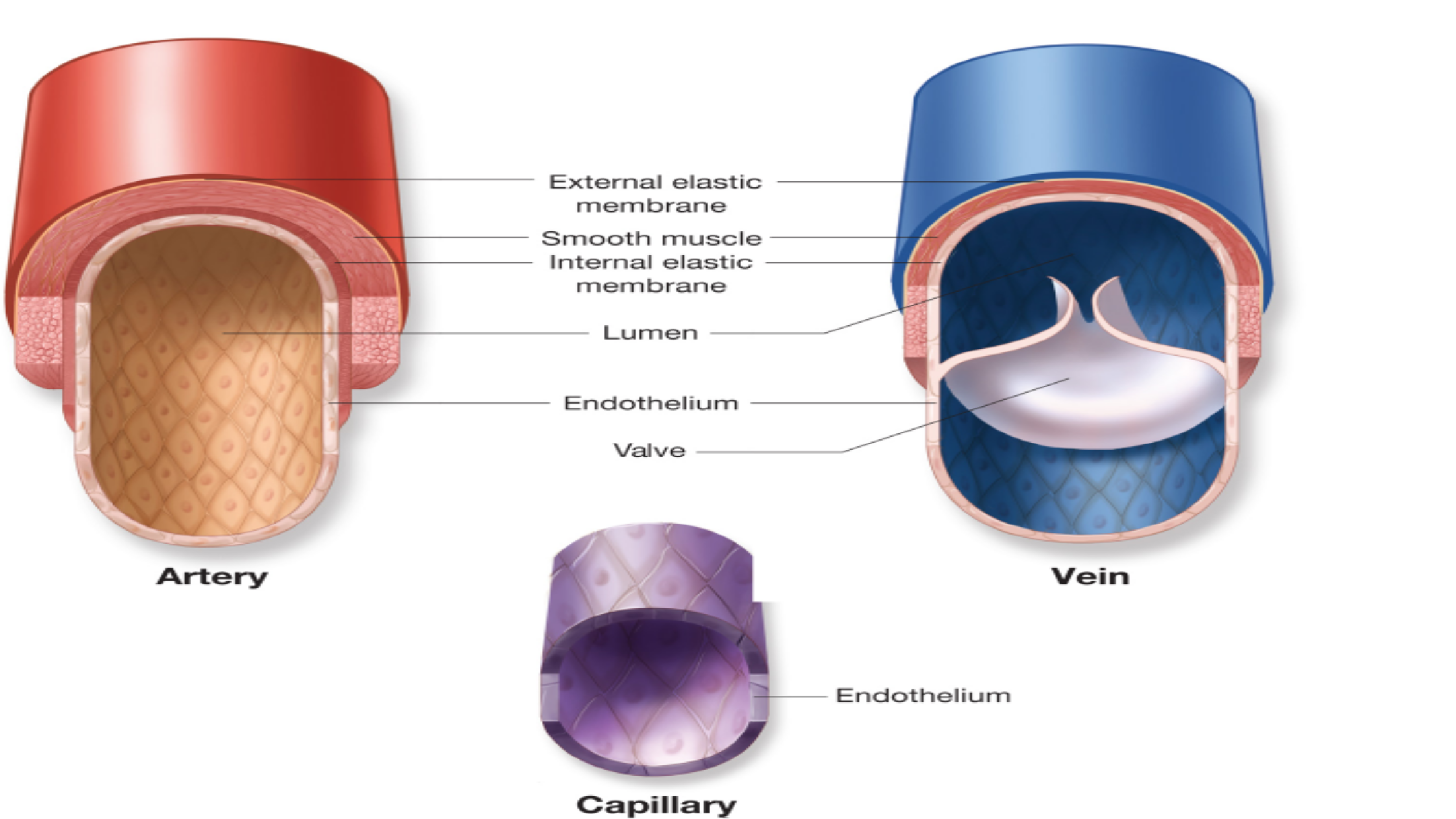
All blood vessels contain a hollow area called the lumen through
which blood is able to flow.
Around the lumen is the wall of the vessel, which may be thin in
the case of capillaries or very thick in the case of arteries.
Allblood vesselsare lined with a thin layer of simple squamous
epithelium known as the endothelium that keeps blood cells
inside of the blood vessels and prevents clots from forming.

•
•
•
•
•
The endothelium lines the entire circulatory system, all the way
to the interior of the heart, where it is called the endocardium.
There are three major types of blood vessels: arteries, capillaries
and veins.
Blood vessels are often named after either the region of the body
through which they carry blood or for nearby structures. For
example, thebrachiocephalic arterycarries blood into the
brachial (arm) and cephalic (head) regions.
One of its branches, the subclavian artery, runs under the clavicle;
hence the name subclavian.
The subclavian artery runs into the axillary region where it
becomes known as the axillary artery.
Arteries and Arterioles
•
•
•
•
•
Arteries are blood vessels that carry blood away from the heart.
Blood carried by arteries is usually highly oxygenated, having just
left the lungs on its way to the body’s tissues.
The pulmonary trunk and arteries of the pulmonary circulation
loop provide an exception to this rule — these arteries carry
deoxygenated blood from the heart to the lungs to be oxygenated.
Arteries face high levels of blood pressure as they carry blood
being pushed from the heart under great force.
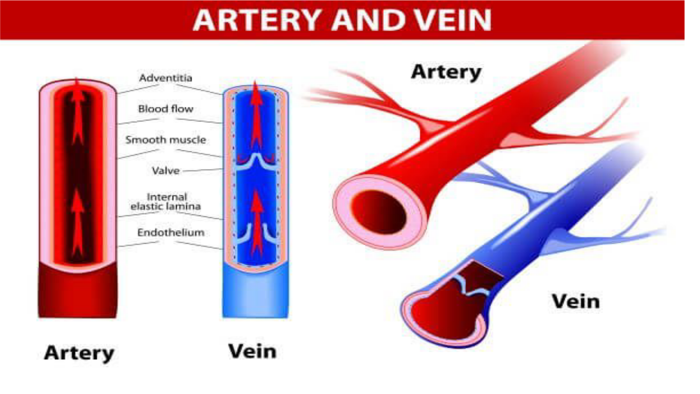
To withstand this pressure, the walls of the arteries are thicker,
more elastic, and more muscular than those of other vessels.
The largest arteries of the body contain a high percentage of
elastic tissue that allows them to stretch and accommodate the
pressure of the heart.

•
•
•
Smaller arteries are more muscular in the structure of their walls.
The smooth muscles of the arterial walls of these smaller
arteries contract or expand to regulate the flow of blood through
their lumen.
In this way, the body controls how much blood flows to different
parts of the body under varying circumstances.
The regulation of blood flow also affects blood pressure, as
smaller arteries give blood less area to flow through and
therefore increases the pressure of the blood on arterial walls.

Arterioles
•
•
•
Arterioles are narrower arteries that branch off from the ends of
arteries and carry blood to capillaries.
They face much lower blood pressures than arteries due to their
greater number, decreased blood volume, and distance from the
direct pressure of the heart.
Thus arteriole walls are much thinner than those of arteries.
Arterioles, like arteries, are able to use smooth muscle to control
their aperture and regulate blood flow and blood pressure.
Capillaries
Thick
outer
layer
of
collagen
fibres
Thick
inner
layer
of
muscle
and
elastin
Narrow
central
lumen
PV
Geka
PAA
Artery
Adventitia
ee
Mood
flaw
Smooch
muscle
Valve
————"}5
Internal
|
elastic
lamina
:
—
Ta
Endothelium
Artery
Vein
External
elastic
membrane
Smooth
muscle
Internal
elastic
membrane
Lumen
——
Endothelium
Valve
._
Endothelium
Capillary

Capillaries
•
•
•
•
•
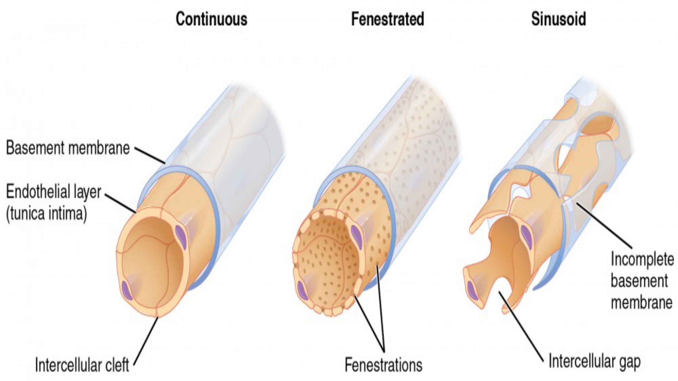
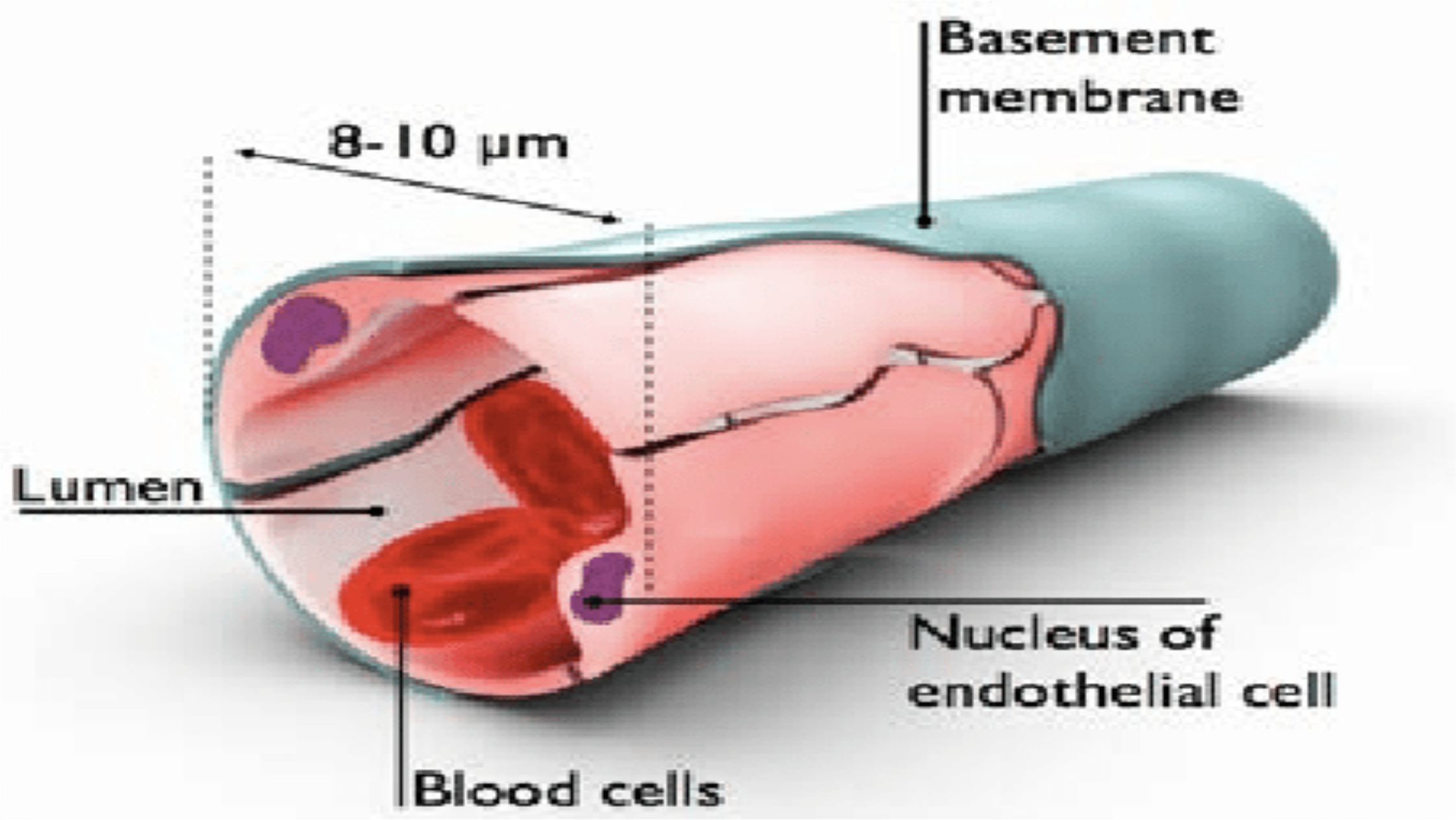
Capillaries are the smallest and thinnest of the blood vessels in
the body and also the most common.
They can be found running throughout almost every tissue of the
body and border the edges of the body’s avascular tissues.
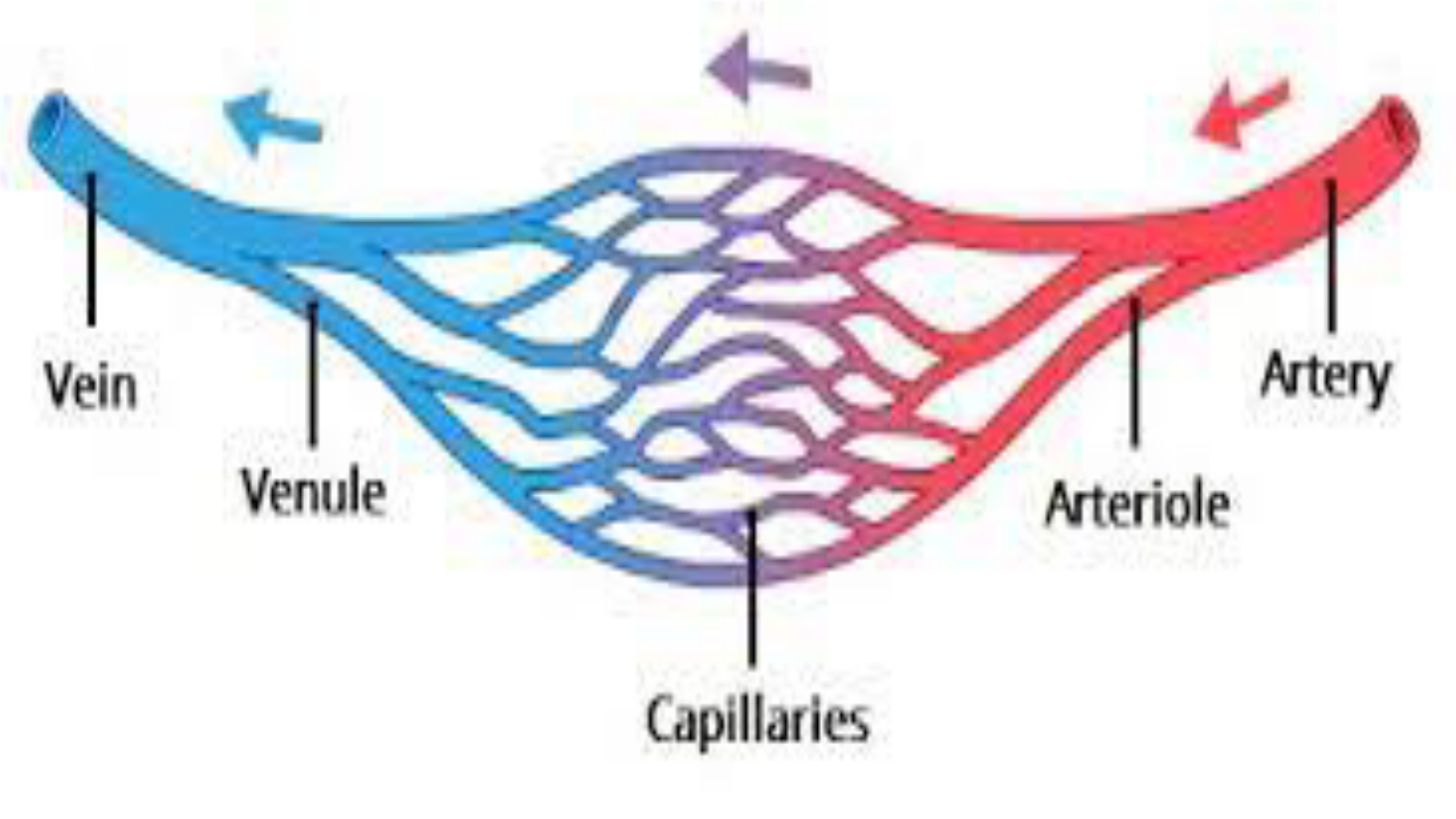
Capillariesconnect to arterioles on one end and venules on the
other.
Capillaries carry blood very close to the cells of the tissues of the
body in order to exchange gases, nutrients, and waste products.
The walls of capillaries consist of only a thin layer of
endothelium so that there is the minimum amount of structure
possible between the blood and the tissues.

Capillaries
•
•
•
•
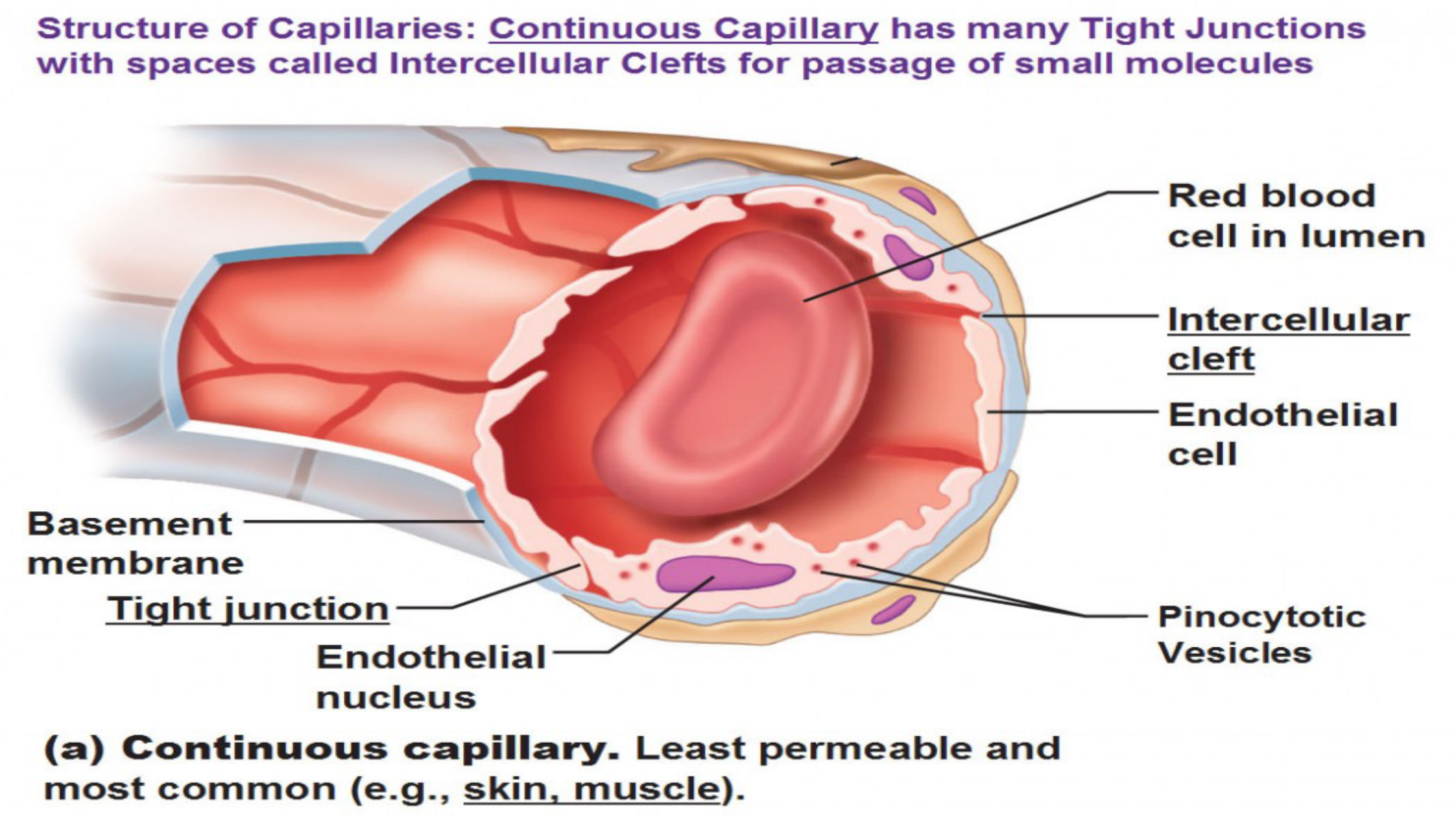
The endothelium acts as a filter to keep blood cells inside of the
vessels while allowing liquids, dissolved gases, and other
chemicals to diffuse along their concentration gradients into or
out of tissues.
Precapillary sphincters are bands of smooth muscle found at the
arteriole ends of capillaries.
These sphincters regulate blood flow into the capillaries.
Since there is a limited supply of blood, and not all tissues have
the same energy and oxygen requirements, the precapillary
sphincters reduce blood flow to inactive tissues and allow free
flow into active tissues.
Continuous
Fenestrated
Sinusoid
Basement
membrane
Nf
Endothelial
layer.
.
(tunica
intima)
g
Incomplete
basement
membrane
Intercellular
gap
Intercellular
cleft
Fenestrations
Basement
membrane
Mucleus
of
endothelial
cell
Blood
cells
Structure
of
Capillaries:
Continuous
Capillary
has
many
Tight
Junctions
with
spaces
called
Intercellular
Clefts
for
passage
of
small
molecules
Red
blood
cell
in
lumen
Intercellular
cleft
9
=
_
2
Endothelial
Fe
/
cell
Basement
membrane
Tight
junction
Pinocytotic
Endothelial
Vesicles
nucleus
(a)
Continuous
capillary.
Least
permeable
and
most
common
(e.g.,
skin,
muscle).
Veins and Venules
•
•
•
•
Veins are the large return vessels of the body and act as the
blood return counterparts of arteries.
Because the arteries, arterioles, and capillaries absorb most of
the force of the heart’s contractions, veins and venules are
subjected to very low blood pressures.
This lack of pressure allows the walls of veins to be much
thinner, less elastic, and less muscular than the walls of arteries.
Veins rely on gravity, inertia, and the force of skeletal muscle
contractions to help push blood back to the heart.
Veins and Venules
•
•
•
•
•
To facilitate the movement of blood, some veins contain many
one-way valves that prevent blood from flowing away from the
heart.
As skeletal muscles in the body contract, they squeeze nearby
veins and push blood through valves closer to the heart.
When the muscle relaxes, the valve traps the blood until another
contraction pushes the blood closer to the heart.
Venules are similar to arterioles as they are small vessels that
connect capillaries, but unlike arterioles, venules connect to
veins instead of arteries.
Venules pick up blood from many capillaries and deposit it into
larger veins for transport back to the heart.
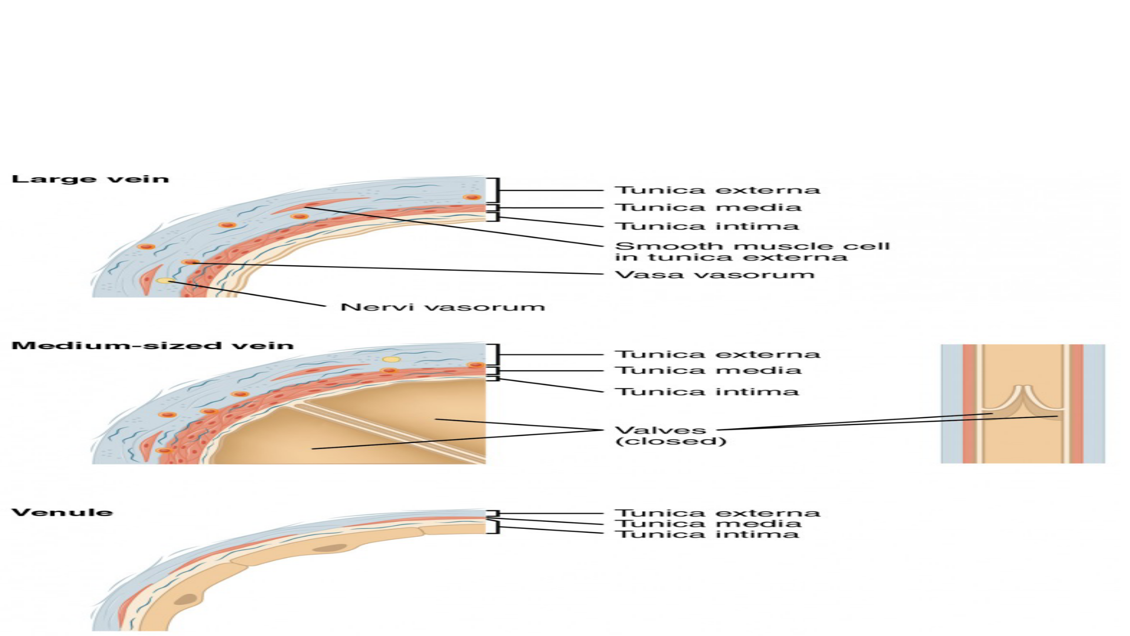
Lares
SS
Ve
Si
——
|
2
=
hai
cae
oe
tS
rma
——
—<bee
nic
aS
OM
eS
sdiS
Temi
SS
imetirn®n
aS
Ss
oe
Ma
Se
eS
oe
=
mm
tunica
Om
tern
a
Wa
Sa
ea
SO
re
Mare
wa
Sao
root
Ws
eB
ie
es
oe
ee
=
—
==
rau
iica
©&xterna
SS
SS
:
=
runMmica
rmmecdia
—
—
EE
ae
Thirties
irmtimmna
=
aS
C4
=
o
VWalwes
A
Cc
Ss
&
cl»
%
(\
al
ae
oe
oe
rics
Om
te
rn
SS
ha
ie
=a
Mm
eS
Sie
Thuinmiaca
mmstinrnmn=aa

LYMPHATIC SYSTEM
•
•
•
•
•
Lymph is a clear and colorless fluid; the word "lymph" comes from the
Latin wordlympha, which means "connected to water.
Lymphatic system, a subsystem of thecirculatory systemin
thevertebratebody that consists of a complex network of vessels,tissues,
andorgans.
The lymphatic system helps maintainfluid balancein the body by
collecting excess fluid and particulate matter from tissues and depositing
them in the bloodstream.
It also helps defend the body against infection by supplying disease-
fightingcellscalledlymphocytes.
The lymphatic system is a network of tissues and organs that help rid the
body of toxins, waste and other unwanted materials.
LYMPHATIC SYSTEM
•
•
•
•
•
The primary function of the lymphatic system is to transport lymph, a
fluid containing infection-fighting white blood cells, throughout the
body.
The lymphatic system primarily consists of lymphatic vessels, which
are similar to the veins and capillaries ofthe circulatory system.
The vessels are connected to lymph nodes, where the lymph is
filtered.
The tonsils, adenoids,spleenand thymus are all part of the lymphatic
system.
Thespleen, which is located on the left side of the body just above the
kidney, is the largest lymphatic organ, acts as a blood filter; it controls
the amount of red blood cells and blood storage in the body, and helps
to fight infection.
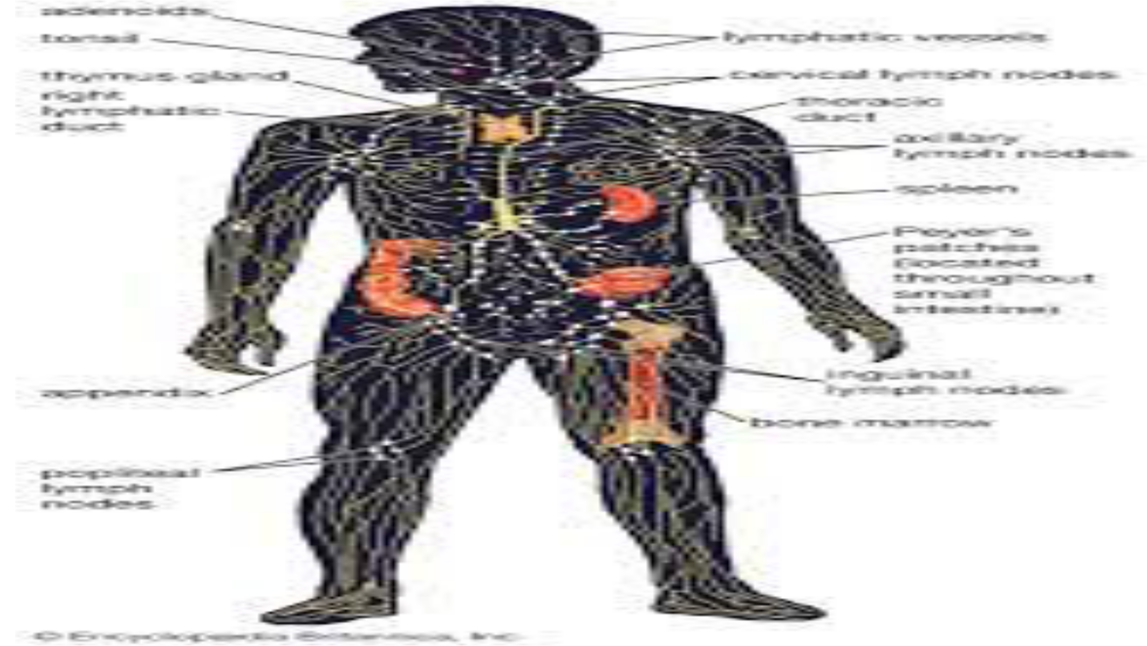
7:
=
ee
=e
ne
ee
ae
ee
a
oe
es
Le
ee
ee
eS
i
es
ee
Se
ae
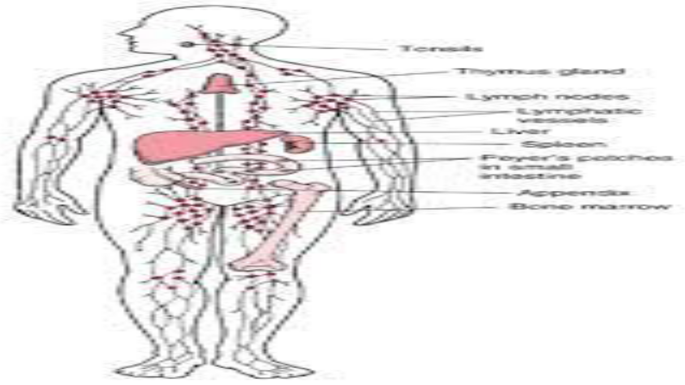
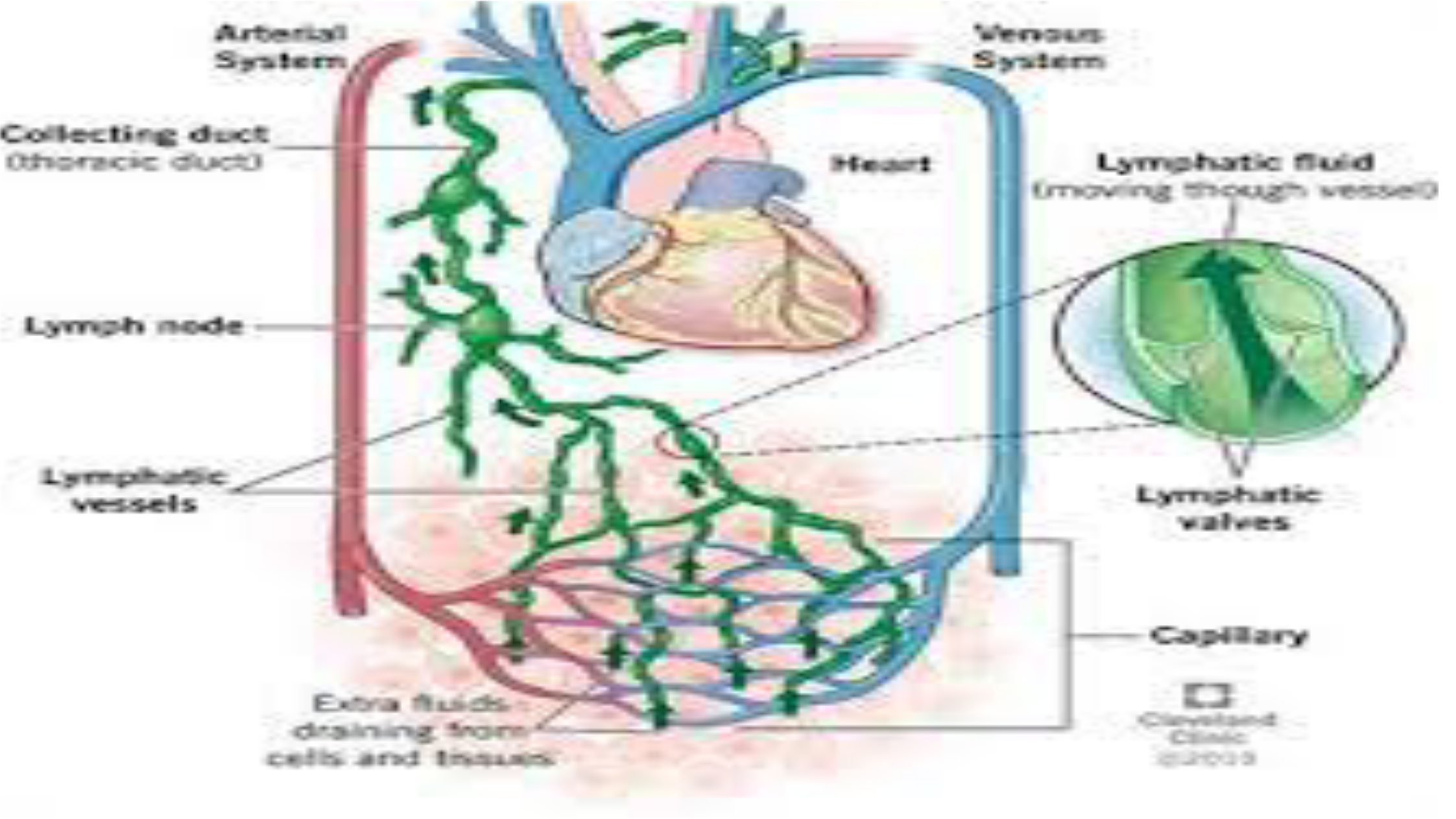

Spleen
•
•
•
If the spleen detects potentially dangerous bacteria, viruses, or
other microorganisms in the blood, it — along with the lymph
nodes — creates white blood cells called lymphocytes, which act
as defenders against invaders.
The lymphocytes produce antibodies to kill the foreign
microorganisms and stop infections from spreading.
Humans can live without a spleen, although people who have
lost their spleen to disease or injury are more prone to infections.
Thymus and Tonsils
•
•
•
•
The thymus is located in the chest just above the heart.
This small organ stores immature lymphocytes (specialized
white blood cells) and prepares them to become active T cells,
which help destroy infected or cancerous cells.
Tonsils are large clusters of lymphatic cells found in the pharynx.
They sample bacteria and viruses that enter the body through the
mouth or nose. it is still among the most common operations
performed and typically follows frequent throat infections.

Formation of Lymph
•
•
•
•
•
Plasma leaves the body's cells once it has delivered its nutrients
and removed debris.
Most of this fluid returns to the venous circulation through tiny
blood vessels called venules and continues as venous blood.
The remainder becomes lymph.
Unlike blood, which flows throughout the body in a continue loop,
lymph flows in only one direction — upward toward the neck.
Lymphatic vessels connect to two subclavian veins, which are
located on either sides of the neck near the collarbones, and the
fluid re-enters the circulatory system.
Diseases and disorders of the lymphatic system
•
•
•
•
Diseases and disorders of the lymphatic system are typically
treated by immunologists.
Vascular surgeons, dermatologists, oncologists and physiatrists
also get involved in treatment of various lymphatic ailments.
There are also lymphedema therapists who specialize in the
manual drainage of the lymphatic system.
The most common diseases of the lymphatic system are
enlargement of the lymph nodes (also known
aslymphadenopathy), swelling due to lymph node to lymph node
blockage (also known aslymphedema) and cancers involving the
lymphatic system.
Diseases and disorders of the lymphatic system
•
•
•
When bacteria are recognized in the lymph fluid, the lymph nodes
make more infection-fighting white blood cells, which can cause
swelling.
The swollen nodes can sometimes be felt in the neck, underarms
and groin.
Lymphadenopathy is usually caused by infection, inflammation,
or cancer. Infections that cause lymphadenopathy include
bacterial infections such as strep throat, locally infected skin
wounds, or viral infections such as mononucleosis or HIV
infection.

THE SKELETAL SYSTEM
•The skeletal system is Human beings body’s central framework.
It consists of bones and connective tissue, including cartilage,
tendons, and ligaments. It’s also called the musculoskeletal
system.
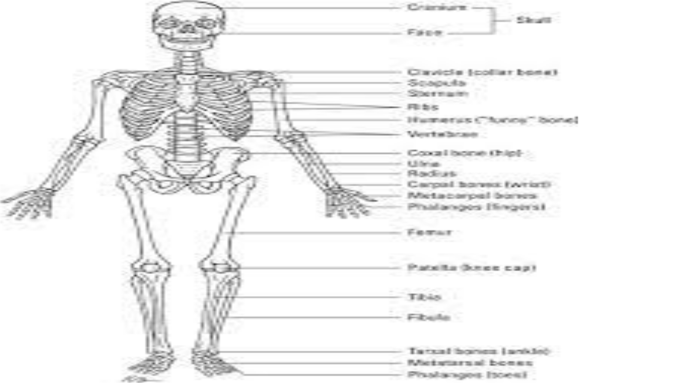
Pid
3
i
au
La
i
"
vt
(s
ie
m
Brey
Ae
a
gh
AU
STALL
fat
|
AE
te
mish
Pte
ns
Nis
othe
—
May
fi
—
Pe
cco
Sere
|
ee
oe
=
He
IrpeTaet-ss
fi
Pl
i
hh
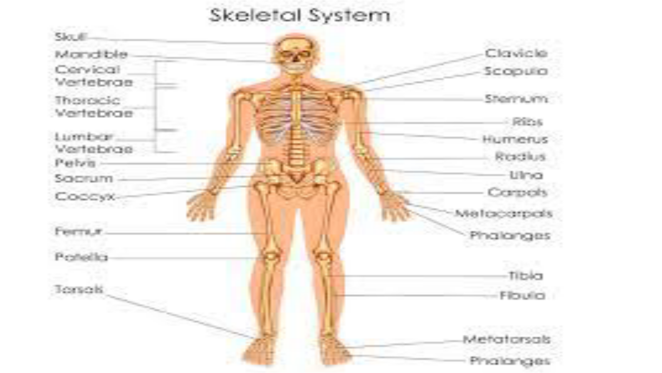
Skeletal
System

FUNCTIONS OF SKELETAL SYSTEM
•
•
•
•
•
The skeletal system has many functions. Besides giving us our
human shape and features, it:
i.Allows movement:Human beingsr skeleton supports Human
beingsr body weight to help Human beings stand and move.
Joints, connective tissue and muscles work together to make
Human beingsr body parts mobile.
ii.Produces blood cells:Bones contain bone marrow. Red and
white blood cells are produced in the bone marrow.
iii.Protects and supports organs:Human beingsr skull shields
Human beingsr brain, Human beingsr ribs protect Human beings
heart and lungs, and Human beingsr backbone protects Human
beingsr spine.
iv.Stores minerals:Bones hold Human beingsr body’s supply of

Parts of the skeletal system
•
•
•
•
•
The skeletal system is a network of many different parts that
work together to help Human beings move.
The main part of Human beingsr skeletal system consists of
Human beingsr bones, hard structures that create Human
beingsr body’s framework — the skeleton.
There are 206 bones in an adult human skeleton. Each bone has
three main layers:
Periosteum:The periosteum is a tough membrane that covers
and protects the outside of the bone.
Compact bone:Below the periosteum, compact bone is white,
hard, and smooth. It provides structural support and protection.

Parts of the Skeletal system
•
•
•
•
Spongy bone:The core, inner layer of the bone is softer than
compact bone. It has small holes called pores to store marrow.
The other components of Human beings skeletal system include:
Cartilage:This smooth and flexible substance covers the tips of
Human beingsr bones where they meet. It enables bones to
move without friction (rubbing against each other). When
cartilage wears away, as in arthritis, it can be painful and cause
movement problems.
Joints:A joint is where two or more bones in the body come
together. There are three different joint types. The types of joints
are:
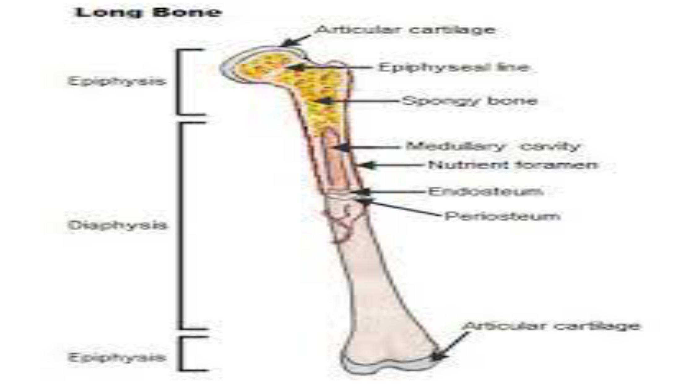
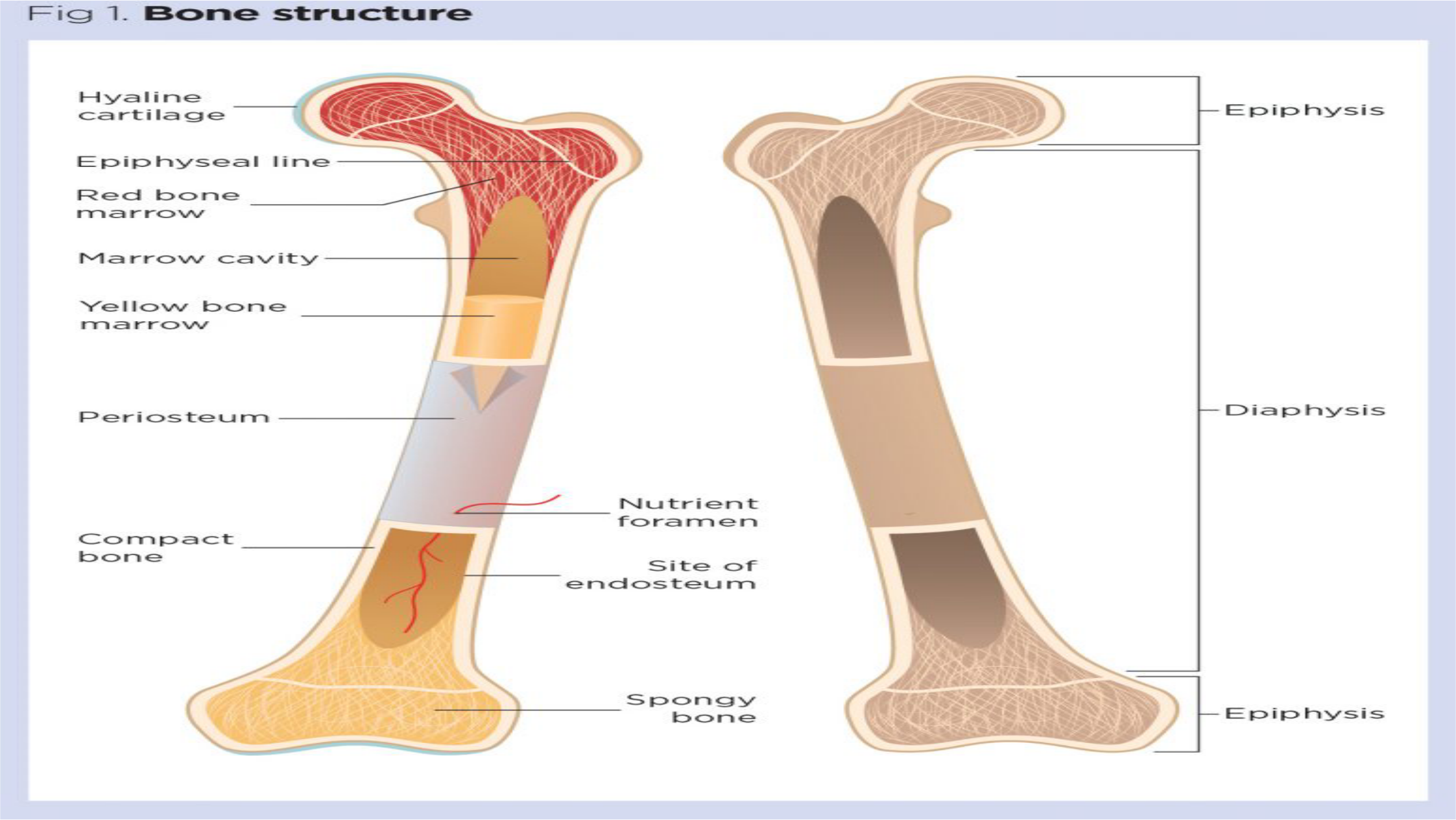
Hyalhine
cartilage
Epiphnhyseal
line
Reed
hone
moa
reow-
Marrow
cawit
w
we
Powe
Eso
&
ro
arrow
Periostewurm®,
Mutrieant
foramen
Site
oF
endosteuirm
SpPponcgy
Pon
=
H—-
Epeipohy
ysis
—_—_
Dmiaphysis
_
FE
moiph
ysis

The Digestive system
•
•
•
•
The gastrointestinal tract is an organ system that enables us to
ingest food via the mouth, digest it by breaking it down, absorb it,
and then expel the remaining waste as faeces via the anus.
The gastrointestinal tract is made up of a series of hollow organs
joined together in a long tube with many folds from the mouth to
the anus.
The hollow organs that make up the gastrointestinal tract include
the mouth, oesophagus, stomach, small intestine, cecum, colon
(large intestine), rectum and anal canal.
The organs of the gastrointestinal tract contain layers of muscles,
enabling their walls to move food through the tract by a process
called peristalsis, allowing for the breaking down and absorption of
food to take place.

•
•
•
•
The oesophagus is a muscular tube, transporting food and liquid
from the pharynx (part of the throat behind the mouth and nasal
cavity) to the stomach. It descends down into the thorax and enters
the abdomen, where it then joins the stomach.
The stomach is located between the oesophagus and the
duodenum. After food enters the stomach, the muscular walls of
the stomach act to mix the food and liquid with digestive juices.
The contents of the stomach, called chyme, are then emptied into
the small intestine.
The small intestine is approximately 6.5m long and extends from
the stomach to the large intestine. Anatomically, the small intestine
can be divided into three parts: the duodenum, jejunum and ileum.

•
•
The colon extends from the cecum to the anal canal and receives
digested food from the small intestine and absorbs water and
electrolytes to form faeces.
In this section, learn more about the physiology of organs within
the gastrointestinal tract- the mouth, stomach, small intestine,
large intestine, the pancreas, liver, gallbladder and spleen.

•Digestion is the chemical breakdown of the ingested food into absorbable
molecules. Absorption refers to the movement of nutrients, water and
electrolytes from the lumen of the small intestine into the cell, then into the
blood.
Carbohydrates Digestion
•
•
•
There are threecarbohydrateproducts which are absorbed by the
small intestine;glucose,galactoseandfructose.
Digestion of starch is initiated in the mouth, facilitated by salivary
amylase. The majority of carbohydrate digestion occurs in the
small intestine.
The main enzyme ispancreatic amylase, which yields
disaccharides from starch by digesting the alpha 1-4 glycosidic
bonds. The disaccharides produced (maltose, maltotriose, and α-
dextrins) are all converted to glucose by various specific
enzymes.

•
•
•
Disaccharides occurring naturally in food do not require amylase
to break them down. Brush border enzymes (lactase, sucrase,
galactase) hydrolyse these compounds into molecules of
glucose, galactose and fructose.
Glucose and galactose are absorbed across the apical
membrane by secondary active transport (along with Na+)
through the Sodium-Glucose cotransporter (SGLT1).
Both glucose and galactose exit the cell via GLUT2 receptors
across the cell membrane into the blood. Fructose enters the cell
by facilitated diffusion and is transported into the blood
Protein Digestion
•
•
•
•
•
Protein digestion begins in the stomach with the action ofpepsin,
which breaks protein into amino acids and oligopeptides.
The process of digestion is completed in the small intestine with
brush border and pancreatic enzymes. They split the oligopeptides
into amino acids, dipeptides and tripeptides.
Absorption
Amino acids are absorbed via a Sodium cotransporter, in a similar
mechanism to the monosaccharides. They are then transported
across the basolateral membrane viafacilitated diffusion.
Di and tripeptides are absorbed via separate H+dependent co-
transporters and once inside the cell are hydrolysed to amino acids.
Lipids Digestion
•
•
•
•
Lipids are hydrophobic, and thus are poorly soluble in the
aqueous environment of the digestive tract.
Their digestion is started by lingual and gastriclipases, but this
only digests 10% of ingested lipids.
The remainder of the lipids are digested in the small intestine.
Here, bile aids digestion byemulsifyingthe fat goblets into
smaller chunks, called micelles, which have a much larger
surface area.
Pancreatic lipase, phospholipase A2 and cholesterol ester
hydrolase (3 major enzymes involved in lipid digestion) hydrolyse
themicelles,breaking them down into fatty acids,
monoglycerides, cholesterol and lysolecithin.
Absorption of fatty acids
•
•
•
The products from digestion are released at the apical
membrane and diffuse into the enterocyte.
Inside the cell, the products are re-esterified to form the original
lipids, triglycerides, cholesterol and phospholipids.
The lipids are then packaged inside apoproteins to form
achylomicron. The chylomicrons are too large to enter
circulation, so they enter lymphatic system via lacteals.
Water Absorption
•
•
•
•
The average adult usually ingests 1-2 L of water each day, but the
fluid load to the small intestine is 9 to 10 L, 8 to 9 L being added
by secretions of the GI system.
Most absorption of water and electrolytes occurs in thesmall
intestine, with some water absorbed in thecolonas well.
Water absorption is dependent on the absorption of solutes such
as(Na+and Cl−).
Na+is absorbed from the intestinal lumen, most use the
cotransport with glucose and amino acids and the Na+/H
exchange, which allow Na+ions to move from the lumen into the
enterocyte.

•Na+ is rapidly removed from the enterocyte via the Na+pumps, allow
water via osmosis to follow either transcellularly or paracellularly (between
the tight junctions of the enterocytes). Water as well as Na+ions can then
diffuse into the capillaries.


