



Cell Biology Final Study Guide
Chapter 15: Cell walls, the extracellular matrix and
cell interactions.!
Introduction!
Many cells are surrounded by insoluble secreted macromolecules. Cells of bacteria, fungi,
algae, and higher plants are surrounded by rigid cell walls. !
Most animal cells are embedded in an extracellular matrix of proteins and polysaccharides.!
Basal laminae: thin layers on which epithelial cells rest. Also surrounds muscle cells, adipose
cells and peripheral nerves. !
Extracellular matrix is most abundant in connective tissues. Connective tissues (loose
connective tissue, bone, tendon, and cartilage) consist mostly of extracellular matrix with cells
distributed throughout. Extracellular matrixes have di erent amounts of each component: ff !
-Tendons: high proportion of fibrous proteins.!
-Cartilage: high level of polysaccharides that form a compression-resistant gel. !
-Bone matrix is hardened by calcium phosphate crystals. !
The major structural protein is collagen. Collagens form triple helices: three polypeptide chains
are wound together. The triple helix domains consist of repeats of the amino acid sequence
Gly-X-Y (meaning a glycine in every third position).!
Glycine is the smallest amino acid. It allows polypeptides to pack closely together. Proline is
frequently found in the X position and hydroxyproline in the Y position. They stabilize the
helices. !
Hydroxyproline is formed in the ER by modification of proline in proline polypeptide chains.
Hydroxyl groups are thought to stabilize the triple helix by forming hydrogen bonds. !
Type I collagen is the most abundant type. It forms collagen fibrils in which the triple helical
molecules form regular staggered arrays. Assembly of fibrils occurs outside the cell from
soluble precursor procollagens. !
Covalent cross-links between side chains of lysine and hydroxylysine. Resides help strengthen
the fibrils. Fibrils can come together to form collagen fibers, which can be several um in
diameter. !
Some types of collagen do not form fibrils. Fibril-associated collagens bind to collagen fibrils
and link them to one another and to other matrix components. !
Basal laminae are mostly type IV collagen, but also VI and XVIII. All are network-forming
collagens. The Gly-X-Y repeats are interrupted by short non helical sequences, making them
more flexible. They form 2-D cross-linked networks instead of fibrils.!
Connective tissue with elastic fibers is common in organs that stretch and return to shape,
such as the lungs. Elastic fibers are made of elastin which is cross-linked into a network. It
behaves like a rubber band. !
1

Extracellular matrix gels are formed from polysaccharides called glycosaminoglycans (GAGs).
These are repeating units of disaccharides. Except for hyaluronan, the sugars are modified with
sulfate groups. The addition of sulfate groups make GAGs highly negatively charged. They bind
positively charged ions and trap water molecules to form hydrated gels. !
Hyaluronan is the only GAG that is a single long polysaccharide chain. It is synthesized at the
plasma membrane by a transmembrane hyaluronan synthase. All of the other GAGs are linked
to proteins to form proteoglycans.!
Proteoglycans interact with hyaluronan to form large complexes in the extracellular matrix.
Aggrecan, the major proteoglycan of cartilage, has about 100 chains of chondroitin sulfate
attached to a core protein. !
Matrix adhesion proteins link matrix components to one another and to cell surfaces.
Fibronectin is the main adhesion protein of connective tissues, which is often cross-linked into
fibrils. It has binding sites for both collagen and GAGs. !
Fibronectin is recognized by cell surface receptors and is responsible for the attachment of
cells to the extracellular matrix.!
Fibronectins vary greatly from tissue to tissue, but all are derived by alternative splicing of
mRNA of a single gene. !
Basal laminae contain adhesion proteins of the lamin family. They consist of three polypeptide
chains, each with rod-like domains with interspersed globular domains. The subunits have
binding sites for di erent receptors. Laminins can self-assemble into networks.ff !
Laminins are tightly associated with , an adhesion protein that also binds to type IV nidogen
collagen. As a result of multiple interactions, laminin, nidogen, collagen and proteoglycans
form cross-linked networks within basal laminae. !
Integrins are cell surface receptors that attach cells to the extracellular matrix. They are a
family of transmembrane proteins that bind to many components of the extracellular matrix,
including collagen, fibronectin and laminin. !
Integrins also anchor the cytoskeleton to the extracellular matrix. There are two types of cell-
matrix junctions: !
1. Focal adhesions: bundles of actin filaments anchored to subunits of integrins via other β
proteins: -actinin, talin and vinculin. α !
2. Hemidesmosomes: anchor epithelial cells to basal laminae. 4 integrins link basal lamina α6β
layer of the extracellular matrix to intermediate filaments via pectin and BP230. Plectin and
BP230 are members of the plakin family. !
Integrins were first identified by immunofluorescence and immunoelectron microscopy of
transmembrane glycoproteins at points of cell adhesion in the matrix. The scientists were then
able to isolate a molecular clone encoding one of these glycoproteins (integrin) from a cDNA
library of chick embryo fibroblasts.!
Animal cells modify the extracellular matrix via enzymes that modify glycosaminoglycans and
proteases that digest collagen. The matrix metalloprotease family digest a variety of matrix
proteins, including collagens, laminin, and perlecan, cell surface receptors, and adhesion
molecules.!
2

Cell-Cell Interactions !
Interactions between cells are critical for development and function of multicellular organisms.
Some cell-cell interactions are transient; others are stable and play a key role in the
organization of tissues. Some types of junctions allow rapid communication between cells.!
Cell-cell adhesion is mediated by four groups of cell adhesion molecules:!
-Selectins!
-Integrins!
-Immunoglobulin (Ig) superfamily !
-Cadherins !
Many adhesions depend on divalent cations, requiring Ca , Mg and Mn
2+ 2+ 2+!
Selectins
Mediate transient interactions between leukocytes and endothelial cells or blood platelets. !
Selectins recognize cell surface carbohydrates and initiate interactions between leukocytes
and endothelial cells during migration of leukocytes. !
Binding of to integrins is an example of ICAMs heterotrophilic interaction: an adhesion
molecule on one cell recognizes a di erent molecule on another cell. ff !
Homophilic interactions: an adhesion molecule on one cell binds to the same molecule on
another cell. Homophilic binding between neural cell adhesion molecules (N-CAMs) contributes
to associations between nerve cells during development. !
Cadherins
Mediate homophilic interactions.Stable adhesion junctions linking the cytoskeletons of
adjacent cells are usually based on cadherins:!
Adherens junctions: include -catenin, p120 and -catenin, and classical β α & & &
transmembrane cadherins. -catenin and p120 are members of the armadillo protein β
family. &They bind cadherin and help maintain stability.!
Desmosomes
Tight junctions in epithelial cell sheets form a seal that prevents free passage of molecules
and ions between cells. They also separate apical and basolateral domains of the plasma
membrane by preventing the free di usion of lipids and membrane proteins. ff !
Tight junctions provide minimal adhesive strength between the cells, so they are usually
associated with adherens junctions and desmosomes in a junctional complex.!
Tight junctions are formed by a network of protein strands that continues around the entire
circumference of the cell. Each strand in the network is composed of transmembrane proteins
of the occludin, claudin, and junctional adhesion molecule (JAM) families. !
Within a tissue, cells are often linked by , open channels through the plasma gap junctions
membrane. They allow ions and small molecules to di use freely between neighboring cells. ff
Proteins and nucleic acids cannot pass through. !
In electrically excitable cells, such as heart muscle, passage of ions through gap junctions
couples and synchronizes the contractions of neighboring cells. Gap junctions also allow
passage of signaling molecules such as cAMP and Ca , potentially coordinating responses of
2+
cells in tissues. !
Gap junctions consists of trans-membrane proteins in the connexin family. Six connexins form
a connexon, a cylinder with an open aqueous pore in its center. Connexons of adjacent cells
align, forming open channels between the two cytoplasms.!
3

Specialized gap junctions occur on specific nerve cells and form an electrical synapse.
Individual connexons can be opened or closed. When open, they allow rapid passage of ions
between the two nerve cells. Several human diseases are associated with connexons
mutations.'
4

Chapter 16: Cell Signaling !
Introduction !
Communication is accomplished by signaling molecules from one cell that bind to receptors on
other cells. This initiates a series of reactions that regulate virtually all aspects of cell behavior. !
Signaling molecules range in complexity from simple gases to proteins. Some carry signals
over long distances, whereas others act locally.!
They also di er in modes of action: some cross the plasma membrane and bind to intracellular ff
receptors; others bind to receptors on the cell surface. !
Modes of signaling include:!
-Direct cell-cell signaling: direct interaction of a cell with its neighbor (via integrins and
cadherins).!
-Signaling by secreted molecules three categories are based on the distance over which
signals are transmitted. !
Endocrine signaling: signaling molecules (hormones) are secreted by specialized endocrine
cells and carried through the circulation to target cells at distant body sites. Like estrogen.!
Paracrine signaling: molecules released by one cell act on neighboring target cells. Like
neurotransmitters.!
Autocrine signaling: cells respond to signaling molecules that they themselves produce. Like
T lymphocytes that respond to antigens by making a growth factor that drives their own
proliferation, thereby amplifying the immune response. !
Receptors may be located on the cell surface or inside the cell. Intracellular receptors respond
to small hydrophobic molecules that can di use across the plasma membrane. For example: ff
steroid hormones, thyroid hormone, vitamin D3 and retinoic acid. !
Steroid hormones are synthesized from cholesterol:!
-Testosterone, estrogen and progesterone are the , produced by the gonads. sex steroids !
-Corticosteroids are produced by the adrenal glands.!
-Glucocorticoids stimulate the production of glucose.!
-Mineralocorticoids act on the kidney to regulate salt and water balance.!
Thyroid hormone: synthesized from tyrosine in the thyroid gland, important in development and
metabolism. Vitamin D3 regulates calcium metabolism and bone growth. Retinoic acid and
retinoids are synthesized from vitamin A, important in vertebrate development.!
Receptors for these molecules are members of the nuclear receptor superfamily. They are
transcription factors that have domains for ligand binding, DNA binding and transcriptional
activation. Ligand binding regulates their function as activators or repressors of genes. !
5

Ligand binding has di erent e ects on di erent receptors. Some nuclear receptors are inactive ff ff ff
in the absence of hormone. !
-Glucocorticoid receptor is bound to Hsp90 chaperones in the absence of hormone.!
-Glucocorticoid binding displaces Hsp90 and leads to binding of regulatory DNA sequences.!
Hormone binding can alter the activity of a receptor: !
-In the absence of hormone, thyroid hormone receptor is associated with a co-repressor
complex and represses the transcription of target genes. Hormone binding results in the
activation of transcription. !
Nitric oxide is a paracrine signaling molecule in the nervous, immune, and circulatory systems.
It can cross the plasma membrane and alter the activity of enzymes. Nitric oxide (NO) is
synthesized from arginine. Its action is restricted to local e ects, because it is extremely ff
unstable, with a half-life only of a few seconds.!
The main target of NO is guanylyl cyclase. NO binding stimulates synthesis of cyclic GMP (a
second messenger). NO can signal dilation of blood vessels: neurotransmitters act on
endothelial cells to stimulate NO synthesis. NO di uses to smooth muscle cells and stimulates ff
cGMP production. cGMP induces muscle cell relaxation and blood vessel dilation. !
Carbon monoxide (CO), also functions as a signaling molecule in the nervous system. It is
related to NO and acts similarly as a neurotransmitter and mediator of blood vessel dilation. !
Neurotransmitters
Carry signals between neurons or from neurons to other target cells. Neurotransmitters are
released when an action potential arrives at the end of a neuron. The neurotransmitters then
diffuse across the synaptic cleft and bind to receptors on the target cell surface. !
Examples:'
-acetylcholine !
-glycine!
-glutamate!
-dopamine!
-norepinephrine!
-epinephrine!
-serotonin!
-histamine!
-γ '-aminobutiric acid
Because neurotransmitters are hydrophilic, they cant cross the plasma membranes and must
bind to cell surface receptors. Many neurotransmitter receptors are ligand-gated ion channels.
Neurotransmitter binding opens the channels.!
Peptide signaling molecules include peptide hormones, neuropeptides, and polypeptide
growth factors. !
Peptide hormones: include insulin, glucagon, and pituitary gland hormones like the growth
hormone, follicle-stimulating hormone and prolactin.!
Neuropeptides: secreted by some neurons instead of small-molecule neurotransmitters.!
Enkephalins and endorphins: act as neurotransmitters and as neurohormones, which are
natural analgesics that decrease pain responses; they bind to the same receptors on brain cells
as morphine does. !
Nerve growth factor (NGF) is a member of the neurotrophin family that regulates development
and survival of neurons. !
Epidermal growth factor (EGF) stimulates cell proliferation. It is the prototype for the study of
growth factors. !
Platelet-derived growth factor (PDGF) is stored in blood platelets and released during blood
clotting at the site of a wound. It stimulates proliferation of fibroblasts, contributing to regrowth
of the damaged tissue.!
6

Cytokines regulate development and di erentiation of blood cells and activities of ff
lymphocytes during the immune response. !
Membrane-anchored growth factors remain associated with the plasma membrane and
function as signaling molecules in direct cell-cell interactions. !
Eicosanoids lipid signaling molecules that include prostaglandins, prostacyclin, thromboxanes
and leukotrienes. These break down rapidly, acting in autocrine or paracrine pathways. !
Eicosanoids are synthesized from arachidonic acid, which is formed by phospholipids.
Arachidonic acid is converted to prostaglandin H2, catalyzed by cyclooxygenase. This enzyme
is the target of aspirin and other non-steroidal anti-inflammatory drugs (NSAIDs).!
Inhibiting synthesis of the prostaglandins reduces inflammation and pain. By inhibiting
synthesis of thromboxane, Aspirin reduces platelet aggregation and blood clotting. Thus, small
daily doses of aspirin are often prescribed for prevention of strokes. !
Aspirin and NSAIDs have also been found to reduce the frequency of colon cancer, apparently
by inhibiting the synthesis of prostaglandins that stimulate cell proliferation. !
Most ligands responsible for cell-cell signaling bind to surface receptors on their target cells.
This initiates a chain of intracellular reactions, ultimately reaching the nucleus and resulting in
programmed changes in gene expression. !
G protein-coupled receptors are the largest family of cell surface receptors. Signals are
transmitted via guanine nucleotide-binding proteins (G proteins). The receptors have seven
membrane-spanning -helices. α !
Binding of a ligand induces a conformational change that allows the cytosolic domain to
activate a G protein on the inner face of the plasma membrane. The activated G protein then
dissociates from the receptor and carries the signal to an intracellular target. !
G proteins have three subunits designated , and . α β γ !
They are called heterotrimeric G proteins to distinguish them from other guanine nucleotide-
binding proteins, such as the Ras proteins.!
The subunit binds guanine nucleotides, which regulate G protein activity. In the inactive state, α
α is bound to GDP in a complex with β γ ! and .
Hormone binding to the receptor causes exchange of GTP for GDP. The and complex then α βγ
dissociate from the receptor and interact with their targets. !
A large array of G proteins connect receptors to distinct targets. In addition to enzyme
regulation, G proteins can also regulate ion channels. Example: action of the neurotransmitter
acetylcholine on heart muscle.!
Heart muscle cells have a di erent acetylcholine receptor than nerve and skeletal muscle cells, ff
which is G protein-coupled. !
The subunit of this G protein (Gi) inhibits adenylyl cyclase. The Gi- subunits open K+ ion α βγ
channels in the plasma membrane, which slow heart muscle contraction. !
Other cell surface receptors are directly linked to intracellular enzymes. The largest family of
these is the receptor protein-tyrosine kinases which phosphorylate their substrates on tyrosine
residues. The family includes receptors for most polypeptide growth factors. !
The human genome encodes 59 receptor protein-tyrosine kinases, including receptors for EGF,
NGF, PDGF, insulin and other growth factors. All have an N-terminal extracellular ligand-binding
domain, a single transmembrane a helix and a cytosolic C-terminal domain with protein-
tyrosine kinase activity. '
7

Functions of cell surface receptors!
Binding of ligands (growth factors) to the extracellular domains activates their cytosolic kinase
domains. This results in phosphorylation of both the receptors and intracellular target proteins
that propagate the signal. !
The first step is ligand-induced receptor dimerization. This results in receptor
autophosphorylation, as the two polypeptide chains cross-phosphorylate each other.!
Autophosphorylation has two roles: !
-Phosphorylation of tyrosine in the catalytic domain increases protein kinase activity. !
-Phosphorylation of tyrosine outside the catalytic domain creates binding sites for other
proteins that transmit signals downstream from the activated receptors. !
The downstream signaling molecules have domains that bind to specific phosphotyrosine-
containing peptides. SH2 domains were the first to be characterized, initially recognized in
protein tyrosine kinases related to Src, the oncogenic protein of Rous sarcoma virus. !
The cytokine receptor superfamily includes receptors for most cytokines and some polypeptide
hormones. Structure is similar to receptor protein-tyrosine kinases, but the cytosolic domains
have no catalytic activity. !
Cytokine receptors function in association with non receptor protein-tyrosine kinases. Ligand
binding induces dimerization of receptors, and cross-phosphorylation of associated non
receptor protein tyrosine kinases.!
The activated kinases then phosphorylate the receptor. This provides phosphotyrosine-binding
sites for recruitment of downstream signaling molecules with SH2 domains. !
The kinases associated with cytokine receptors belong to the Janus kinase (JAK) family.!
Members of the JAK family are required for signaling from cytokine receptors. !
Additional non receptor protein-tyrosine kinases belong to the Src family:!
-first identified as the oncogenic protein of Rous sarcoma virus!
-first protein shown to have protein-tyrosine kinase activity !
-Important in experiments leading to current understanding of cell signaling. !
Some enzyme-linked receptors are associated with other enzymatic activities: !
•Protein-tyrosine phosphatase remove phosphate groups from phosphotyrosine,
counterbalancing the e ects of protein-tyrosine kinases.ff !
•Transforming growth factor B (TGF-B) receptors protein kinases that phosphorylate serine
or threonine on substrates. TGF-B is the prototype of a receptor family with a cytosolic
protein-serine/threonine kinase domain that control cell proliferation and di erentiation. ff !
•Receptor guanylyl cyclases have a cytosolic domain that catalyzes formation of cyclic
GMP. !
Pathways of intracellular signal transduction !
Intracellular signal transduction chain of reactions that transmits signals from the cell surface
to intracellular targets. Targets often include transcription factors that regulate gene expression. !
Intracellular signaling was first studied in hormones like epinephrine, which signals the
breakdown of glycogen to glucose. In 1958 Sutherland discovered that epinephrine action was
mediated by an increase in cyclic AMP (cAMP), leading to the concept of cAMP as a second
messenger.!
CAMP is formed from ATP by adenylyl cyclase and degraded to AMP by cAMP
phosphodiesterase. !
8

CAMP e ects are mediated by cAMP dependent protein kinase, or protein kinase A. Inactive ff
form has two regulatory subunits. cAMP binds to the regulatory subunits, which dissociate. !
The free catalytic subunits can then phosphorylate serine on target proteins. !
In glycogen metabolism, protein kinase A phosphorylates two enzymes: !
-Phosphorylase kinase is activated, and in turn activated glycogen phosphorylase. !
-glycogen synthase is inactivated!
So, glycogen breakdown is stimulated and glycogen synthesis blocked. !
Signal amplification: each molecule of epinhephrine activates one receptor. Each receptor may
activate up to 100 molecules of Gs, which then stimulates adenylyl cyclase. This catalyzes
synthesis of many cAMP. Each molecule of protein kinase A phosphorylates many molecules of
phosphorylase kinase, and so forth. !
cAMP can activate transcription of genes with the cAMP response element or CRE regulatory
sequence. The free catalytic subunit of protein kinase A goes to the nucleus and
phosphorylates the transcription factor CREB (CRE-binding protein). This leads to expression
of cAMP-inducible genes.!
Protein phosphorylation is rapidly reversed by protein phosphatases, which terminates
responses initiated by receptor activation of protein kinases. !
cAMP can also directly regulate ion channels because: !
It is a second messenger in sensing smells —> odorant receptors are G protein-coupled. They
stimulate adenylyl cyclase, leading to increased cAMP. cAMP opens Na+ channels in the
plasma membrane, leading to initiation of a nerve impulse. !
Cyclic GMP (cGMP) is also an important second messenger. cGMP is formed from GTP by
guanylyl cyclases and degraded to GMP by a phosphodiesterase. cGMP mediates biological
responses such as blood vessel dilation. !
In the vertebrate eye, cGMP is the second messenger that converts visual signals to nerve
impulses. The photoreceptor in retinal rod cells is a G protein-coupled receptor called
Rhodopsin. Rhodopsin is activated when light is absorbed by the associated small molecule
retinal. Rhodopsin then activates the G protein transducer. The subunit stimulates cGMP α
phosphodiesterase, leading to decreased levels of cGMP. cGMP levels are translated to nerve
impulses by a direct e ect of cGMP on ion channels. ff !
Two pathways use second messengers derived from the membrane phospholipid
phosphatidylinositol 4,5-bisphosphate (PIP2). Hydrolysis of this phospholipid by
phospholipase C produces two second-diacylglycerolmessengers: !
-diacylglycerol (DAG) in membrane!
-inositol 1,4,5-trisphosphate (IP3) in cytosol!
Two-forms of phospholipase C: !
-PLC-β is stimulated by G proteins!
-PLC-γ has SH2 domains that associate with receptor protein-tyrosine kinases. Tyrosine
phosphorylation increases PLC-y activity, stimulating hydrolysis of PIP2. !
DAG remains associated with the plasma membrane and activates protein-serine/threonine
kinases of the protein kinase C family. !
9

IP3 is a small polar molecule that is released to the cytosol, where it signals release of Ca
2+
from the ER.!
Cytosol concentration of Ca is maintained at very low levels by Ca pumps. IP3 stimulates
2+ 2+
release of Ca from the ER by binding to receptors that are ligand-gated Ca channels.
2+ 2+ !
Increased Ca a ects activity of several proteins, including protein kinases and phosphatases.
2+ ff !
One of the major Ca binding proteins that mediates the e ects of Ca is . Which
2+ ff2+ calmodulin
is activated when Ca concentration increases Ca /calmodulin then binds to target proteins,
2+ 2+
including protein kinases? !
Members of the CaM kinase family are also activated by Ca /calmodulin. They phosphorylate
2+
metabolic enzymes, ion channels and transcription factors. One form of CaM kinase regulates
synthesis and the release of neurotransmitters. !
CREB is a transcription factor that is phosphorylated by CaM kinase and also by protein kinase
A. This illustrates one of many intersections between the Ca and cAMP signaling pathways.
2+
These pathways regulate function coordinately to regulate many cellular responses. !
Ca2+ is also increased by uptake of extracellular Ca by regulated channels in the plasma
2+
membrane. In electrically excitable cells of nerve and muscle, voltage gated Ca channels are
2+
opened by membrane depolarization. The resulting increase in intracellular Ca signals further
2+
release of Ca from the ER by opening Ca channels called ryanodine receptors in the ER
2+ 2+
membrane. Ca is a versatile second messenger that controls a wide range of extracellular
2+
processes. !
PIP2 is also the start of another signaling pathway. PIP2 is phosphorylated by
phosphatidylinositide (PI) 3-kinase. This yields the second messenger phosphatidylinositol
3,4,5-trisphosphate (PIP3). PIP3 targets a protein-serine/threonine kinase called Akt and it also
binds the protein kinase PDK1. !
Activation of Akt also requires protein kinase mTOR (in the mTORC2) complex, which is also
stimulated by growth factors. !
Activated Akt phosphorylates several target proteins, including transcription factors and other
protein kinases. Transcription factors include members of the Forkhead of FOXO family.!
Akt phosphorylation of FOXO sequesters it in inactive form. !
If growth factors are not present, Akt is not active and FOXO travels to the
nucleudownregulations, where it stimulates transcription of genes that inhibit cell proliferation
or induce cell death.!
Protein kinase GSK-3 is also inhibited by Akt phosphorylation. GSK-3 targets include the
translation initiation factor eIF2B. Phosphorylation of eIF2B leads to a global downregulation of
translational initiation. !
SLIDE 171:!
Multiple signal transduction pathways interact with one another to form signaling networks
within the cell. Junctions can be positive (one pathway stimulates the other) or negative (one
pathway inhibits the other). Feedforward relays: activity of one component stimulates a distant
downstream component. '
10

Chapter 17: The Cell Cycle!
Introduction!
Self reproduction is perhaps the most fundamental characteristic of cells. All cells reproduce by
dividing in two, each parental cell gives rise to two daughter cells on completion of a cycle of
cell division. The process of cell division must be carefully regulated and coordinated. !
In eukaryotic cells, progression through the cell cycle is controlled by protein kinases that have
been conserved from yeasts to mammals. Defects in cell cycle regulation are a common cause
of the abnormal proliferation of cancer cells.!
The Eukaryotic Cell Cycle!
It has four phases: M, G1, S and G2. !
M Phase: mitosis (nuclear division), usually ending with cell division (cytokinesis).!
Interphase: period between mitoses, divided into G1, S and G2. !
-G1 Phase (gap1): it is the interval between mitosis and DNA replication. The cell is
metabolically active and growing. !
-S Phase (synthesis): DNA replication takes place!
-G2 Phase (gap2): cell growth continues and proteins are synthesized in preparation for
mitosis. !
Duration of phases varies considerably in di erent kinds of cells. Budding yeasts can progress ff
through all four phases in 90 minutes. Early embryos may have cell cycles of only 30 minutes,
but there is no growth (no G1 or G2). !
In contrast, some cells in adult animals cease division altogether (like nerve cells). Others may
divide only occasionally, to replace cells that have been lost. !
Cell cycle analysis requires identification of the phases. Phase of the interphase must be
identified biochemically, usually by DNA content. Animal cells in G1 are diploid. Their DNA
contents is 2n. In G2 it would be 4n. !
During S phase, replication increases the DNA content to 4n. DNA content can be determined
by incubation of cells with a fluorescent dye that binds to DNA.!
Fluorescence intensity of individual cells is measured in a flow cytometer or fluorescence
activated cell sorter. !
Progression of cells through the division cycle is regulated by both extracellular and internal
signals.!
Cellular processes, such as growth, DNA replication, and mitosis, are regulated by a series of
control points. !
11
START
A major control point, called START controls progression from G1 to S, it was first defined in
yeast cells. Once cells pass START, they are committed to entering S phase and undergoing
one division cycle. !
Passage through START is highly regulated by external signals such as nutrient availability and
cell size. If there is a shortage of nutrients, cells can arrest the cycle at START and enter a
resting phase. !
In order to maintain constant size, yeast cells must reach a minimum size to pass START. The
small daughter cells of budding yeasts spend a longer time in G1 and grow more than the large
mother cell. !
Restriction Point
In most animal cells, the restriction point in late G1 functions like START. Passage through the
restriction point is regulated by extracellular growth factors. Once it has passed the restriction
point, the cell is committed to proceed through S phase and the rest of the cell cycle. !
If appropriate growth factors are not present in G1, progression stops at the restriction point
and cells enter a stage called G0.!
Skin fibroblasts are arrested in G0 until stimulated by platelet-derived growth factor (PDGF) to
proliferate and repair wound damage. !
Some cell cycles are controlled principally in G2. The fission yeast Schizosaccharomyces
pombe cell cycle is controlled by transition from G2 to M, the point at which cell size and
nutrient availability are monitored.!
Cell cycle control in G2 also occurs in animal oocytes. Vertebrate oocytes can remain arrested
in G2 for long periods (decades in humans). Progression to M phase is triggered by hormonal
stimulation (from G2 to M). !
Events in di erent stages of the cell cycle must be coordinated so they occur in the ff
appropriate order. It is critically important, for example, that the cell does not begin mitosis until
replication of the genome has been completed. Coordination of the cell cycle phases is
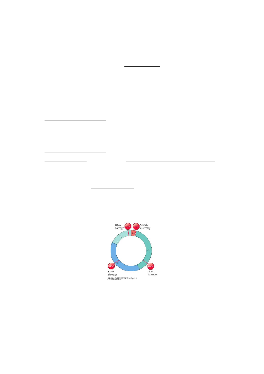
dependent on a series of cell cycle checkpoints. !
DNA damage checkpoints ensure that damaged DNA is not replicated and passed on to
daughter cells. The cell cycle is arrested until DNA is repaired or replicated. !
Spindle assembly checkpoint stops mitosis at metaphase if chromosomes are not properly
aligned on the spindle. '
12

It is also important to ensure that the genome is replicated only once per cell cycle. !
After replication in S phase, control mechanisms prevent re-initiation of DNA replication until
the cell cycle has been completed. !
Regulators of cell cycle progression!
MCM helicase proteins bind to ORC proteins and form origin recognition complex. These are
required for the initiation of DNA replication. !
Once initiation has occurred, the MCM proteins are displaced from the origin so that replication
cannot initiate again until after mitosis. !
Recent studies have revealed that eukaryote cell cycles are controlled by a conserved set of
protein kinases which trigger the major cell cycle transitions.!
Three experimental approaches contributed to identification of the molecules responsible for
cell cycle regulation (protein kinases): !
1. Studies of frog oocytes which are arrested in G2 until hormonal stimulation triggers entry
into M phase. In 1971, researchers found that oocytes could be induced to enter M phase
by microinjection of cytoplasm from ooccytes that had been hormonally stimulated. The
cytoplasmic factor responsible was called maturation promoting factor (MPF). Later work
showed that MPF is also present in somatic cells, where it induces entry into M phase.
MPF thus appeared to act as a general regulator of the transition from G2 to M.!
2. Genetic analyses of yeasts: investigators found temperature-sensitive mutants that were
defective in cell cycle progression (Cdc for cell division cycle mutants). Cdc genes are
required for passage through START and entry into mitosis, they encode for protein
kinases. The protein kinase has since been shown to be a cell cycle regulator conserved in
all eukaryotes, known as Cdk1. !
3. Protein synthesis in early sea urchin embryos: In 1983 Hunt and colleagues identified two
proteins that accumulate throughout interphase but are rapidly degraded at the end cyclins
of each mitosis, suggesting a role in inducing mitosis. Later studies showed that
microinjection of cyclin A into frog oocytes is su cient to trigger the G2 to M transition.ffi !
The three experimental approaches converged in 1988, when MPF was purified and shown to
be composed of Cdk1 and Cyclin B. Cyclin B is a regulatory subunit required for catalytic
activity of the Cdk1 protein kinase. !
Further studies demonstrated the regulation of MPF by phosphorylation and
dephosphorylation of Cdk1. During G2, cyclin B is synthesized and forms complexes with
Cdk1. Cdk1 is phosphorylated and inhibited, leading to accumulation of inactive Cdk1/cyclin B
complexes during G2. !
13
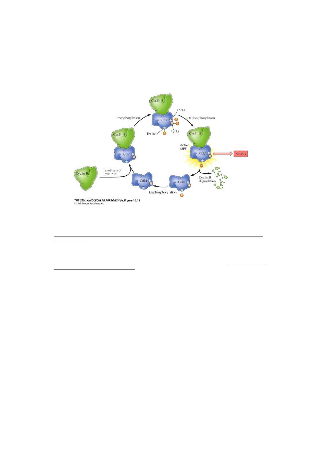
Dephosphorylation activates Cdk1, which phosphorylates several proteins that initiate
the events of M phase Destruction . Cyclin B is degraded by ubiquitin-mediated proteolysis.
of Cyclin B inactivates Cdk1, leading the cell to exit mitosis, undergo cytokinesis, and
return to interphase.!
Further research established that Cdk1 and cyclin B are members of protein families. Di erent ff
members of these families control progression through the phases of the cell cycle. !
Cdk1 controls passage through START and entry into mitosis in yeasts, in association with G1
cyclins of CIn’s. In higher eukaryotes, there are multiple cyclins and multiple Cdk1-related
protein kinases, known as Cdks for cyclin-dependent kinases. !
Studies of Cdks and cyclins in genetically modified mice reveal a higher level of plasticity,
allowing di erent cyclins and Cdks to compensate for the loss of one another. Cdk1 is capable ff
of substituting for all the other Cdks.!
14
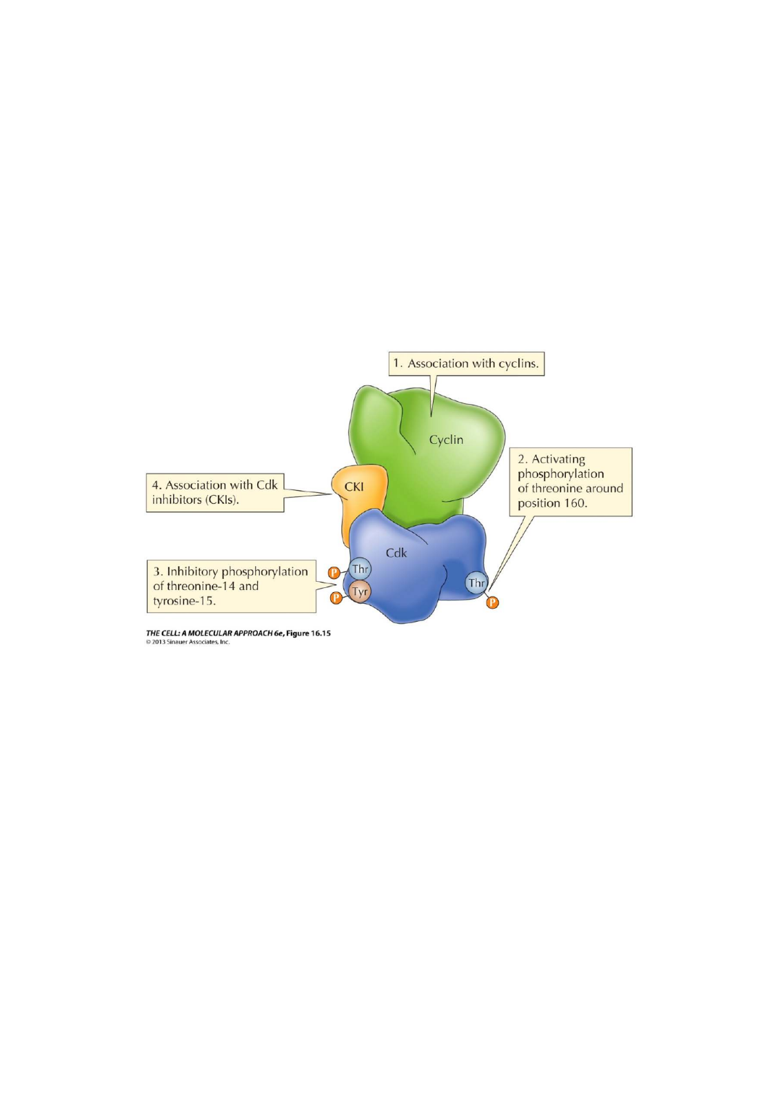
The activity of Cdks is regulated by four mechanisms:
1. Association of Cdks and cyclin partners: formation of specific Cdk/cyclin complexes is
controlled by cyclin synthesis and degradation. !
2. Activation of Cdk/cyclin complexes requires phosphorylation of threonine at position 160.
This is catalyzed by CAK (Cdk-activating kinase), which is composed of Cdk7/cyclin H!
3. Inhibitory phosphorylation of tyrosine near the Cdk amino terminus, catalyzed by Wee1
protein kinase. The Cdks are then activated by dephosphorylation by Cdc25 protein
phosphatases. !
4. Binding of inhibitory proteins Cdk inhibitors (CKIs). In mammalian cells, two families of
inhibitors Ink4 and Cip/Kip!
!
The combined e ects of these multiple modes of Cdk regulation are responsible for controlling ff
cell cycle progression in response to checkpoint controls and to extracellular stimuli.!
Proliferation of animal cells is regulated by extracellular growth factors that control progression
through the restriction point in late G1. This implies that intracellular signaling pathways
ultimately act to regulate components of the cell cycle machinery. !
Progression through the restriction point is mediated by activation of Cdk2/cyclin E complexes.
In G0 and early G1, Cdk2/cyclin E is inhibited by p27 (one of the inhibitors) (Cip/Kip family). !
15
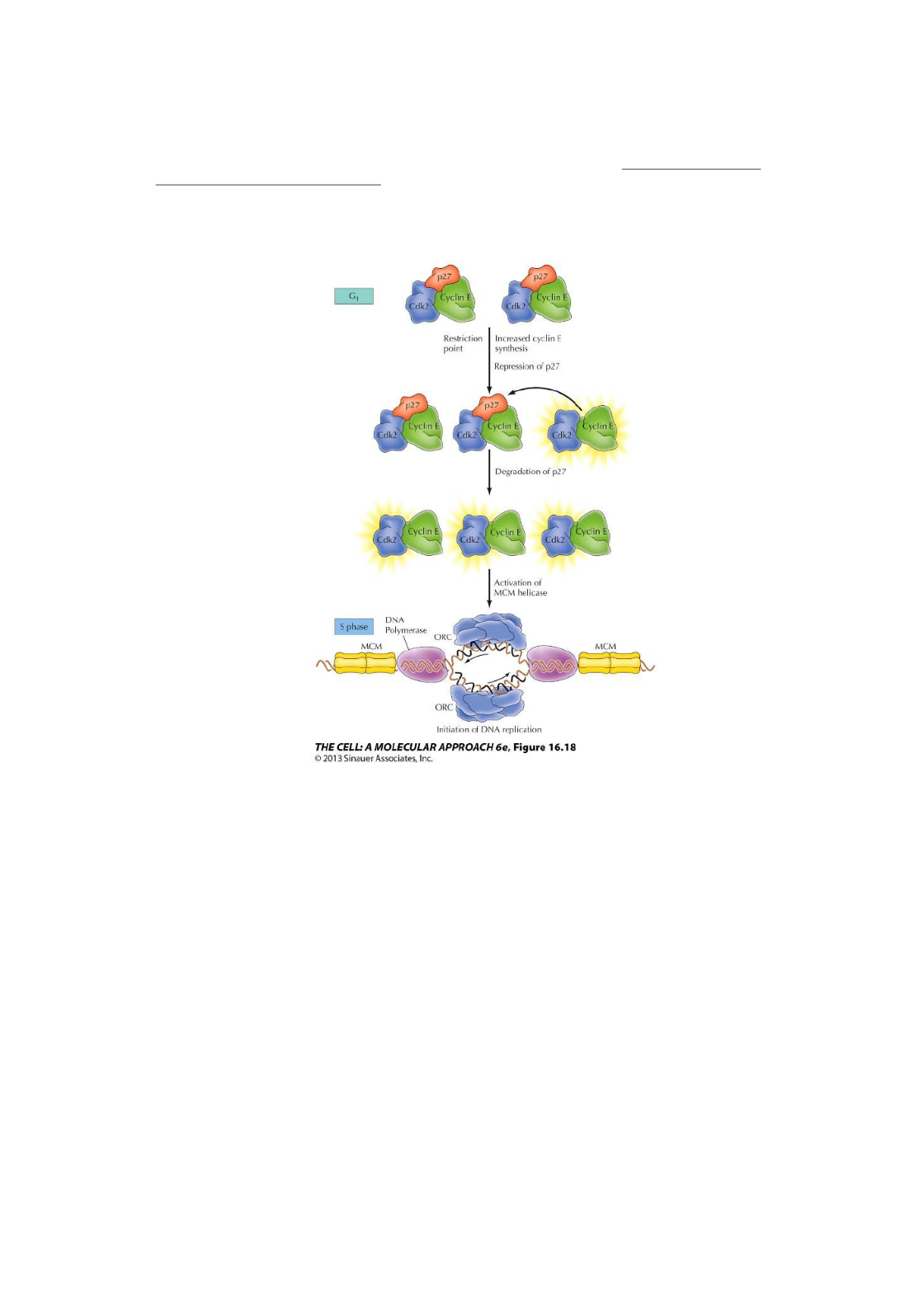
Cyclin E synthesis is stimulated by E2F after phosphorylation of Rb, and transcription of p27 is
inhibited by growth factor signaling. The resulting activation of Cdk2/cyclin E leads to
activation of MCM helicase and initiation of DNA replication. !
!
16
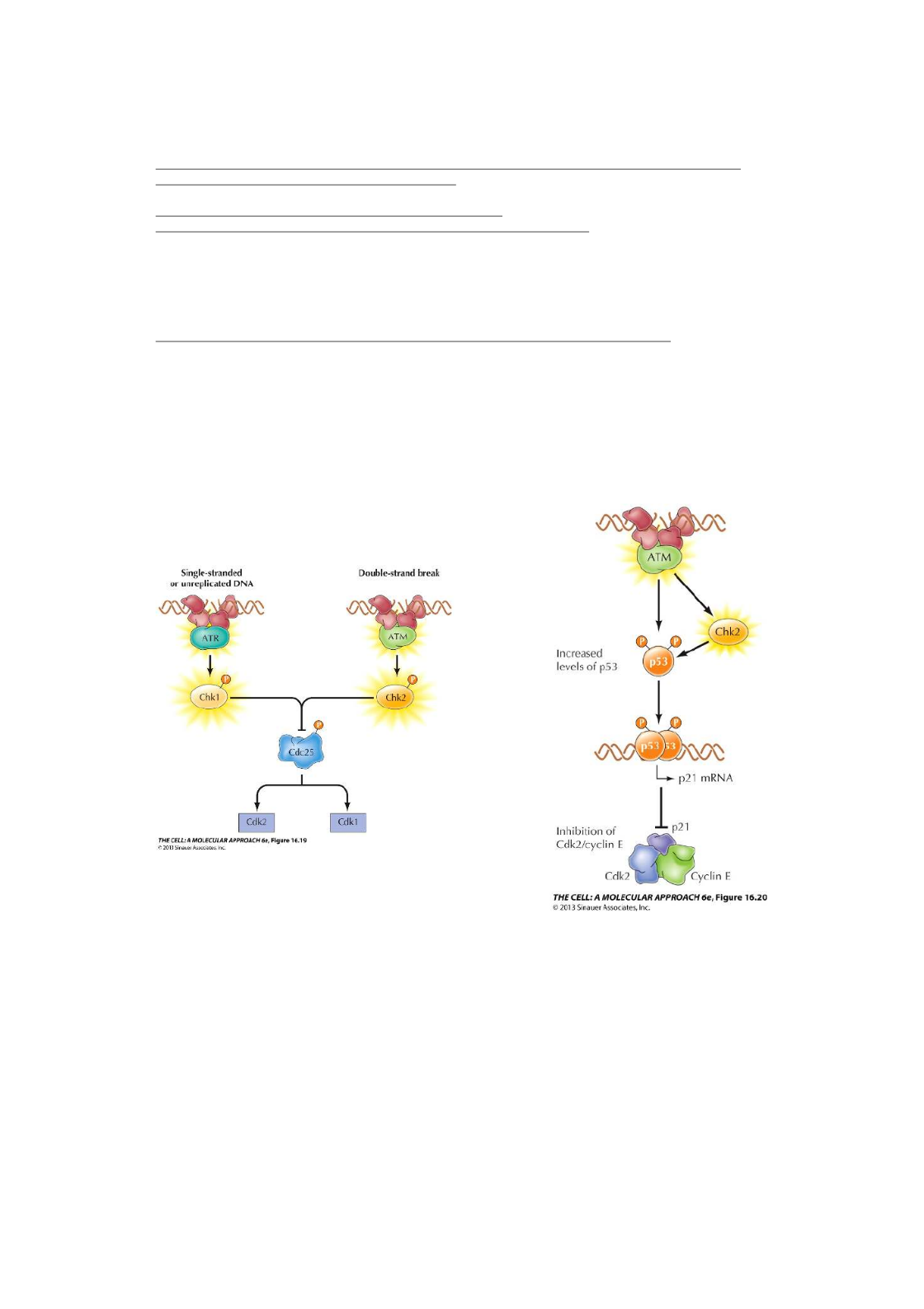
Cell cycle arrest at DNA damage checkpoints is mediated by protein kinases ATM and ATR,
that are activated in response to DNA damage. They then activate a signaling pathway, that
leads to cell cycle arrest, DNA repair and sometimes, programmed cell death. !
ATM recognizes double-strand breaks, activates Chk2!
ATR recognizes single-stranded or unreplicated DNA, activate Chk1!
They phosphorylate and activate the checkpoint kinases Chk1 and Chk2.!
Chk1 and Chk2 phosphorylate and inhibit Cdc25 phosphatases, which are required to activate
Cdk1 and Cdk2. Inhibition of Cdk2 results in cell cycle arrest in G1 and S. Inhibition of Cdk1
results in arrest of G2. !
In mammalian cells, arrest at the G1 checkpoint is also mediated by protein p53, which is
phosphorylated by both ATM and Chk2. p53 is a transcription factor, and its increased
expression leads to induction of Cdk inhibitor p21 (Cip/Kip family). !
p21 inhibits Cdk2/cyclin E complexes, leading to cell cycle arrest in G1. !
The p53 gene is frequently mutated in human cancers. Loss of p53 (inhibitor) prevents G1
arrest in response to DNA damage, so the damaged DNA is replicated and passed on daughter
cells. '
17

The events of M phase!
M phase involves a major reorganization of virtually all cell components: !
Chromosomes condense, nuclear envelope breaks down, cytoskeleton reorganizes to form the
mitotic spindle, and chromosomes move to opposite poles. !
Cell division (cytokinesis) usually follows. !
Mitosis is divided into four stages: !
1. Prophase
Appearance of condensed chromosomes (two sister chromatids). The chromatids are attached
at the centromere, where proteins bind to form the kinetochore (site of eventual spindle
attachment). The centrosomes (which duplicated during interphase) separate and move to
opposite sides of the nucleus. They serve as the two poles of the mitotic spindle, which begins
to form during late prophase. In higher eukaryotes prophase ends when the nuclear envelope
breaks down (open mitosis). In yeasts, the nuclear envelope remains intact (closed mitosis).
Spindle pole bodies are embedded in the nuclear envelope; the nucleus divides after migration
of daughter chromosomes. !
Pro-metaphase: transition between prophase and metaphase. Spindle microtubules attach to
kinetochores of condensed chromosomes. The chromosomes shu e back and forth until they ffl
align on the metaphase plate. The cell is then at metaphase.
2. Metaphase
Most cells remain briefly at metaphase, before proceeding to anaphase. Formation of the
mitotic spindle (where chromosomes align at the metaphase plate).!
3. Anaphase
The links between sister chromatids break, and they separate and move to opposite poles of
the spindle. !
4. Telophase
Nuclei re-form and chromosomes decondense. Cytokinesis usually begins during late
anaphase and is most complete by the end of telophase. !
18
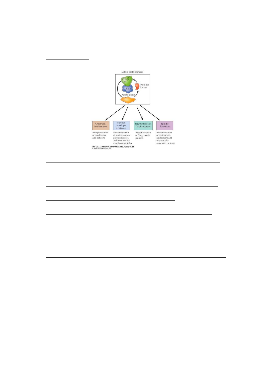
Cdk1/cyclin B protein kinase (MPF) is the master regulator of M phase transition. It activates
other mitotic protein kinases and directly phosphorylates structural proteins involved in the
cellular reorganization. !
Condensation of chromatin by nearly a thousand fold is a key event in mitosis. Transcription
ceases during condensation. The mechanism of condensation is not fully understood, but it is
driven by , structural maintenance of chromatin (SMC) proteins.condensins !
Condensins and cohesins contribute to chromosome segregation. !
Cohesins bind to DNA in S phase (as DNA is being replicated) and maintain links between
sister chromatids. !
Condensins are activated by Cdk1/cyclin B phosphorylation; they replace most of the
cohesins, leaving sister chromatids linked at only at the centromere.!
Breakdown of the nuclear envelope involves changes in all components: nuclear membranes
fragment, nuclear pore complexes dissociate and nuclear lamina depolymerizes, due to
phosphorylation of lamins by Cdk1.!
The Golgi apparatus fragments into vesicles at mitosis, which are absorbed into the ER or
distributed to daughter cells at cytokinesis. Golgi breakdown is mediated by phosphorylation of
proteins by Cdk1 and other protein kinases. !
Centrosome maturation and spindle assembly involve protein kinases of the Aurora and
Polo-like families, located at the centrosome. Microtubule turnover rate increases, resulting
in depolymerization and shrinkage of the interphase microtubules. The number of microtubules
radiating from the centrosomes also increases. !
Breakdown of the nuclear envelope allows spindle microtubules to attach to chromosomes at
the kinetochores. Chromosomes in prometaphase shu e back and forth due to activity of ffl
microtubule motors at the kinetochore and centrosomes. !
The balance of forces acting on the chromosomes leads to their alignment on the metaphase
plate. The spindle consists of kinetochore and chromosomal microtubules, attached to the
chromosomes; and polar microtubules, which overlap with one another in the center of the cell. !
19

At the spindle assembly checkpoint (last check), progression to anaphase is mediated by
activation of anaphase-promoting complex/cyclosome (APC/C) ubiquitin ligase. !
The checkpoint is mediated by the Mad/Bub proteins that inhibit Cdc20, a required component
of the APC/C. !
Activation of APC/C results in ubiquitination and degradation of securin, a regulatory
subunit of separase. Separase degrades cohesin, which breaks the link between sister
chromatids, allowing them to segregate and move to opposite spindle poles.
Cytokinesis usually starts shortly after anaphase starts. It is triggered by the inactivation of
Cdk1. Cytokinesis of yeast and animal cells is mediated by a contractile ring of actin and
myosin II filaments that form beneath the plasma membrane. !
Ring formation is activated by Aurora and Polo-like kinases.
The cell is cleaved in a plane that passes through the metaphase plate. Cleaveage proceeds as
contraction of the actin-myosin filaments pulls the plasma membrane inward, eventually
pinching the cell in half. !
Meiosis and Fertilization!
Meiosis is a specialized cell cycle that reduces the chromosome number by half, resulting in
haploid daughter cells. Unicellular eukaryotes, such as yeasts, can undergo meiosis as well as
reproduce by mitosis. !
In multicellular plants and animals, meiosis is restricted to the germ cells. !
Meiosis results in haploid progeny, each with only one member of the pair of homologous
chromosomes that were present in the diploid parent cell. Two rounds of nuclear and cell
division (meiosis I and meiosis II) follow a single round of DNA replication. !
During meiosis I, homologous chromosomes pair with one another and segregate to di erent ff
daughter cells. Sister chromatids remain together, so the daughter cells contain a single
member of each chromosome pair (two sister chromatids). !
Meiosis II resembles mitosis in that the sister chromatids separate and segregate to
different daughter cells. The result is four haploid daughter cells; each has only one copy of
each chromosome. !
Recombination of homologous chromosomes occurs during prophase of meiosis I.
!
Prophase I has five stages, based on chromosome morphology:'
-leptotene!
-zygotene!
-pachytene!
-diplotene!
-diakinesis'
Recombination occurs at high rates during meiosis. In leptotene, double-strand breaks are
initiated by the highly conserved endonuclease Spo11. Double-strand breaks lead to single-
strand regions that invade a homologous chromosome by complimentary base pairing. !
Close association of homologous chromosomes (synapsis) begins during zygotene.!
A zipper-like protein structure, the synaptonemal complex, forms along the length of the paired
chromosomes. This keeps the homologous chromosomes closely associated and aligned with
one another. !
20

Recombination is complete by the end pachytene, leaving the chromosomes linked at
sites of crossing over (chiasmata). The synaptonemal complex disappears at diplotene,
except at the chiasmata. Each chromosome pair (a bivalent) consists of four chromatids with
chiasmata.!
Diakinesis is the transition to metaphase during which chromosomes become fully condensed.!
Metaphase I: bivalent chromosomes align on the spindle. Kinetochores of sister chromatids are
oriented in the same direction; kinetochores of homologous chromosomes are pointed toward
opposite spindle poles. !
Microtubules from the same pole of the spindle attach to sister chromatids, while microtubules
from opposite poles attach to homologous chromosomes. !
Anaphase I: the chiasmata are disrupted and homologous chromosomes separate, sister
chromatids remain attached. !
Meiosis II starts immediately after cytokinesis, usually before the chromosomes have fully
decondensed. Meiosis II resembles mitosis, because of separation of sister chromatids.
Vertebrate oocytes have been useful models in cell cycle research because they are large and
easy to manipulate in the laboratory. Meiosis of these oocytes is regulated at two unique points
in the cell cycle. !
Oocytes can remain arrested in the diplotene stage of meiosis I for long periods of up to 50
years. During this arrest, chromosomes decondense and are actively transcribed. Oocytes
grow very large and stockpile materials for early embryonic growth. !
In some animals, oocytes remain arrested at diplotene until they are fertilized. Oocytes of most
vertebrates resume meiosis in response to hormonal stimulation and proceed through meiosis I
prior to fertilization. !
Cell division after meiosis I is asymmetric, resulting in small polar body and an oocyte that
retains the large size. The oocyte enters meiosis II without having re-formed a nucleus or
decondensed its chromosomes. !
Most vertebrate oocytes are arrested again at metaphase II, until fertilization. !
Meiosis of oocytes is controlled by Cdk1/cyclin B complexes: !
Hormonal stimulation activates Cdk1/cyclin B, resulting in progression to metaphase I. !
Levels of Cdk1/cyclin B determine progression to the next stages. !
The factor responsible for metaphase II arrest was identified in 1971, in the same series of
experiments that lead to discovery of MPF. !
Cytoplasm from an egg arrested at metaphase II was injected into an early embryo cell,
causing it to arrest at metaphase. !
Because this factor acted to arrest mitosis, it was called cytostatic factor (CSF).!
A protein-serine/threonine kinase (Mos) is an essential component of CSF. It is synthesized in
oocytes at completion of meiosis I and is required for maintenance of Cdk1/cyclin B activity. !
The action of Mos results from activation of ERK MAP kinase, but ERK plays a di erent role in ff
oocytes. It activates another protein kinase, Rsk, which maintains the activity of MPF by
inhibiting cyclin B degradation. !
21

Fertilization
The sperm binds to a receptor on the egg surface and fuses with the egg plasma membrane.
Fertilization mixes paternal and maternal chromosomes and induces changes in the egg
cytoplasm important for further development. !
Binding of a sperm to its receptor signals an increase in Ca levels in the egg cytoplasm,
2+
probably from hydrolysis of PIP2.!
Secretory vesicles release materials that coat the egg and block entry of additional sperm. This
ensures a normal diploid embryo. !
Increased Ca also signals the completion of meiosis. Asymmetric cytokinesis gives rise to a
2+
second small polar body. After completion of meiosis, the fertilized egg (zygote) contains two
haploid nuclei (pronuclei), one derived from each parent.!
!
The pro-nuclei replicate their DNA as they migrate toward each other. As they meet, the zygote
enters M phase of the first mitotic division. Chromosomes align on the spindle. !
Completion of mitosis gives rise to two embryonic cells, each containing a new diploid
genome. !
22

Long Lab Review!
General Objectives
1. Does Ca a ect release of cytochrome c?
2+ ff !
2. If it does, what is the mechanism by which this is occurring? Is this mechanism tissue
specific?!
3. Is this dose-specific? Dose dependent? !
Extra information
Cytochrome c can trigger apoptosis by two mechanisms. Two mechanisms, rupture and MTPT
channels. !
Found in the inter-membrane space (electron carrier). !
Procedures
1. Tissue prep/mitochondria isolation!
2. Protein Assay: to quantify how much protein there is (if prep was bad, protein assay could
have given it away).!
3. Calcium Incubation (setting up the process by which increased Ca can lead to apoptosis)!
4. Cytochrome c oxidase assay (expected is decrease in absorbance) looking at outer
membrane integrity !
5. JC-1 assay (looks at both outer and inner membrane integrity). !
6. Western Blot!
7. Protein Purification Table/Data Table
23

