


CELLULAR ADAPTATION
Cells adapt to their environment to escape and protect themselves from injury. An adapted cell is
neither normal nor injured—its condition lies somewhere between these two states. Cellular
adaptations, however, are a common and central part of many disease states. In the early stages of a
successful adaptive response, cells may have enhanced function; thus it is hard to know whether the
response is pathologic or an extreme adaptation to an excessive functional demand. The most
significant adaptive changes in cells include atrophy (decrease in cell size), hypertrophy (increase in cell
size), hyperplasia (increase in cell number), and metaplasia (reversible replacement of one mature cell
type by another less mature cell type). Dysplasia (deranged cellular growth) is not considered a true
cellular adaptation but rather an atypical hyperplasia.
Atrophy
Atrophy is a decrease or shrinkage in cellular size. If atrophy occurs in a sufficient number of an organ’s
cells, the entire organ shrinks or becomes atrophic. Atrophy can affect any organ, but it is most common
in skeletal muscle, the heart, secondary sex organs, and the brain. Atrophy can be classified as
physiologic or pathologic. Physiologic atrophy occurs with early development. For example, the thymus
gland undergoes physiologic atrophy during childhood. Pathologic atrophy occurs as a result of
decreases in workload, pressure, use, blood supply, nutrition, hormonal stimulation, and nervous
stimulation. Individuals immobilized in bed for a prolonged time exhibit a type of skeletal muscle
atrophy called disuse atrophy. Aging causes brain cells to become atrophic and endocrine-dependent
organs, such as the gonads, to shrink as hormonal stimulation decreases. Whether atrophy is caused by
normal physiologic conditions or by pathologic conditions, atrophic cells exhibit the same basic changes.
The atrophic muscle cell contains less endoplasmic reticulum and fewer mitochondria and myofilaments
(part of the muscle fiber that controls contraction) than found in the normal cell. In muscular atrophy
caused by nerve loss, oxygen consumption and amino acid uptake are immediately reduced. The
biochemical changes of atrophy are just beginning to be understood. The mechanisms probably include
decreased protein synthesis, increased protein catabolism, or both. The primary pathway of protein
catabolism is the ubiquitin-proteosome pathway and catabolism involves proteosomes (protein
degrading complexes). Proteins degraded in this pathway are first conjugated to ubiquitin (another
small protein) and then degraded by proteosomes. Muscles atrophy can occur because of this pathway.
Deregulation of this pathway often leads to abnormal cell growth and is associated with cancer and
other diseases. Atrophy as a result of chronic malnutrition is often accompanied by a “self-eating”
process called autophagy that creates autophagic vacuoles. These vacuoles are membrane-bound
vesicles within the cell that contain cellular debris and hydrolytic enzymes, which function to break
down substances to the simplest units of fat, carbohydrate, or protein. The level of hydrolytic enzymes
rises rapidly in atrophy. The enzymes are isolated in autophagic vacuoles to prevent uncontrolled
cellular destruction. Thus the vacuoles form as needed to protect uninjured organelles from the injured
organelles and are eventually engulfed and destroyed by lysosomes. Certain contents of the autophagic
vacuole may resist destruction by lysosomal enzymes and persist in membrane-bound residual bodies.
An example of this is granules that contain lipofuscin, the yellow-brown age pigment. Lipofuscin
accumulates primarily in liver cells, myocardial cells, and atrophic cells.
Hypertrophy

Hypertrophy is an increase in the size of cells and consequently in the size of the affected organ. The
cells of the heart and kidneys are particularly prone to enlargement. The increased cellular size is
associated with an increased accumulation of protein in the cellular components (plasma membrane,
endoplasmic reticulum, myofilaments, mitochondria) and not with an increase in cellular fluid.
Hypertrophy can be physiologic or pathologic and is caused by specific hormone stimulation or by
increased functional demand. The triggers for hypertrophy include two types of signals: (1) mechanical
signals, such as stretch, and (2) trophic signals, such as growth factors, hormones, and vasoactive
agents. For example, in skeletal muscles, physiologic hypertrophy occurs in response to heavy work.
Muscular hypertrophy tends to diminish if the excessive workload diminishes. When a diseased kidney is
removed, the remaining kidney adapts to the increased workload with an increase in both the size and
the number of cells. The major contributing factor to this renal enlargement is hypertrophy. Another
example of normal or physiologic hypertrophy is the increased growth of the uterus and mammary
glands in response to pregnancy. A pathologic example is pathophysiologic hypertrophy in the heart
secondary to hypertension or diseased heart valves.
Hyperplasia
Hyperplasia is an increase in the number of cells resulting from an increased rate of cellular division.
Hyperplasia, as a response to injury, occurs when the injury has been severe and prolonged enough to
have caused cell death. Loss of epithelial cells and cells of the liver and kidney triggers deoxyribonucleic
acid (DNA) synthesis and mitotic division. Increased cell growth is a multistep process involving the
production of growth factors, which stimulate the remaining cells to synthesize new cell components
and, ultimately, to divide. Hyperplasia and hypertrophy often occur together, and both take place if the
cells can synthesize DNA; however, in nondividing cells (e.g., myocardial fibers) only hypertrophy occurs.
Two types of normal, or physiologic, hyperplasia are (1) compensatory and (2) hormonal. Compensatory
hyperplasia is an adaptive mechanism that enables certain organs to regenerate. For example, removal
of part of the liver leads to hyperplasia of the remaining liver cells (hepatocytes) to compensate for the
loss. Even with removal of 70% of the liver, regeneration is complete in about 2 weeks. Several growth
factors and cytokines (chemical messengers) are induced and play critical roles in liver regeneration.1
Some cells—such as nerve, skeletal muscle, and myocardial cells and the lens cells of the eye—are
classically known not to regenerate. Additional skeletal muscle cells, however, can be made by the
fusion of myoblasts.2 Much new research also is being done with the peripheral nervous system (PNS).
PNS nerve regeneration enables severed limbs to be reattached and continue growing. Significant
compensatory hyperplasia occurs in epidermal and intestinal epithelia, hepatocytes, bone marrow cells,
and fibroblasts, and some hyperplasia is noted in bone, cartilage, and smooth muscle cells. Another
example of compensatory hyperplasia is the callus, or thickening, of the skin as a result of hyperplasia of
epidermal cells in response to a mechanical stimulus.
Hormonal hyperplasia occurs chiefly in estrogen-dependent organs, such as the uterus and breast. After
ovulation, for example, estrogen stimulates the endometrium to grow and thicken in preparation for
receiving the fertilized ovum. If pregnancy occurs, hormonal hyperplasia, as well as hypertrophy,
enables the uterus to enlarge.
Pathologic hyperplasia is the abnormal proliferation of normal cells, usually in response to excessive
hormonal stimulation or growth factors on target cells. The most common example is pathologic
hyperplasia of the endometrium (caused by an imbalance between estrogen and progesterone

secretion, with over secretion of estrogen). Pathologic endometrial hyperplasia, which causes excessive
menstrual bleeding, is under the influence of regular growth-inhibition controls. If these controls fail,
hyperplastic endometrial cells can undergo malignant transformation.
Dysplasia: Not a True Adaptive Change
Dysplasia refers to abnormal changes in the size, shape, and organization of mature cells. Dysplasia is
not considered a true adaptive process but is related to hyperplasia and is often called atypical
hyperplasia. Dysplastic changes often are encountered in epithelial tissue of the cervix and respiratory
tract, where they are strongly associated with common neoplastic growths and often are found adjacent
to cancerous cells. Importantly, however, the term dysplasia does not indicate cancer and may not
progress to cancer. Dysplasia is often classified as mild, moderate, or severe; yet, because this
classification scheme is somewhat subjective, it has prompted some to recommend the use of either
“low grade” or “high grade” instead. If the inciting stimulus is removed, dysplastic changes often are
reversible.
Metaplasia
Metaplasia is the reversible replacement of one mature cell type by another, sometimes less
differentiated, cell type. It is thought to develop from a reprogramming of stem cells that exist on most
epithelia or of undifferentiated mesenchymal (tissue from embryonic mesoderm) cells present in
connective tissue. These precursor cells mature along a new pathway because of signals generated by
growth factors in the cell’s environment. The best example of metaplasia is replacement of normal
columnar ciliated epithelial cells of the bronchial (airway) lining by stratified squamous epithelial cells.
The newly formed cells do not secrete mucus or have cilia, causing loss of a vital protective mechanism.
Bronchial metaplasia can be reversed if the inducing stimulus, usually cigarette smoking, is removed.
With prolonged exposure to the inducing stimulus, however, dysplasia and cancerous transformation
can occur.
CELLULAR INJURY
Most diseases begin with cell injury. Cellular injury occurs if the cell is unable to maintain homeostasis—
a normal or adaptive steady state—in the face of injurious stimuli. Injured cells may recover (reversible
injury) or die (irreversible injury). Injurious stimuli include chemical agents, lack of sufficient oxygen
(hypoxia), free radicals, infectious agents, physical and mechanical factors, immunologic reactions,
genetic factors, and nutritional imbalances.
The extent of cellular injury depends on the type, state (including level of cell differentiation and
increased susceptibility to fully differentiated cells), and adaptive processes of the cell, as well as the
type, severity, and duration of the injurious stimulus. Two individuals exposed to an identical stimulus
may incur varying degrees of cellular injury. Modifying factors, such as nutritional status, can profoundly
influence the extent of injury. The precise “point of no return” that leads to cellular death is a
biochemical puzzle, and the exact mechanisms responsible for the transition from reversible to
irreversible cellular damage are being debated.
General Mechanisms of Cell Injury

Common biochemical themes are important to understanding cell injury and cell death regardless of the
injuring agent. These include ATP (adenosine triphosphate) depletion, mitochondrial damage, oxygen
and oxygen-derived free radicals, membrane damage (depletion of ATP), protein folding defects, DNA
damage defects, and calcium level alterations. Examples of common forms of cell injury are (1) hypoxic
injury, (2) free radicals and reactive oxygen species injury, and (3) chemical injury.
Hypoxic Injury
Hypoxia, or lack of sufficient oxygen, is the single most common cause of cellular injury. Hypoxia can
result from a reduced amount of oxygen in the air, loss of hemoglobin or decreased efficacy of
hemoglobin, decreased production of red blood cells, diseases of the respiratory and cardiovascular
systems, and poisoning of the oxidative enzymes (cytochromes) within the cells. Hypoxia can induce
inflammation and inflamed lesions can become hypoxic.
The cellular mechanisms involved in hypoxia and inflammation are emerging and include activation of
immune responses and oxygen sensing compounds called ptolyl hydroxylases (PHDs) and hypoxia–
inducible transcription factor (HIF). Hypoxia induced signaling involves complicated cross-talk between
hypoxia and inflammation linking hypoxia and inflammation to inflammatory bowel disease, certain
cancers, and infections.
The most common cause of hypoxia is ischemia (reduced blood supply). Ischemic injury often is caused
by the gradual narrowing of arteries (arteriosclerosis) and complete blockage by blood clots
(thrombosis). Progressive hypoxia caused by gradual arterial obstruction is better tolerated than the
acute anoxia (total lack of oxygen) caused by a sudden obstruction, as with an embolus (a blood clot or
other plug in the circulation). An acute obstruction in a coronary artery can cause myocardial cell death
(infarction) within minutes if the blood supply is not restored, whereas the gradual onset of ischemia
usually results in myocardial adaptation. Myocardial infarction and stroke, which are common causes of
death in the United States, generally result from atherosclerosis (a type of arteriosclerosis) and
consequent ischemic injury.
Cellular responses to hypoxic injury caused by ischemia have been demonstrated in studies of the heart
muscle. Within 1 minute after blood supply to the myocardium is interrupted, the heart becomes pale
and has difficulty contracting normally. Within 3 to 5 minutes, the ischemic portion of the myocardium
ceases to contract because of a rapid decrease in mitochondrial phosphorylation, causing insufficient
ATP production. Lack of ATP leads to increased anaerobic metabolism, which generates ATP from
glycogen when there is insufficient oxygen. When glycogen stores are depleted, even anaerobic
metabolism ceases. A reduction in ATP levels causes the plasma membrane’s sodium potassium (Na+-
K+) pump and sodium-calcium exchange mechanism to fail, which leads to an intracellular accumulation
of sodium and calcium and diffusion of potassium out of the cell. Sodium and water then can enter the
cell freely, and cellular swelling, as well as early dilation of the endoplasmic reticulum, results. Dilation
causes the ribosomes to detach from the rough endoplasmic reticulum, reducing protein synthesis. With
continued hypoxia, the entire cell becomes markedly swollen, with increased concentrations of sodium,
water, and chloride and decreased concentrations of potassium. These disruptions are reversible if
oxygen is restored. If oxygen is not restored, however, vacuolation (formation of vacuoles) occurs within
the cytoplasm and swelling of lysosomes and marked mitochondrial swelling result from damage to the
outer membrane. Continued hypoxic injury with accumulation of calcium subsequently activates
multiple enzyme systems resulting in membrane damage, cytoskeleton disruption, DNA and chromatin

degradation, ATP depletion, and eventual cell death. Structurally, with plasma membrane damage,
extracellular calcium readily moves into the cell and intracellular calcium stores are released. Increased
intracellular calcium levels activate cell enzymes (caspases) that promote cell death by apoptosis. If
ischemia persists, irreversible injury is associated structurally with severe swelling of the mitochondria,
severe damage to plasma membranes, and swelling of lysosomes. Restoration of oxygen, however, can
cause additional injury called reperfusion injury. Reperfusion injury results from the generation of highly
reactive oxygen intermediates (oxidative stress), including hydroxyl radical (OH−), superoxide radical
(O−˙2 ), and hydrogen peroxide (H2O2). These radicals can all cause further membrane damage and
mitochondrial calcium overload. The white blood cells (neutrophils) are especially affected with
reperfusion injury, including neutrophil adhesion to the endothelium. Antioxidant treatment not only
reverses neutrophil adhesion but also can reverse neutrophil-mediated heart injury. Other potential and
current treatments may include blockage of inflammatory mediators and inhibition of certain cell death
pathways.
Free Radicals and Reactive Oxygen Species—Oxidative Stress Species—Oxidative Stress
An important mechanism of cellular injury is injury induced by free radicals, especially by reactive
oxygen species (ROS); this form of injury is called oxidative stress. Oxidative stress occurs when excess
ROS overwhelm endogenous antioxidant systems. A free radical is an electrically uncharged atom or
group of atoms that has an unpaired electron. Having one unpaired electron makes the molecule
unstable; the molecule becomes stabilized either by donating or by accepting an electron from another
molecule. When the attacked molecule loses its electron, it becomes a free radical. Therefore it is
capable of injurious chemical bond formation with proteins, lipids, and carbohydrates—key molecules in
membranes and nucleic acids. Free radicals are difficult to control and initiate chain reactions. They are
highly reactive because they have low chemical specificity, meaning they can react with most molecules
in their proximity. Free radicals may be initiated within cells by (1) absorption of extreme energy sources
(e.g., ultraviolet light, radiation); (2) activation of endogenous reactions by systems involved in electron
and oxygen transport; for example, reduction of oxygen to water (redox reactions); all biologic
membranes contain redox systems important for cell defense (e.g., inflammation, iron uptake, growth
and proliferation, and signal transduction) ; and (3) enzymatic metabolism of exogenous chemicals or
drugs (e.g., CCl · 3, a product of carbon tetrachloride [CCl4]). During normal metabolism, the
mitochondria are the greatest source and target of ROS. These ROS contribute to mitochondria
dysfunction and are related to many human diseases and the aging process. Usually ROS are reduced by
intracellular antioxidant enzymes, including superoxide dismutase (SOD), glutathione peroxidase, and
catalase, as well as antioxidant molecules such as glutathione and vitamin E. In pathologic conditions,
however, the large numbers of ROS overwhelm the balance by antioxidants. This inefficiency of
antioxidants is even more serious in mitochondria because mitochondria in most cells lack catalase.
Consequently, the excessive production of hydrogen peroxide and eventually hydroxyl radical (OH•) in
mitochondria will damage lipid, proteins, and mitochondrial DNA (mDNA), resulting either in cell death
by necrosis or in a specific type of cell suicide called apoptosis. Mitochondrial oxidative stress has been
implicated in heart disease, Alzheimer disease, Parkinson disease, prion diseases, and amyotrophic
lateral sclerosis (ALS), as well as aging itself.
Free radicals cause several damaging effects by (1) lipid peroxidation, which is the destruction of
polyunsaturated lipids (the same process by which fats become rancid), leading to membrane damage
and increased permeability; (2) protein alterations, causing fragmentation of polypeptide chains; (3)

DNA fragmentation, causing decreased protein synthesis; and (4) mitochondrial damage, causing the
liberation of calcium into the cytosol. Because of the increased understanding of free radicals, a growing
number of diseases and disorders have been linked either directly or indirectly to these reactive species.
It is fortunate that the body can sometimes eliminate free radicals. The oxygen free radical superoxide
may spontaneously decay into oxygen and hydrogen peroxide
Chemical Injury
Mechanisms
Individual sensitivities to chemicals vary because of age (timing of exposure), genetics, and complex
interactions among various pollutants. For example, combinations of chemicals may not be just additive
(1 + 2 = 3) but rather synergistic (1 + 2 = 5). Chemicals can act at the site of entry or at other sites
following transport in the circulation.
Humans are constantly exposed to a variety of compounds termed xenobiotics (Greek xenos, “foreign;”
bios, “life”) that include toxic, mutagenic, and carcinogenic chemicals. Some of these chemicals are
found in the human diet. Most xenobiotics are transported in the blood by lipoproteins and penetrate
lipid membranes. These chemicals can react with cellular macromolecules, such as proteins and DNA, or
can react directly with cell structures to cause cell damage. The body has two defense systems for
counteracting these effects: (1) detoxification enzymes and (2) antioxidant systems. Detoxification
enzymes are located predominantly in the liver and provide clearance of compounds through the portal
circulation, thereby preventing the potentially carcinogenic agent(s) from entering the body through the
gastrointestinal tract and portal circulation. These enzymes also occur in the skin epithelia and can be
induced in other extrahepatic tissue, such as the lung.
CELLULAR DEATH
Cell death has historically been classified as necrosis and apoptosis.
Necrosis is characterized by rapid loss of the plasma membrane structure, organelle swelling,
mitochondrial dysfunction, and the lack of typical features of apoptosis. Apoptosis is known as a
regulated or programmed cell process characterized by the “dropping off” of cellular fragments called
apoptotic bodies. Until recently, necrosis was only considered passive or accidental cell death occurring
after severe and sudden injury. It is the main outcome in several common injuries including ischemia,
exposure to toxins, certain infections, and trauma. It is now understood that under certain conditions,
such as activation of death proteases, necrosis has been proposed to be regulated or programmed in a
well-orchestrated way as a back-up for apoptosis (apoptosis may progress to necrosis).
Necrosis
Cellular death eventually leads to cellular dissolution, or necrosis. Necrosis is the sum of cellular changes
after local cell death and the process of cellular self-digestion, known as autodigestion or autolysis. Cells
die long before any necrotic changes are noted by light microscopy. The structural signs that indicate
irreversible injury and progression to necrosis are dense clumping and progressive disruption both of
genetic material and of plasma and organelle membranes. In later stages of necrosis, most organelles
are disrupted, and karyolysis (nuclear dissolution and lysis of chromatin from the action of hydrolytic
enzymes) is under way. In some cells the nucleus shrinks and becomes a small, dense mass of genetic

material (pyknosis). The pyknotic nucleus eventually dissolves (by karyolysis) as a result of the action of
hydrolytic lysosomal enzymes on DNA. Karyorrhexis means fragmentation of the nucleus into smaller
particles or “nuclear dust”.
Although necrosis still refers to death induced by nonspecific trauma or injury (e.g., cell stress or the
heat shock response), with the very recent identification of molecular mechanisms regulating the
process of necrosis, the study of necrosis has experienced a new twist.
Unlike apoptosis, necrosis has been viewed as passive with cell death occurring in a disorganized and
unregulated manner. Recently, some molecular regulators governing programmed necrosis have been
identified and demonstrated to be interconnected by a large network of signaling pathways. Emerging
evidence suggests that programmed necrosis is associated with pathologic diseases and provides innate
immune response to viral infection.
Different types of necroses tend to occur in different organs or tissues and sometimes can indicate the
mechanism or cause of cellular injury. The four major types of necroses are coagulative, liquefactive,
caseous, and fatty. Another type, gangrenous necrosis, is not a distinctive type of cell death but refers
instead to larger areas of tissue death. These necroses are summarized as follows:
1. Coagulative necrosis. Occurs primarily in the kidneys, heart, and adrenal glands; commonly results
from hypoxia caused by severe ischemia or hypoxia caused by chemical injury, especially ingestion of
mercuric chloride. Coagulation is caused by protein denaturation, which causes the protein albumin to
change from a gelatinous, transparent state to a firm, opaque state.
2. Liquefactive necrosis. Commonly results from ischemic injury to neurons and glial cells in the brain .
Dead brain tissue is readily affected by liquefactive necrosis because brain cells are rich in digestive
hydrolytic enzymes and lipids and the brain contains little connective tissue. Cells are digested by their
own hydrolases, so the tissue becomes soft, liquefies, and segregates from healthy tissue, forming cysts.
This can be caused by bacterial infection, especially Staphylococci, Streptococci, and Escherichia coli.
3. Caseous necrosis. Usually results from tuberculous pulmonary infection, especially by Mycobacterium
tuberculosis. It is a combination of coagulative and liquefactive necroses. The dead cells disintegrate, but
the debris is not completely digested by the hydrolases. Tissues resemble clumped cheese in that they
are soft and granular. A granulomatous inflammatory wall encloses areas of caseous necrosis.
4. Fat necrosis. Fat necrosis is cellular dissolution caused by powerful enzymes, called lipases, that occur
in the breast, pancreas, and other abdominal structures. Lipases break down triglycerides, releasing free
fatty acids that then combine with calcium, magnesium, and sodium ions, creating soaps
(saponification). The necrotic tissue appears opaque and chalk-white.
5. Gangrenous necrosis. Refers to death of tissue and results from severe hypoxic injury, commonly
occurring because of arteriosclerosis, or blockage, of major arteries, particularly those in the lower leg.
With hypoxia and subsequent bacterial invasion, the tissues can undergo necrosis. Dry gangrene is
usually the result of coagulative necrosis. The skin becomes very dry and shrinks, resulting in wrinkles,
and its color changes to dark brown or black. Wet gangrene develops when neutrophils invade the site,
causing liquefactive necrosis. This usually occurs in internal organs, causing the site to become cold,
swollen, and black. A foul odor is present, and if systemic symptoms become severe, death can ensue.

6. Gas gangrene. Refers to a special type of gangrene caused by infection of injured tissue by one of
many species of Clostridium. These anaerobic bacteria produce hydrolytic enzymes and toxins that
destroy connective tissue and cellular membranes and cause bubbles of gas to form in muscle cells. This
can be fatal if enzymes lyse the membranes of red blood cells, destroying their oxygen-carrying capacity.
Death is caused by shock.
Cellular Injury: Alcoholism
Chronic alcoholism causes structural alterations in practically all organs and tissues in the body because
most tissues contain enzymes capable of ethanol oxidation or nonoxidative metabolism. The most
significant activity, however, occurs in the liver. The following alterations occur in the liver: fatty liver,
alcoholic hepatitis, and cirrhosis. Cirrhosis is associated with portal hypertension and an increased risk
for hepatocellular carcinoma. Acute gastritis is a direct toxic effect and chronic use can lead to acute and
chronic pancreatitis. Cellular damage is increased by reactive oxygen species (ROS) and oxidative stress.
Activation of proinflammatory cytokines from neutrophils and lymphocytes mediates liver damage.
Oxidative stress is associated with cell membrane phospholipid depletion, which alters the fluidity and
function of cell membranes as well as intercellular transport. Chronic alcoholism is related to several
disorders, including injury to the myocardium (alcoholic cardiomyopathy), increased tendency to
hypertension, and regressive changes in skeletal muscle.
Ethanol is implicated in the onset of a variety of immune defects, including effects on the production of
cytokines involved in inflammatory responses (tumor necrosis factor, interleukin-1, interleukin- 6). The
deleterious effects of prenatal alcohol exposure can cause mental retardation and neurobehavioral
disorders, as well as fetal alcohol syndrome. Fetal alcohol syndrome includes growth retardation, facial
anomalies, cognitive impairment, and ocular malformations. Alcohol crosses the placenta, reaching the
fetus rapidly. Research has demonstrated an unimpeded bidirectional movement of alcohol between
the fetus and the mother. The fetus may completely depend on maternal hepatic detoxification because
the activity of alcohol dehydrogenase (ADH) in fetal liver is less than of that in the adult liver.28
Additionally, the amniotic fluid acts as a reservoir for alcohol, prolonging fetal exposure. The specific
mechanisms of injury are unknown; however, acetaldehyde can alter fetal development by disrupting
differentiation and growth; DNA and protein synthesis; modification of carbohydrates, proteins, and
fats; and the flow of nutrients across the placenta.
Alcohol also may cause fetal disturbances, even preconceptual effects, epigenetically. Whatever the
cause, persons with chronic alcoholism have a significantly shortened life span related mainly to damage
to the liver, stomach, brain, and heart. Alcohol is a well-known cause of hepatic injury, terminating in
cirrhosis, yet moderate amounts (e.g., 20 to 30 g/day or 250 ml of wine) of alcohol may decrease the
incidence of coronary heart disease.
ALTERATIONS OF THYROID FUNCTION
Disorders of thyroid function develop as a result of primary dysfunction or disease of the thyroid gland
or, secondarily, as a result of pituitary or hypothalamic alterations. Primary thyroid disorders result in
alterations of thyroid hormone (TH) levels with secondary feedback effects on pituitary thyroid-
stimulating hormone (TSH). For example, when there are primary elevations in TH level, TSH level will
secondarily decrease because of negative feedback. When TH level is decreased because of a condition
affecting the thyroid gland, TSH level will be elevated. Thyroid disease also can present with minimal or

no symptoms but with abnormal laboratory values, known as subclinical thyroid disease. Central
(secondary) thyroid disorders are related to disorders of pituitary gland TSH production. When there is
excessive TSH production, TH level is elevated secondary to the primary elevation of TSH concentration.
The reverse is true with inadequate TSH production.
Hypothyroidism
Primary hypothyroidism accounts for 99% of all cases. Causes of central (secondary) hypothyroidism are
less common and are related to either pituitary or hypothalamic failure.
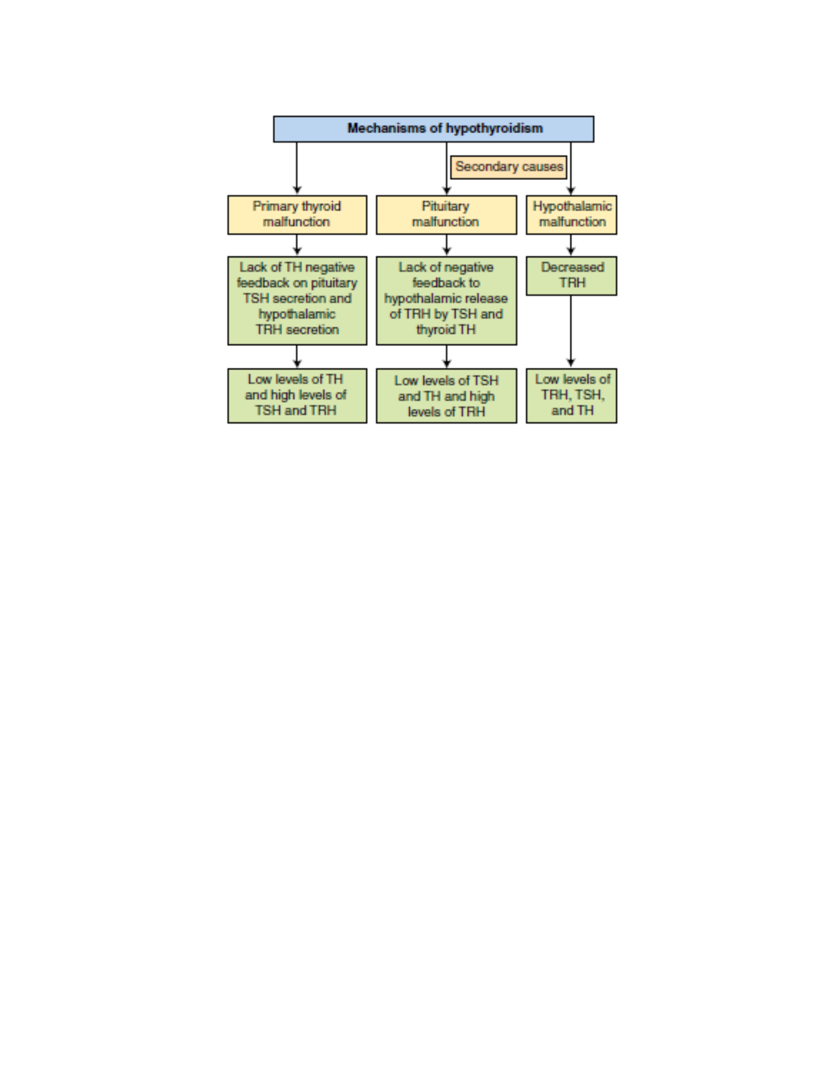
PATHOPHYSIOLOGY In primary hypothyroidism, loss of thyroid function leads to decreased production
of TH and increased secretion of TSH and TRH. The most common causes of primary hypothyroidism in
adults include autoimmune thyroiditis (Hashimoto disease), iatrogenic loss of thyroid tissue after
surgical or radioactive treatment for hyperthyroidism or after head and neck radiation therapy,
medications, and endemic iodine deficiency. Infants and children may present with hypothyroidism
because of congenital defects. Central (secondary) hypothyroidism is caused by the pituitary’s failure to
synthesize adequate amounts of TSH or a lack of TRH. Pituitary tumors that compress surrounding
pituitary cells or the consequences of their treatment are the most common causes of central
hypothyroidism. Other causes include traumatic brain injury, subarachnoid hemorrhage, or pituitary
infarction. Hypothalamic dysfunction results in low levels of TH, TSH, and TRH.
CLINICAL MANIFESTATIONS Hypothyroidism generally affects all body systems and occurs insidiously
over months or years. The decrease in TH level lowers energy metabolism and heat production. The
individual develops a low basal metabolic rate, cold intolerance, lethargy, and slightly lowered basal
body temperature. The decrease in the level of TH can lead to excessive TSH production, which
stimulates thyroid tissue and causes goiter.
The characteristic sign of severe or long-standing hypothyroidism is myxedema, which results from the
altered composition of the dermis and other tissues. The connective tissue fibers are separated by large
amounts of protein and mucopolysaccharide. This complex binds water, producing nonpitting, boggy
edema, especially around the eyes, hands, and feet and in the supraclavicular fossae. The tongue and
laryngeal and pharyngeal mucous membranes thicken, producing thick, slurred speech and hoarseness.
Myxedema coma, a medical emergency, is a diminished level of consciousness associated with severe
hypothyroidism. Signs and symptoms include hypothermia without shivering, hypoventilation,
hypotension, hypoglycemia, and lactic acidosis. Older individuals with severe vascular disease and with
moderate or untreated hypothyroidism are particularly at risk for developing myxedema coma. It also
may occur after overuse of narcotics or sedatives or after an acute illness in hypothyroid individuals.
Symptoms of hypothyroidism in older adults should not be attributed to normal aging changes.
EVALUATION AND TREATMENT The diagnosis of primary hypothyroidism is made by documentation of
the clinical symptoms of hypothyroidism, and measurement of increased levels of TSH and decreased
levels of TH (total T3 and both total and free T4). When hypothyroidism is caused by pituitary
deficiencies, serum TSH levels and basal metabolic rate (BMR) decrease. Hormone replacement therapy
with the hormone levothyroxine is the treatment of choice. The restoration of normal TH levels should
be timed appropriately; a regimen of hormonal therapy depends on the individual’s age, the duration
and severity of the hypothyroidism, and the presence of other disorders, particularly cardiovascular
disorders.
COMPLICATIONS OF DIABETES MELLITUS
Acute Complications of Diabetes Mellitus
The major acute complications of diabetes mellitus are hypoglycemia, diabetic ketoacidosis, and
hyperosmolar hyperglycemic nonketotic syndrome. Somogyi phenomenon and dawn phenomenon also
may be seen.
Hypoglycemia in diabetes is sometimes called insulin shock or insulin reaction. Individuals with type 2
diabetes are at less risk for hypoglycemia than those with type 1 diabetes because they retain relatively
intact glucose counterregulatory mechanisms. However, hypoglycemia does occur in type 2 diabetes
when treatment involves insulin secretogogues (e.g., sulfonylureas) or exogenous insulin. Symptoms
include pallor, tremor, anxiety, tachycardia, palpitations, diaphoresis, headache, dizziness, irritability,
fatigue, poor judgment, confusion, visual disturbances, hunger, seizures, and coma. Treatment requires
immediate replacement of glucose either orally or intravenously. Prevention is achieved with
individualized management of medications and diet, blood glucose monitoring, and education.
Diabetic ketoacidosis (DKA) is a serious complication related to a deficiency of insulin and an increase in
the levels of insulin counterregulatory hormones (catecholamines, cortisol, glucagon, growth hormone).
The American Diabetes Association criteria for the diagnosis of DKA are (1) a serum glucose level >250
mg/dl, (2) a serum bicarbonate level <18 mg/dl, (3) a serum pH <7.30, (4) the presence of an anion gap,
and (5) the presence of urine and serum ketones.75 DKA is much more common in type 1 diabetes
because insulin is more deficient. Insulin normally stimulates lipogenesis and inhibits lipolysis, thus
preventing fat catabolism. With insulin deficiency, lipolysis is enhanced and there is an increase in the
amount of nonesterified fatty acids delivered to the liver. The consequence is increased glyconeogenesis
contributing to hyperglycemia and production of ketone bodies (acetoacetate, hydroxybutyrate, and
acetone) by the mitochondria of the liver at a rate that exceeds peripheral use. Accumulation of ketone
bodies causes a drop in pH, resulting in metabolic acidosis. Symptoms of diabetic ketoacidosis include
Kussmaul respirations (hyperventilation in an attempt to compensate for the acidosis), postural
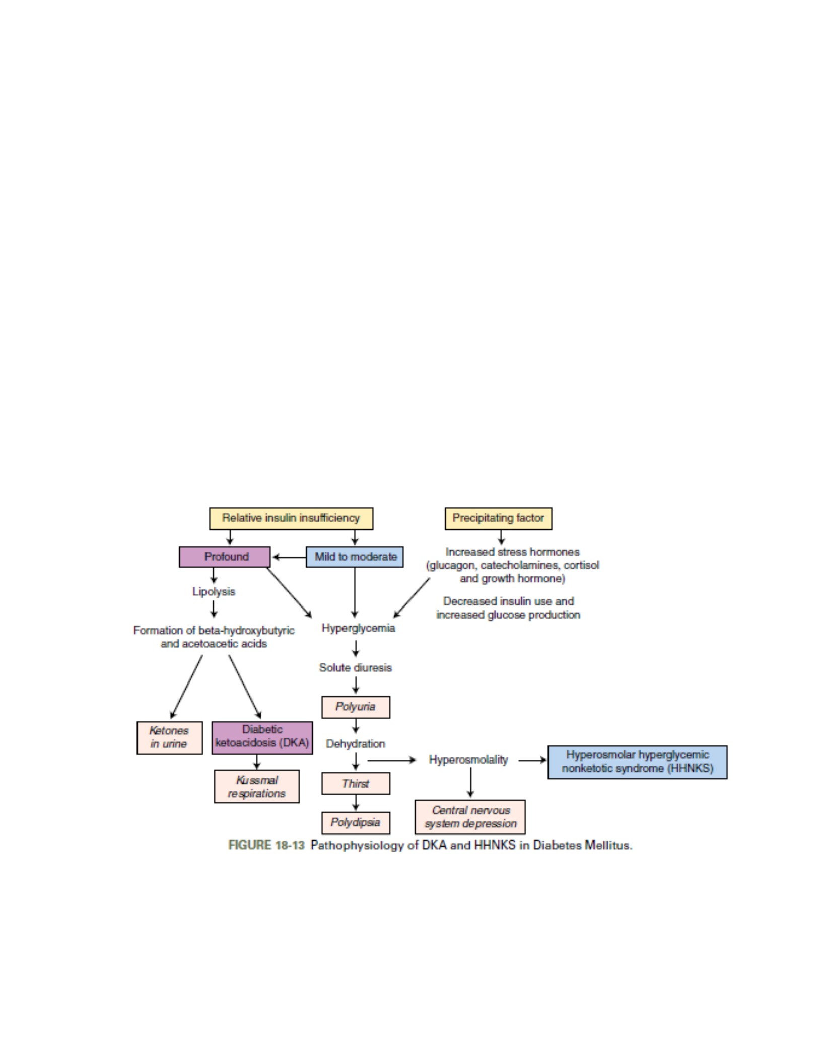
dizziness, central nervous system depression, ketonuria, anorexia, nausea, abdominal pain, thirst, and
polyuria. Hyperosmolar hyperglycemic nonketotic syndrome (HHNKS) is an uncommon but significant
complication of type 2 diabetes mellitus with a high overall mortality. It occurs more often in elderly
individuals who have other comorbidities, including infections or cardiovascular or renal disease. HHNKS
differs from DKA in the degree of insulin deficiency (which is more profound in DKA) and the degree of
fluid deficiency (which is more marked in HHNKS). The clinical features of HHNKS include a serum
glucose level >600 mg/dl, a serum pH >7.30, a serum bicarbonate level >15 mg/dl, aserum osmolarity
>320 mOsm/L, and either absent or small numbers of ketones in the urine and serum.75 Glucose levels
are considerably higher in HHNKS than in DKA because of volume depletion. Because the amount of
insulin required to inhibit fat breakdown is less than that needed for effective glucose transport, insulin
levels are sufficient to prevent excessive lipolysis and ketosis. Clinical manifestations include severe
dehydration; loss of electrolytes, including potassium; and neurologic changes, such as stupor. The
Somogyi effect is a unique combination of hypoglycemia followed by rebound hyperglycemia. The rise in
blood glucose concentration occurs because of counterregulatory hormones (epinephrine, GH,
corticosteroids), which are stimulated by hypoglycemia. They produce gluconeogenesis. Excessive
carbohydrate intake may contribute to the rebound hyperglycemia. The clinical occurrence of Somogyi
effect is controversial. The dawn phenomenon is an early morning rise in blood glucose concentration
with no hypoglycemia during the night. It is related to nocturnal elevations of GH, which decrease
metabolism of glucose by muscle and fat. Increased clearance of plasma insulin also may be involved.
Altering the time and dose of insulin administration manages the problem.
Embolism
Embolism is the obstruction of a vessel by an embolus—a bolus of matter circulating in the
bloodstream. The embolus may consist of a dislodged thrombus; an air bubble; an aggregate of amniotic
fluid; an aggregate of fat, bacteria, or cancer cells; or a foreign substance. An embolus travels in the
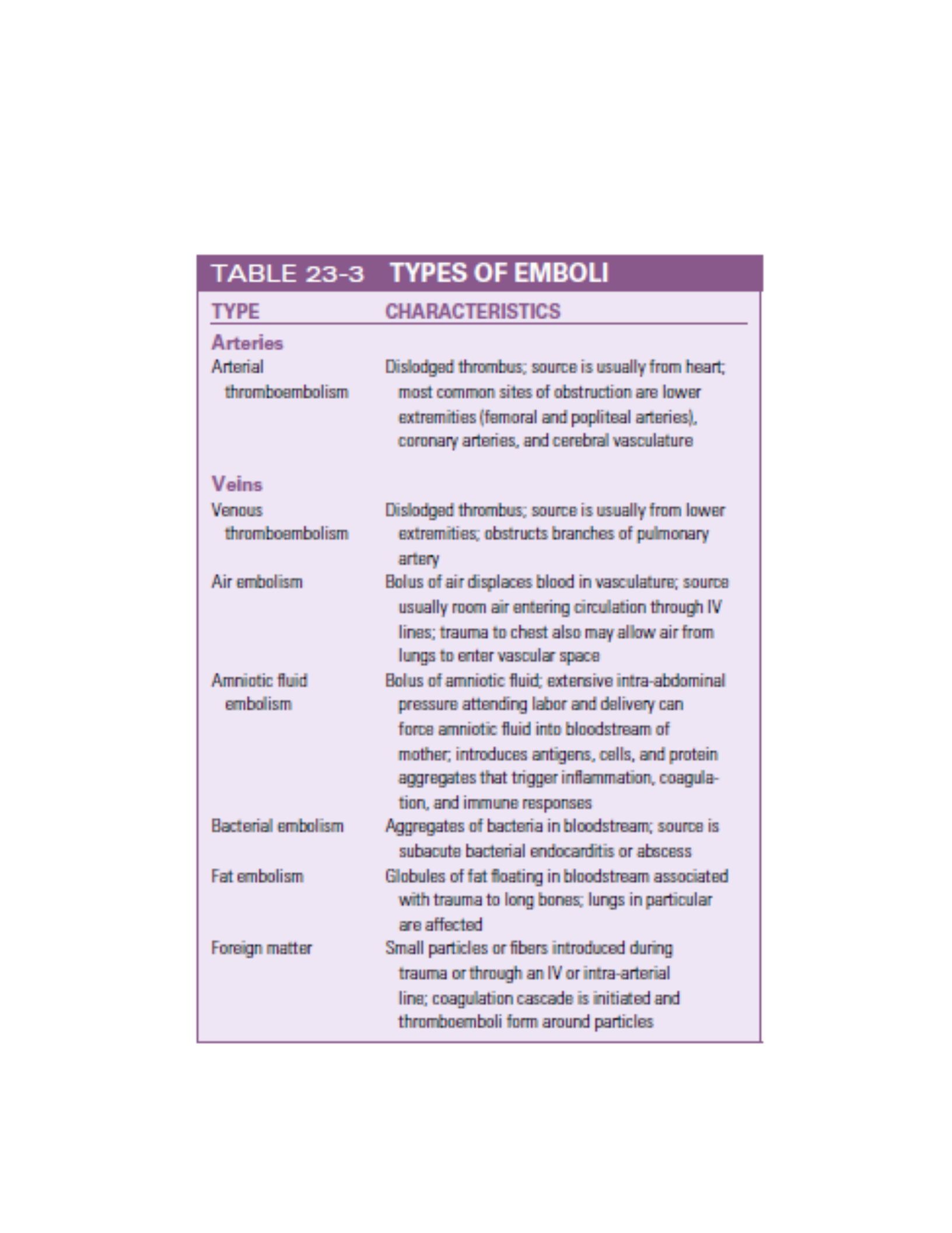
bloodstream until it reaches a vessel through which it cannot fit. No matter how tiny it is, an embolus
will eventually lodge in a systemic or pulmonary vessel determined by its source. Pulmonary emboli
originate on the venous side (mostly from the deep veins of the legs) of the systemic circulation or in the
right heart; arterial emboli most commonly originate in the left heart and are associated with thrombi
after myocardial infarction, valvular disease, left heart failure, endocarditis, and dysrhythmias.

DISORDERS OF BONES
Metabolic Bone Diseases
Metabolic bone disease is characterized by abnormal bone structure that is caused by altered or
inadequate biochemical reactions, which may be attributable to genetics, diet, or hormones.
Osteoporosis
Osteoporosis, or porous bone, is a complex, multifactorial, chronic disease that often progresses silently
for decades until fractures occur. It is the most common disease that affects bone but is not necessarily
a consequence of the aging process because some elderly people retain strong, relatively dense bones.
In osteoporosis, old bone is being resorbed faster than new bone is being made, causing the bones to
lose density, becoming thinner and more porous. A progressive loss of bone mass may continue until the
skeleton is no longer strong enough to support itself. Eventually, bones can fracture spontaneously. As
bone becomes more fragile, falls or bumps that would not have caused a fracture previously now cause
bone to break. Osteoporosis appears to be most severe in the femoral neck, thoracic and lumbar
spine,19 and wrist.
Osteomalacia
Osteomalacia is a metabolic disease characterized by inadequate and delayed mineralization of osteoid
in mature compact and spongy bone. In osteomalacia, the remodeling cycle proceeds normally through
osteoid formation, but mineral calcification and deposition do not occur. Bone volume remains
unchanged, but the replaced bone consists of soft osteoid instead of rigid bone. Rickets is similar to
osteomalacia in pathogenesis, but it occurs in the growing bones of children, whereas osteomalacia
occurs in adult bone. Many factors contribute to the development of osteomalacia, but the most
important is a deficiency of vitamin D. The major risk factors in vitamin D deficiency are diets deficient in
vitamin D, decreased endogenous production of vitamin D, intestinal malabsorption of vitamin D, renal
tubular diseases, certain types of tumors (particularly of mesenchymal origin), and anticonvulsant
therapy.
Paget Disease
Paget disease of bone (PDB, or osteitis deformans) is a state of increased metabolic activity in bone
characterized by abnormal and excessive bone remodeling, both resorption and formation. Chronic
accelerated remodeling eventually enlarges and softens the affected bones. Paget disease can occur in
any bone but most often affects the vertebrae, skull, sacrum, sternum, pelvis, and femur. The disease
process may occur in one or more bones without causing significant clinical manifestations.
PATHOPHYSIOLOGY: Paget disease begins with excessive resorption of spongy bone and deposition of
disorganized bone. The trabeculae diminish, and bone marrow is replaced by extremely vascular fibrous
tissue. The resorption phase of Paget disease is followed by the formation of abnormal new bone at an
accelerated rate. The collagen fibers are disorganized, and glycoprotein levels in the matrix decrease.
Mineralization may extend into the bone marrow. Bone formation is excessive around partially resorbed
trabeculae, causing them to thicken and enlarge. The net result of this accelerated remodeling process is
increased bone fragility and an increased risk for bone tumors.
Infectious Bone Disease: Osteomyelitis

Osteomyelitis is a bone infection most often caused by bacteria; however, fungi, parasites, and viruses
also can cause bone infection. It is further categorized according to the pathogen’s mode of entry into
bone tissue. The most common type is exogenous osteomyelitis, an infection that enters from outside
the body, for example, through open fractures, penetrating wounds, or surgical procedures. In
exogenous osteomyelitis, the infection also can spread from soft tissue into adjacent bone. An example
of this is a diabetic foot infection. Endogenous osteomyelitis is caused by pathogens carried in the blood
from sites of infection elsewhere in the body; the infection can then spread to adjacent soft tissue.
Hematogenous osteomyelitis (a common form of osteomyelitis) is usually found in infants, children, and
elderly persons. In infants, incidence rates among males and females are approximately equal. In
children and older adults, however, males are most commonly affected. Osteomyelitis is a common
complication of sickle cell anemia and low oxygen tension. Staphylococcus aureus remains the primary
microorganism responsible for osteomyelitis. Other microorganisms include group B streptococcus,
Haemophilus influenzae, Salmonella, and gram-negative bacteria. Group B streptococcus and H.
influenzae tend to infect young children; Salmonella infection is associated with sickle cell anemia; and
gram-negative infections are most common in older adults and immunocompromised individuals with
impaired immunity. Mycobacterial, viral, and fungal infections occur in immunocompromised
individuals. Cutaneous, sinus, ear, and dental infections are the primary sources of bacteria in
hematogenous bone infections. Soft tissue infections, disorders of the gastrointestinal tract, infections
of the genitourinary system, and respiratory tract infections are also sources of bacterial contamination.
In addition, infections that occur after total joint replacement procedures are sometimes the cause. The
vulnerability of specific bone depends on the anatomy of its vascular supply. In adults, hematogenous
osteomyelitis is more common in the spine, pelvis, and small bones. Microorganisms reach the
vertebrae through arteries, veins, or lymphatic vessels. The spread of infection from pelvic organs to the
vertebrae is well documented. Vaginal, uterine, ovarian, bladder, and intestinal infections can lead to
iliac or sacral osteomyelitis. Exogenous osteomyelitis can be caused by human bites or fist blows to the
mouth. Superficial animal or human bites inoculate local soft tissue with bacteria that later spread to
underlying bone. Deep bites can introduce microorganisms directly onto bone. The most common
infecting organism in human bites is S. aureus. In animal bites, the most common infecting organism is
Pasteurella multocida, which is part of the normal mouth flora of cats and dogs. Direct contamination of
bones with bacteria can also occur in open fractures or dislocations with an overlying skin wound.
Intervertebral disk surgery and operative procedures involving implantation of large foreign objects,
such as metallic plates or artificial joints, are associated with exogenous osteomyelitis. Local injections
and venous punctures are significant causes of exogenous osteomyelitis. Exogenous osteomyelitis of the
arm and hand bones tends to occur in persons who abuse drugs. In general, persons who are chronically
ill, have diabetes or alcoholism, or are receiving large doses of steroids or immunosuppressive drugs are
particularly susceptible to exogenous osteomyelitis or recurring episodes of this disease.

