


NURS 115 TEST TWO STUDY GUIDE
This is a guide for student preparation for testing in NURS 115. It is understood that the student is responsible
for all course material designated for each test including course readings, in/out of class study / activities
and lecture material (in-class and online).
Immunity and AIDS- HIV: Immunity is the ability to resist infection
Antigen and antibody
Antigens are markers of cells that can either determine it as “self” or “non-self” to the host
oForeign or non-self-antigens can cause a healthy immune response to occur
Antibody: also known as immunoglobulins are produced by cells during a healthy immune response to
neutralize, destroy, and remove specific antigens/ pathogens
Immune response- self vs non self (Bs and Ts) (humoral response vs cell-mediated response): in order for both
responses to begin an antigen has to be recognized as “non-self” and lymphocytes need to proliferate.
Humoral response: B-cells are associated with the humoral response (AKA antibody response) and they
produce antibodies against extracellular antigens
Cell-mediated response: T-cells are associated with the cell mediated response because they fight against
intracellular antigens (viruses, intracellular bacteria); they also regulate the antibody mediated immunity
and responses from helper T and regulatory T cells
oRegulatory T-cells: primary function is to maintain tolerance to self-antigens and prevent
autoimmune response
oHelper T-cells: they help activate B-cells to produce more antibodies, activate macrophages to
destroy microbes, and activate cytotoxic T cells to kill infected cells.
Know types of immunity and terminology (innate/non-specific, acquired/adaptive)
Innate Immunity (classical pathway): Primary action is the inflammatory response and phagocytosis.
oBasic WBC’s besides lymphocytes involved
Adaptive immunity: is also known as acquired immunity because your body is making antibodies to fight
foreign bacteria/antigens
oInvolves B& T cells.
oAntibody mediated/ humoral immunity: b-cells make antibodies to fight extracellular antigens
oCell-mediated: T-cells fight against intracellular viruses/ bacteria
Non-specific immunity: responds to any kind of foreign antigens
oMacrophages, neutrophils, and dendritic cells respond to ANY foreign antigen, they don’t target
specific foreign bodies.
oAnatomical barriers: skin, mucous membranes,
oBiochemical barriers: acid in the stomach.
Components of the immune system
WBC’s
oNeutrophils: part of nonspecific immunity because they react to anything foreign that makes its
way into the body
oLymphocytes: T-cells and B-cells
Natural Killer or (NK cells): recognize whole classes of cells not specific types; they go
and lyse tumor cells
oMonocytes: found in the bloodstream and they are responsible for phagocytizing foreign bodies
Macrophages are monocytes that leave the bloodstream to roam in the tissues to help
fight foreign bodies in infected areas of tissue
oEosinophils: mainly serve to kill worm-like parasites; they also break down the chemicals
released by basophils which stimulate inflammation (histamine and heparin)
oBasophils: travel to and accumulate at sites of injury; responsible for releasing chemicals that
stimulate inflammation
Mast cells are basophils that are non-motile and anchored down in certain connective
tissues
Role of lymphocytes and cytokines
Lymphocytes: responsible for recognizing and reacting to antigens and creating antibodies to respond to
those said antigens
Cytokines: are secreted by macrophages, B & T cells, and mast cells and they act as chemical messengers
to promote and regulate the immune response
Immunoglobulins (antibodies) are produced by what cells and where can you find them, how/when do they
Respond
Immunoglobulins are produced by B cells. They respond to antigens that are not “self”.
oTypes of Immunoglobulins
IgG: Most common immunoglobulin; found in breast milk; Can cross the placenta and
can cause Hemolytic disease of the newborn
IgA: Found in secretions such as mucous, oil on the skin, tears; also found in breast milk
to provide immunity to baby
IgM: First Ig factor to respond to antigens; acts as an antigen-antibody receptor on B-
cells; HIGH levels of IgM in newborn means INFECTION
IgD: acts as antigen-antibody receptor on B cells; necessary for the maturation of B-cells;
common in the chest and abdominal cavity
IgE: Binds to basophils and mast cells and causes the release of inflammatory chemicals;
found commonly in the lungs, skin, and mucous membranes
Mediators in allergic response, and from where are they released; Mast cells play a major role in an allergic
response by releasing potent chemical mediators:
Histamine: increases vascular permeability and diameter of vessels to allow the passage of WBC’s and
some proteins into the tissue to combat infections
Prostaglandin: modulates inflammation and causes bronchoconstriction and vasodilation; also attracts
neutrophils
Leukotrienes: RECRUIT neutrophils to areas of tissues that are damaged; PROMOTES production of
cytokines; cause inflammatory response to last longer
Chemokines: help direct immune cells to areas of infection so that they can destroy foreign bodies
Cytokines: function as chemical mediators; promote the proliferation of T-cells; secreted by
macrophages, mast cells, and B & T cells
Test blueprint:
Immunity 40%
Inflammation & Healing 14%
Diabetes Mellitus 20%
Cardiovascular 26%
Types of hypersensitivity reactions: Primary immune response is when the body encounters an infectious agent
the first time and a Secondary immune response is every interaction the body has with that agent again.
-Anaphylaxis: an acute reaction to an antigen which the body is prone to.
Anaphylactic Shock: an extreme reaction to an antigen the body is prone to
*Dizziness
*Loss of consciousness
*Labored breathing due to bronchoconstriction and swelling of breathing tubes
*Cyanosis
*Low BP (Heart failure is another condition experienced)
1. Type 1: Local and Systemic anaphylaxis (involves basophils and mast cells rapidly releasing
histamines); your vessels dilate so severely that your blood pressure drops drastically suffocating tissues.
a. Allergic reaction to for example peanuts, latex, bee sting
b. Primary: (happens QUICKLY after exposure) Vasodilation; vascular leakage; and bronchoconstriction
c. Secondary: Eosinophils get involved and epithelial tissue is destroyed
i. Can occur hours to days after exposure to stimulant
d. Anaphylaxis: rapid antigen-antibody reaction
i. IgE is released and binds to Mast cells and basophils which leads to the release of histamine
e. Treatment
i. Give epinephrine which is a vasoconstrictor and broncho-relaxant
ii. Support w/ O2
iii. Antihistamine (reduce inflammation) drugs and corticosteroids (reduce inflammation in the airways)
iv. BEST TREATMENT IS PREVENTION OF EXPOSURE
2. Type 2: Antibody mediated hypersensitivity
a. IgG & IgM are responsible for the activation of compliment in the blood
b. *Hemolytic disease of the newborn*
c. *Myasthenia Gravis*: serious weakening of skeletal muscles because antibodies are blocking the
receptors for nervous signals to be transmitted to the muscle
3. Type 3: Immune complex mediated (Floating: insoluble antigen-antibody complex is formed)
a. Signs & Symptoms: Fever, rash
b. Serum sickness: similar to an allergic reaction where too many antibodies are formed and they form
too many complexes; occurs when exposed to drugs that contain antigen
c. Arthus reaction: the formation of antigen-antibody complexes
(Antibodies formed to the antibodies: second time receiving antivenom for snake/spider bite)
i. It is a local inflammation of vasculature
d. Treatment
i. Remove drug that initiated this response
ii. Response is local: A.) when antigen is injected into skin (B.) Pain, swelling, and redness
4. Type 4: Cell mediated hypersensitivity or delayed hypersensitivity (associated with T-cells)
a. DELAYED HYPERSENSITIVITY REACTION
b. Graft V.S. Host
i. Whatever you are receiving is the aggressor
1. Transplanted feature has functioning immune component that responds negatively to the antigen
of the host
2. Recipient has compromised immune system
c. TB test: injecting a small amount of special fluid underneath the skin and if a raised hard bump forms
at the site of injection or swelling on the arm occurs TB germs are present and the test returns Positive
Opportunistic pathogens: germs/ pathogens that take advantage of a hose with a weakened immune system
Stages of illness:
1. Incubation: exposed to & pathogen has been obtained by the (now) host
2. Prodromal: Feeling bad & general discomfort= Malaise; no symptoms yet
3. Acute: Body is fighting pathogens; this stage presents the biggest manifestation of symptoms
4. Convalescent: Pathogen has been addressed but the body is weak and physically exhausted from fighting
foreign antigens. This is when the body is most vulnerable because it has a severely low count of
antibodies
5. Resolution: pathogen is no longer present in system and antibody count is back up to normal levels
Chain of infection
A. Etiologic agent: the microorganism or bacteria
B. Reservoir or Source: where the etiologic agent is obtained by a host (i.e. a doorknob or someone else’s
sneeze cloud
C. Portal of Exit: how the etiologic agent leaves a host to be transmitted to another organism
a. Coughing, vomiting, feces, open wounds, blood & other bodily fluids
D. Method of Transmission: how the etiologic agent is transmitted i.e. blood, coughing, and touch
a. Nurses are the largest mode of transmission which can lead to nosocomial infection.
E. Portal of entry: how the etiologic agent enters a new host i.e. touching the face, mouth, nose, eyes, open
wounds
F. Susceptible: a measure of how strained a potential host’s immunity is
a. Low susceptibility: antibodies are already formed against etiologic agent and host is healthy
b. High: Just got done fighting an infection (Convalescent phase of infection), under copious
amounts of stress, old/ young patients
Nosocomial infection: or a hospital acquired infection is an infection that is spread by various means to
susceptible patients
Types and role of WBCs in infection
Neutrophils: promote phagocytosis; promotion of pro-inflammatory cytokines
Lymphocytes: limiting and preventing the expansion of microorganisms
Monocytes: phagocytize foreign bodies and fight at sites of infection
Eosinophils: fight against parasitic infections and consume foreign substances
Basophils: release heparin and histamine to promote inflammation to fight infections; these chemicals
released help promote vascular permeability and blood flow to allow more immune cells to travel to sites
of infection and fight
Major histocompatibility complex (MHC I and MHC II): the system that the body uses to determine whether or
not cells are “self” or “non-self”
MHC 1: When this protein is presented with a self-antigen it is known to other cells that this cell is
healthy. However, when this protein complex presents an antigen that is foreign, it is recognized by other
cells that this cell is infected and cytotoxic T cells or CD8’s come and kill the infected cell.
oFlag saying “kill me now”
MHC 2: Protein complex that is present on antigen presenting cells such as B-cells. This complex binds
to foreign antigens and presents them to helper T cells or CD4 cells. When a CD4 detects a foreign
antigen on a presenting cell by binding to it, it releases cytokines and other chemicals which help
promote costimulation and proliferation of memory b cells, as well as helper, regulatory, and memory T
cells
oLike a cell holding a “wanted” poster for the bad guy antigen.
HIV/AIDS—etiology, manifestations, treatment, prevention******
CD4 levels: 500-1500
HIV: (Human immunodeficiency virus) is a virus that targets and fights cells that help the body fight
infection which makes them vulnerable to other viruses and diseases (opportunistic pathogens)
oCD4 levels: <500
AIDS: chronic condition of HIV which is acquired through transmission or contact with infected bodily
fluids (except for tears and saliva)
oCD4 levels: <200
Cardiovascular
Pathway of blood flow through the heart
1.) Systemic circulation to sup. and inf. Vena cava, (2.) right atria, (3.) tricuspid valve, (4.) right
ventricle, (5.) pulmonary semilunar valve, (6.) pulmonary arteries, (7.) pulmonary circuit (lungs), (8.)
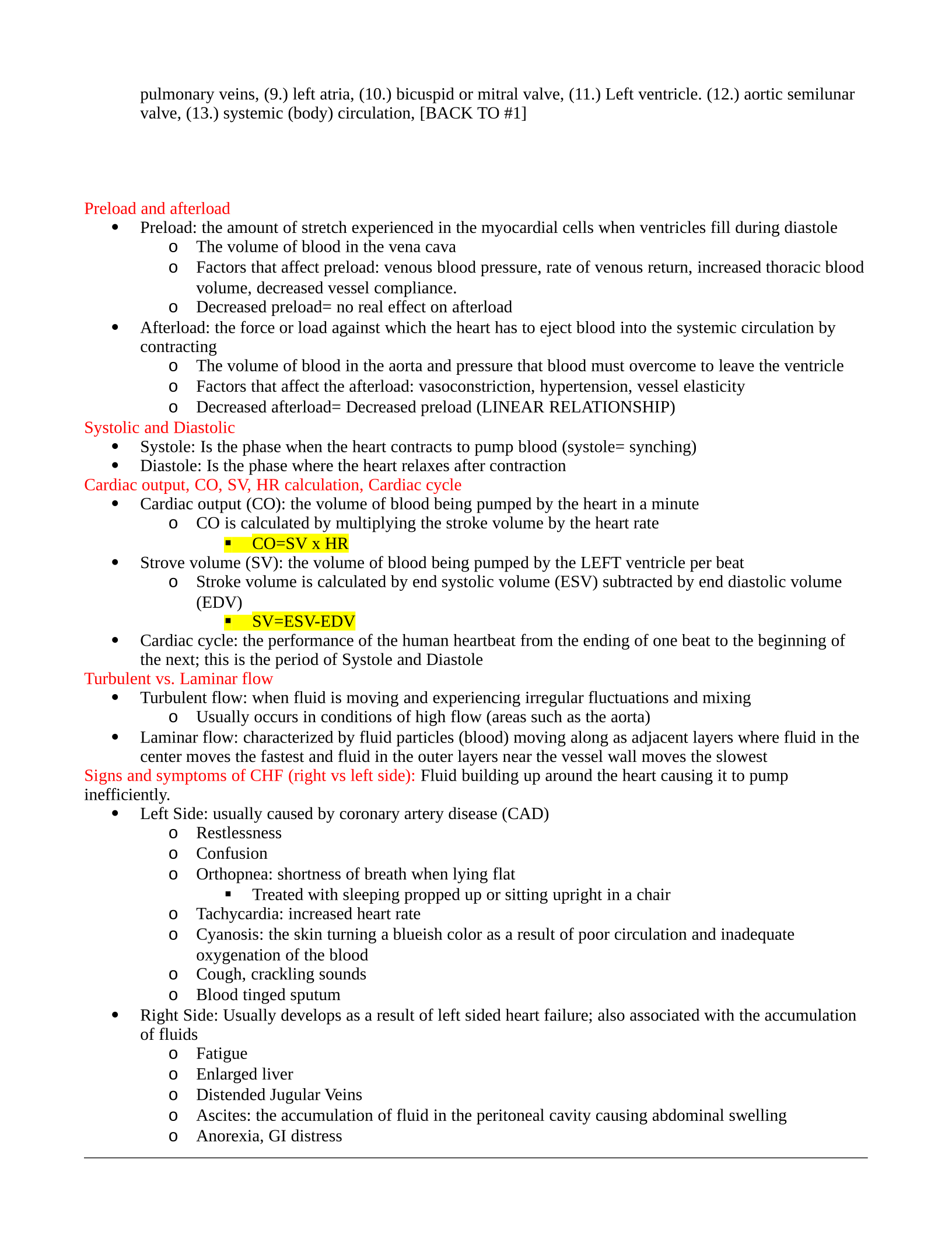
pulmonary veins, (9.) left atria, (10.) bicuspid or mitral valve, (11.) Left ventricle. (12.) aortic semilunar
valve, (13.) systemic (body) circulation, [BACK TO #1]
Preload and afterload
Preload: the amount of stretch experienced in the myocardial cells when ventricles fill during diastole
oThe volume of blood in the vena cava
oFactors that affect preload: venous blood pressure, rate of venous return, increased thoracic blood
volume, decreased vessel compliance.
oDecreased preload= no real effect on afterload
Afterload: the force or load against which the heart has to eject blood into the systemic circulation by
contracting
oThe volume of blood in the aorta and pressure that blood must overcome to leave the ventricle
oFactors that affect the afterload: vasoconstriction, hypertension, vessel elasticity
oDecreased afterload= Decreased preload (LINEAR RELATIONSHIP)
Systolic and Diastolic
Systole: Is the phase when the heart contracts to pump blood (systole= synching)
Diastole: Is the phase where the heart relaxes after contraction
Cardiac output, CO, SV, HR calculation, Cardiac cycle
Cardiac output (CO): the volume of blood being pumped by the heart in a minute
oCO is calculated by multiplying the stroke volume by the heart rate
CO=SV x HR
Strove volume (SV): the volume of blood being pumped by the LEFT ventricle per beat
oStroke volume is calculated by end systolic volume (ESV) subtracted by end diastolic volume
(EDV)
SV=ESV-EDV
Cardiac cycle: the performance of the human heartbeat from the ending of one beat to the beginning of
the next; this is the period of Systole and Diastole
Turbulent vs. Laminar flow
Turbulent flow: when fluid is moving and experiencing irregular fluctuations and mixing
oUsually occurs in conditions of high flow (areas such as the aorta)
Laminar flow: characterized by fluid particles (blood) moving along as adjacent layers where fluid in the
center moves the fastest and fluid in the outer layers near the vessel wall moves the slowest
Signs and symptoms of CHF (right vs left side): Fluid building up around the heart causing it to pump
inefficiently.
Left Side: usually caused by coronary artery disease (CAD)
oRestlessness
oConfusion
oOrthopnea: shortness of breath when lying flat
Treated with sleeping propped up or sitting upright in a chair
oTachycardia: increased heart rate
oCyanosis: the skin turning a blueish color as a result of poor circulation and inadequate
oxygenation of the blood
oCough, crackling sounds
oBlood tinged sputum
Right Side: Usually develops as a result of left sided heart failure; also associated with the accumulation
of fluids
oFatigue
oEnlarged liver
oDistended Jugular Veins
oAscites: the accumulation of fluid in the peritoneal cavity causing abdominal swelling
oAnorexia, GI distress
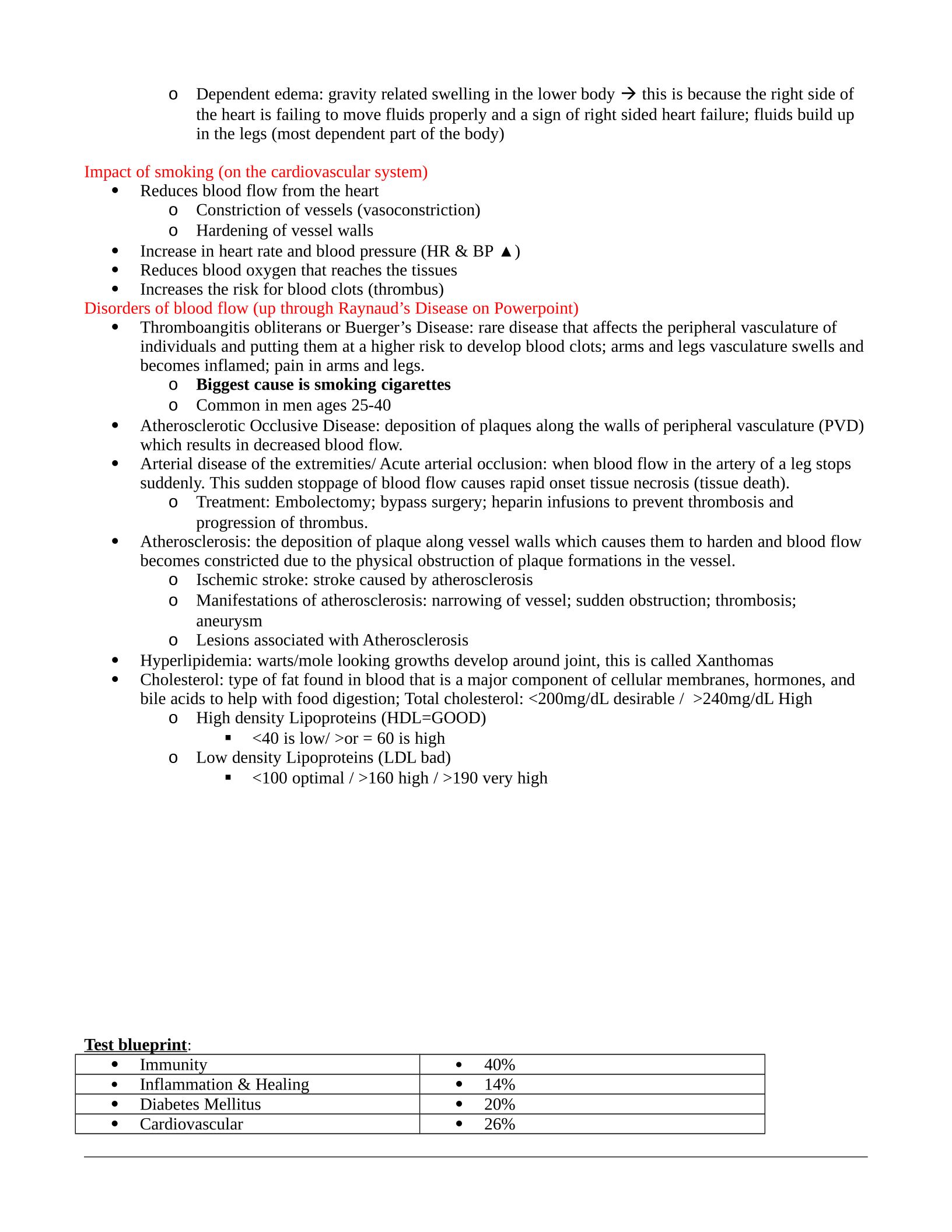
oDependent edema: gravity related swelling in the lower body this is because the right side of
the heart is failing to move fluids properly and a sign of right sided heart failure; fluids build up
in the legs (most dependent part of the body)
Impact of smoking (on the cardiovascular system)
Reduces blood flow from the heart
oConstriction of vessels (vasoconstriction)
oHardening of vessel walls
Increase in heart rate and blood pressure (HR & BP ▲)
Reduces blood oxygen that reaches the tissues
Increases the risk for blood clots (thrombus)
Disorders of blood flow (up through Raynaud’s Disease on Powerpoint)
Thromboangitis obliterans or Buerger’s Disease: rare disease that affects the peripheral vasculature of
individuals and putting them at a higher risk to develop blood clots; arms and legs vasculature swells and
becomes inflamed; pain in arms and legs.
oBiggest cause is smoking cigarettes
oCommon in men ages 25-40
Atherosclerotic Occlusive Disease: deposition of plaques along the walls of peripheral vasculature (PVD)
which results in decreased blood flow.
Arterial disease of the extremities/ Acute arterial occlusion: when blood flow in the artery of a leg stops
suddenly. This sudden stoppage of blood flow causes rapid onset tissue necrosis (tissue death).
oTreatment: Embolectomy; bypass surgery; heparin infusions to prevent thrombosis and
progression of thrombus.
Atherosclerosis: the deposition of plaque along vessel walls which causes them to harden and blood flow
becomes constricted due to the physical obstruction of plaque formations in the vessel.
oIschemic stroke: stroke caused by atherosclerosis
oManifestations of atherosclerosis: narrowing of vessel; sudden obstruction; thrombosis;
aneurysm
oLesions associated with Atherosclerosis
Hyperlipidemia: warts/mole looking growths develop around joint, this is called Xanthomas
Cholesterol: type of fat found in blood that is a major component of cellular membranes, hormones, and
bile acids to help with food digestion; Total cholesterol: <200mg/dL desirable / >240mg/dL High
oHigh density Lipoproteins (HDL=GOOD)
<40 is low/ >or = 60 is high
oLow density Lipoproteins (LDL bad)
<100 optimal / >160 high / >190 very high
Test blueprint:
Immunity 40%
Inflammation & Healing 14%
Diabetes Mellitus 20%
Cardiovascular 26%
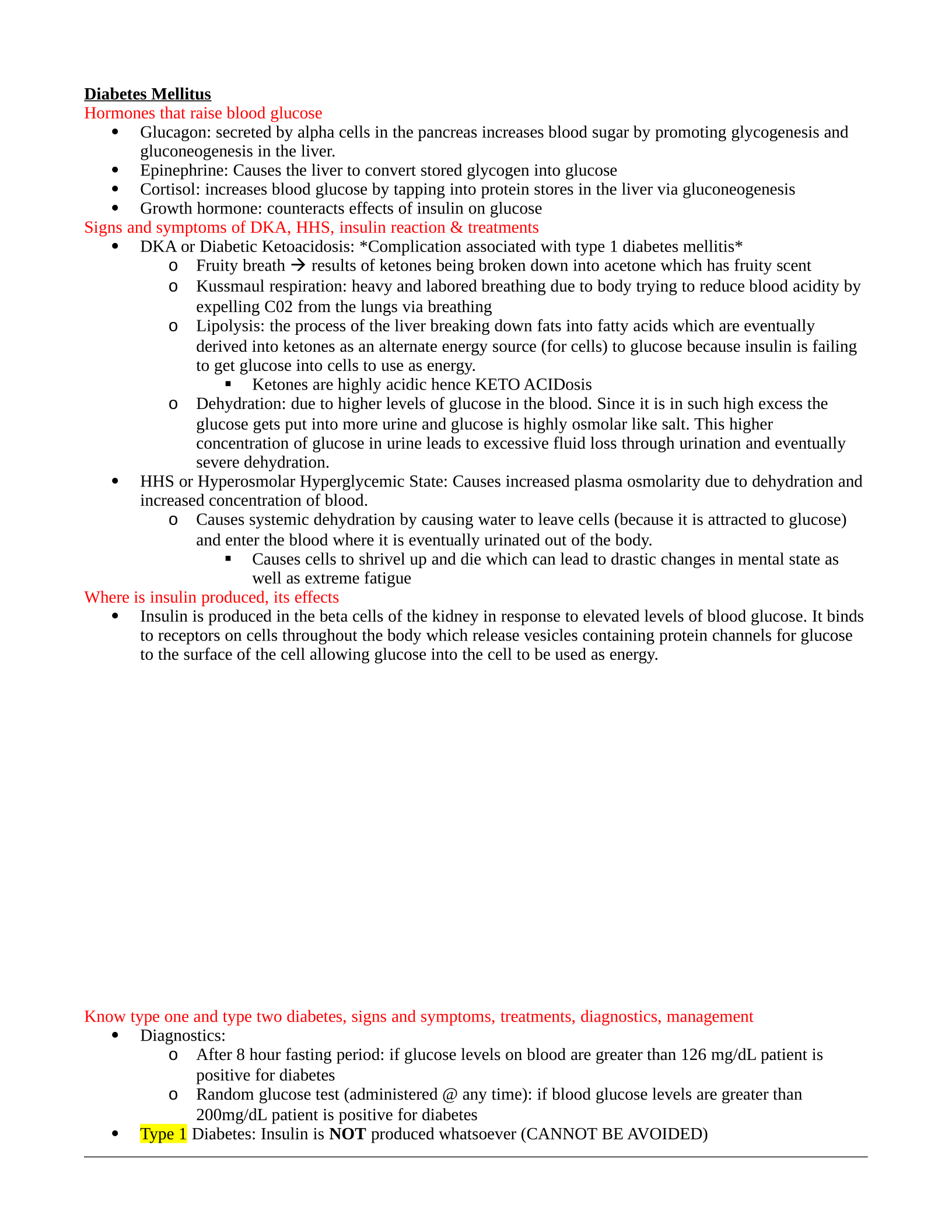
Diabetes Mellitus
Hormones that raise blood glucose
Glucagon: secreted by alpha cells in the pancreas increases blood sugar by promoting glycogenesis and
gluconeogenesis in the liver.
Epinephrine: Causes the liver to convert stored glycogen into glucose
Cortisol: increases blood glucose by tapping into protein stores in the liver via gluconeogenesis
Growth hormone: counteracts effects of insulin on glucose
Signs and symptoms of DKA, HHS, insulin reaction & treatments
DKA or Diabetic Ketoacidosis: *Complication associated with type 1 diabetes mellitis*
oFruity breath results of ketones being broken down into acetone which has fruity scent
oKussmaul respiration: heavy and labored breathing due to body trying to reduce blood acidity by
expelling C02 from the lungs via breathing
oLipolysis: the process of the liver breaking down fats into fatty acids which are eventually
derived into ketones as an alternate energy source (for cells) to glucose because insulin is failing
to get glucose into cells to use as energy.
Ketones are highly acidic hence KETO ACIDosis
oDehydration: due to higher levels of glucose in the blood. Since it is in such high excess the
glucose gets put into more urine and glucose is highly osmolar like salt. This higher
concentration of glucose in urine leads to excessive fluid loss through urination and eventually
severe dehydration.
HHS or Hyperosmolar Hyperglycemic State: Causes increased plasma osmolarity due to dehydration and
increased concentration of blood.
oCauses systemic dehydration by causing water to leave cells (because it is attracted to glucose)
and enter the blood where it is eventually urinated out of the body.
Causes cells to shrivel up and die which can lead to drastic changes in mental state as
well as extreme fatigue
Where is insulin produced, its effects
Insulin is produced in the beta cells of the kidney in response to elevated levels of blood glucose. It binds
to receptors on cells throughout the body which release vesicles containing protein channels for glucose
to the surface of the cell allowing glucose into the cell to be used as energy.
Know type one and type two diabetes, signs and symptoms, treatments, diagnostics, management
Diagnostics:
oAfter 8 hour fasting period: if glucose levels on blood are greater than 126 mg/dL patient is
positive for diabetes
oRandom glucose test (administered @ any time): if blood glucose levels are greater than
200mg/dL patient is positive for diabetes
Type 1 Diabetes: Insulin is NOT produced whatsoever (CANNOT BE AVOIDED)
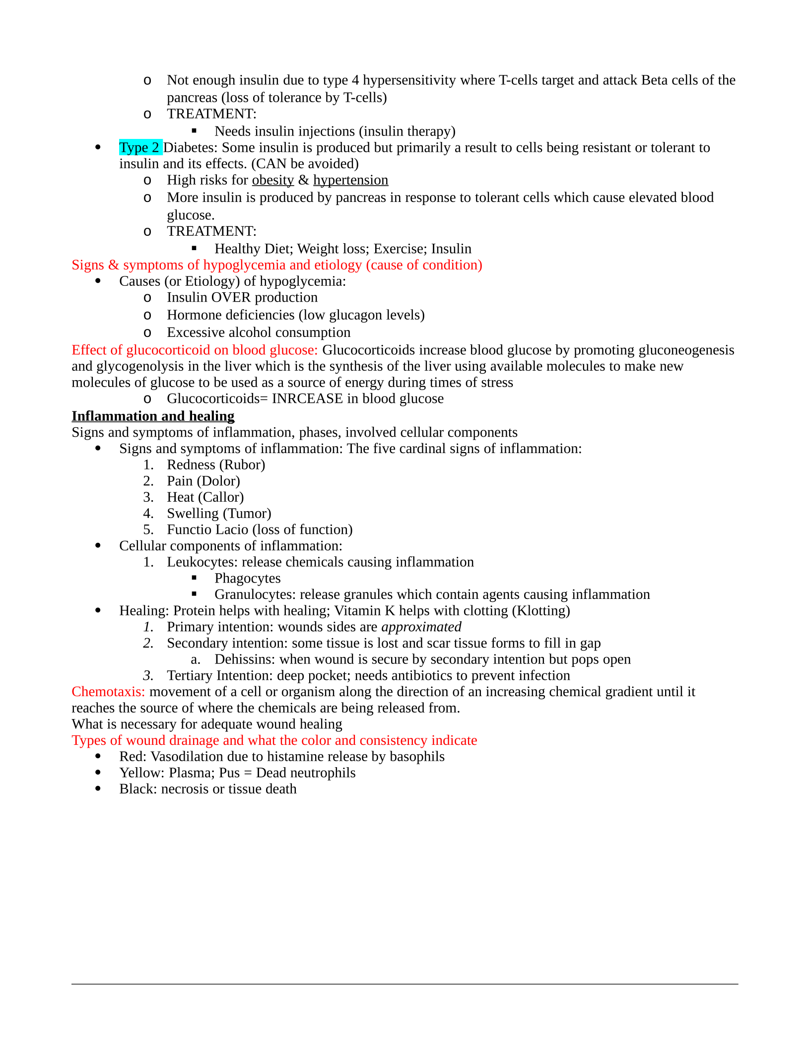
oNot enough insulin due to type 4 hypersensitivity where T-cells target and attack Beta cells of the
pancreas (loss of tolerance by T-cells)
oTREATMENT:
Needs insulin injections (insulin therapy)
Type 2 Diabetes: Some insulin is produced but primarily a result to cells being resistant or tolerant to
insulin and its effects. (CAN be avoided)
oHigh risks for obesity & hypertension
oMore insulin is produced by pancreas in response to tolerant cells which cause elevated blood
glucose.
oTREATMENT:
Healthy Diet; Weight loss; Exercise; Insulin
Signs & symptoms of hypoglycemia and etiology (cause of condition)
Causes (or Etiology) of hypoglycemia:
oInsulin OVER production
oHormone deficiencies (low glucagon levels)
oExcessive alcohol consumption
Effect of glucocorticoid on blood glucose: Glucocorticoids increase blood glucose by promoting gluconeogenesis
and glycogenolysis in the liver which is the synthesis of the liver using available molecules to make new
molecules of glucose to be used as a source of energy during times of stress
oGlucocorticoids= INRCEASE in blood glucose
Inflammation and healing
Signs and symptoms of inflammation, phases, involved cellular components
Signs and symptoms of inflammation: The five cardinal signs of inflammation:
1. Redness (Rubor)
2. Pain (Dolor)
3. Heat (Callor)
4. Swelling (Tumor)
5. Functio Lacio (loss of function)
Cellular components of inflammation:
1. Leukocytes: release chemicals causing inflammation
Phagocytes
Granulocytes: release granules which contain agents causing inflammation
Healing: Protein helps with healing; Vitamin K helps with clotting (Klotting)
1. Primary intention: wounds sides are approximated
2. Secondary intention: some tissue is lost and scar tissue forms to fill in gap
a. Dehissins: when wound is secure by secondary intention but pops open
3. Tertiary Intention: deep pocket; needs antibiotics to prevent infection
Chemotaxis: movement of a cell or organism along the direction of an increasing chemical gradient until it
reaches the source of where the chemicals are being released from.
What is necessary for adequate wound healing
Types of wound drainage and what the color and consistency indicate
Red: Vasodilation due to histamine release by basophils
Yellow: Plasma; Pus = Dead neutrophils
Black: necrosis or tissue death
Powered by TCPDF (www.tcpdf.org)

