


Peris Moraa Mokua
1
CELL BIOLOGY
General introduction
Biology: is the study of living organisms.
Cell biology: is the biological science that entails the study of structure, function, molecular
organization, growth and genetics of cells. The great percentage of study is devoted to the
study of structures and functions of specialized cells.
Cell: is the basic unit of life. They are tiny and complex bodies. Hence it is difficult to see
their structure, understand their molecular composition and further to discern the various
functions of their components.
Life begins with cells
Like ourselves, the individual cells that form our bodies can grow, reproduce, process
information, respond to stimuli, and carry out an amazing array of chemical reactions. These
abilities define life. We and other multicellular organisms contain billions or trillions of cells
organized into complex structures, but many organisms consist of a single cell. Even simple
unicellular organisms exhibit all the hallmark properties of life, indicating that the cell is the
fundamental unit of life.
THE CELL
Cell is the basic structural and functional unit of all known living organisms. It can also be
defined as Basic unit of life
Most plant and animal cells are tiny/small size and are visible only under the microscope:-
1-100µm in size
Prokaryotic: 1-10 μm
Eukaryotic: 10 - 100 μm
Where 1 μm = .001 mm
All living things are made of cells: 1 or >1
Bacteria are unicellular organisms, but humans are multicellular (hierarchy:
body>organ>tissue>cell).
Characteristics of cells
1. Cells as open systems: a cell is an open system which exchanges both matter and
energy with its environment.
2. Cells are self-regulating system: cells functions on maximum economy basis hence
reactions must be self-regulatory and self-controlled.
3. Cells are self-replicating system: reproduction; cell division
PHYSIOLOGICAL SIGNIFICANCE OF CELL SIZE
As cell size increases the volume increases much faster than the surface area. Cells obtain
nutrients, gain information and get rid of waste through their plasma membrane. As cell size
increases, a cell’s ability to exchange with its environment becomes limited by the amount of
membrane area that is available for exchange.

Peris Moraa Mokua
2
Why are cells so small?
To have increased surface area to volume ratio so as to use simple mechanisms to
obtain/get rid of nutrients/wastes respectively
Enable them to penetrate places
Easy to divide
So as many cells can occupy a small volume; tissues, organ size
DISCOVERY OF CELLS
1665- English Scientist, Robert Hooke, discovered cells while looking at a thin slice of cork
(Obtained from a bark of a tree). He observed small boxes and called them cells. He
described the cells as tiny boxes or a honeycomb. He thought that cells only existed in plants
and fungi
Anton van Leuwenhoek
1673- Used a handmade microscope to observe pond scum & discovered single-
celled organisms
He called them “animalcules”
He also observed blood cells from fish, birds, frogs, dogs, and humans
Therefore, it was known that cells are found in animals as well as plants
150-200 Year Gap
Between the Hooke/Leuwenhoek discoveries and the mid-19th century, very little cell
advancements were made.
This is probably due to the widely accepted, traditional belief in Spontaneous
Generation. Examples:
Mice from dirty clothes/corn husks
Maggots from rotting meat
Much doubt existed around Spontaneous Generation
From 1824 to 1830
Works of Dutrochet, Turpin, and Meyers among others not only confirmed the universality of
cellular structure but also hinted at the concept of cellular autonomy in both the
morphological and physiological sense
Discovery of the Nucleus
Robert Brown (1831) was the first to report the discovery the nucleus of the cell.
1832: Dumortier reported on cell division in algae, and from 1835 to 1839, von Mohl
sketched many features of mitosis.
These workers might be considered founders of the specialized aspect of cytology sometimes
known as karyology

Peris Moraa Mokua
3
DEVELOPMENT OF CELL THEORY
1838- German Botanist, Matthias Jakob Schleiden, concluded that all plant parts are
made of cells
1839- German physiologist, Theodor Schwann, who was a close friend of Schleiden, stated
that all animal tissues are composed of cells.
1858- Rudolf Virchow, German physician, after extensive study of cellular pathology, he
concluded that cells must arise from preexisting cells.
Components of the Cell Theory
The 3 Basic Components of the Cell Theory were now complete: that’s
1. All organisms are composed of one or more cells.
2. The cell is the basic unit of life in all living things.
3. All cells are produced by the division of preexisting cells.
Modern Cell Theory
Modern Cell Theory contains 4 statements, in addition to the original Cell Theory:
The cell contains hereditary information (DNA) which is passed on from cell to cell
during cell division.
All cells are basically the same in chemical composition and metabolic activities.
All basic chemical & physiological functions are carried out inside the cells
(movement, digestion, etc.)
Cell activity depends on the activities of sub-cellular structures within the cell
(organelles, nucleus, plasma membrane)
Uses of Cell Theory
The basic discovered truths about cells, listed in the Cell Theory, are the basis for
things such as: Disease/Health/Medical Research and Cures (AIDS, Cancer, Vaccines,
Cloning, Stem Cell Research, etc.)
TYPES OF CELLS
There are two major types of cells: The Prokaryotic cell and The Eukaryotic cells
Prokaryotic and eukaryotic. "Karyose" comes from a Greek word which means "kernel," as in
a kernel of grain. In biology, one use this word root to refer to the nucleus of a cell. "Pro"
means "before," and "eu" means "true," or "good." So "Prokaryotic" means "before a
nucleus," and "eukaryotic" means "possessing a true nucleus."
Prokaryotic cells have no nuclei, while eukaryotic cells do have true nuclei. This is far
from the only difference between these two cell types.
Peris Moraa Mokua
4
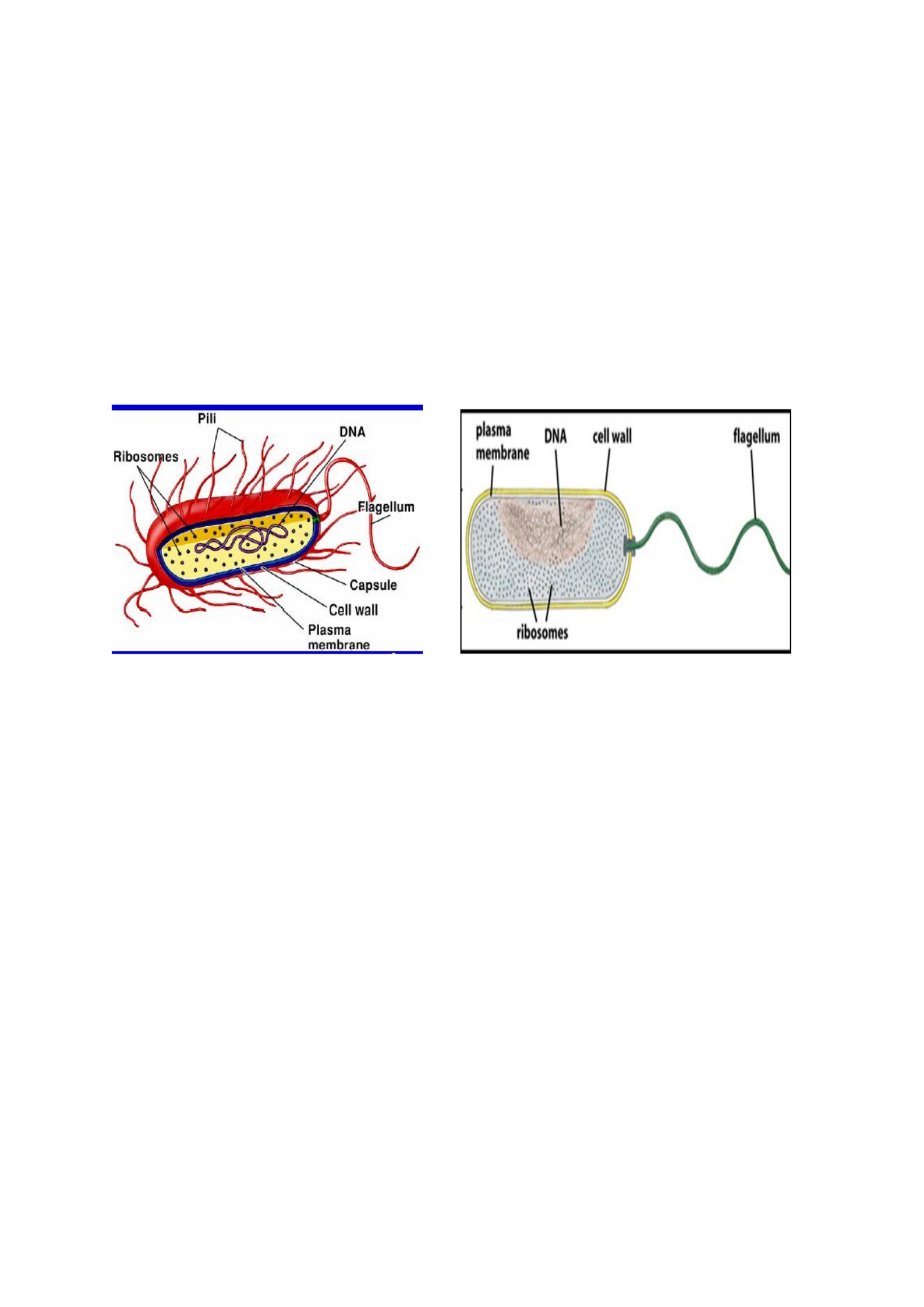
Prokaryotic cells
Consist of simple closed compartment that is surrounded by the plasma membrane
They lack a defined nucleus
Their internal organization is simple with no organelles
Prokaryotes with this kind of organization include:
Bacteria
Cyano-bacteria
Blue green algae
Prokaryotic cell structure
Fig. 1: Structure of prokaryotic cell
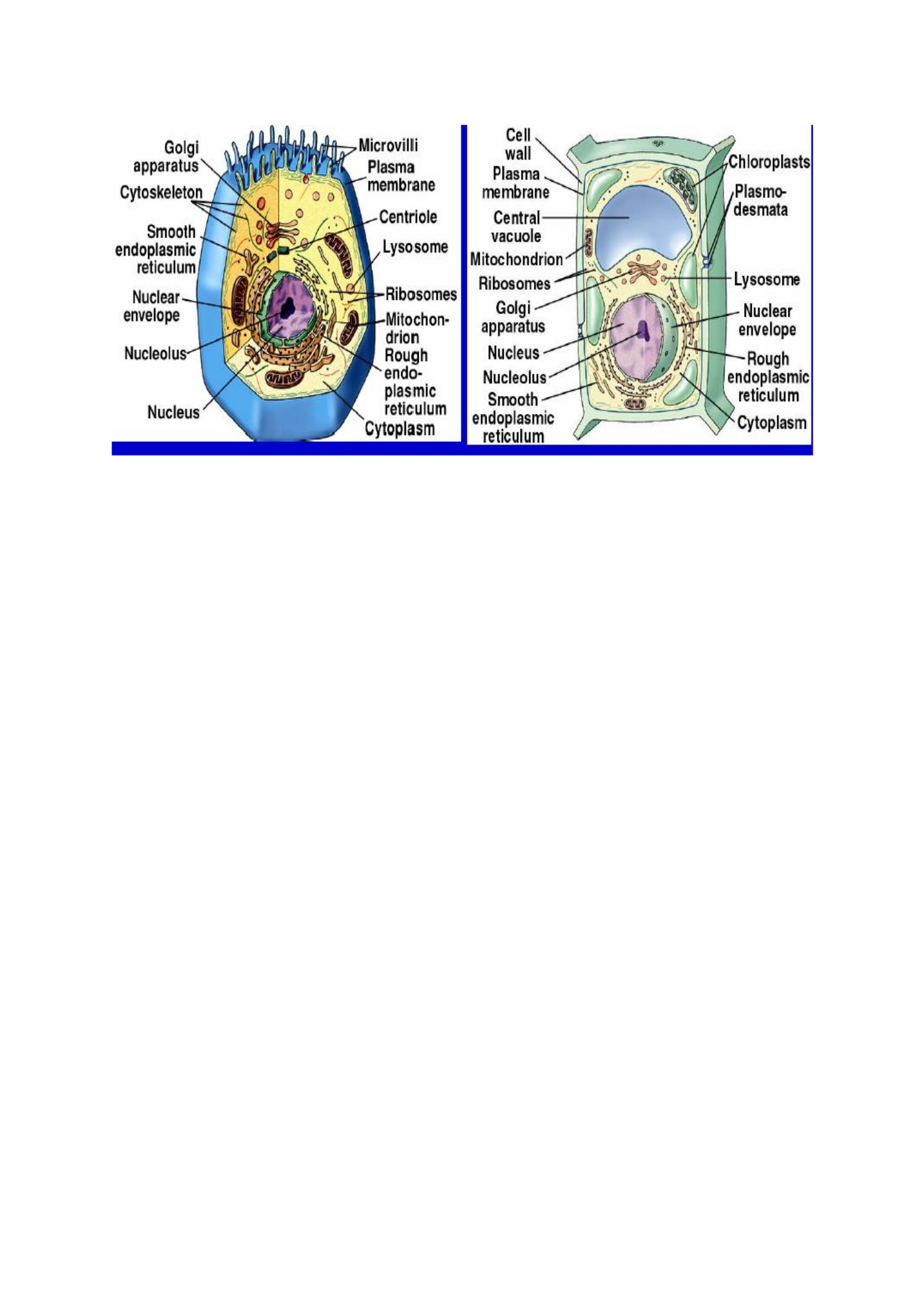
Eukaryotic cells
Are larger, with a typical plasma membrane - some with a cell wall
Have many organelles and other interior spaces enclosed by membranes:
Examples of organelles: Nucleus, Endoplasmic reticulum, Golgi apparatus, Mitochondria,
Chloroplasts, Lysosomes,Vacuoles, Vesicles
They have a cytoplasm with a cytoskeleton - protein tubules and fibers
Eukaryotic plant cells have a ell wall made of cellulose, fungi (chitin), some protists.
Animal cell Plant cell
Peris Moraa Mokua
5
Fig.2 : Structure of eukaryotic cells
Similarities between Prokaryotic and Eukaryotic cells
Both are enclosed by plasma membranes, filled with cytoplasm, and loaded with
small structures called ribosomes.
Both have DNA which carries the archived instructions for operating the cell.
And the similarities go far beyond the visible-physiologically they are very similar in
many ways. For example, the DNA in the two cell types is precisely the same kind of
DNA, and the genetic code for a prokaryotic cell is exactly the same genetic code
used in eukaryotic cells.
Some things which seem to be differences aren't. For example, the prokaryotic cell has a cell
wall, and this animal cell does not. However, many kinds of eukaryotic cells do have cell
walls.
Peris Moraa Mokua
6
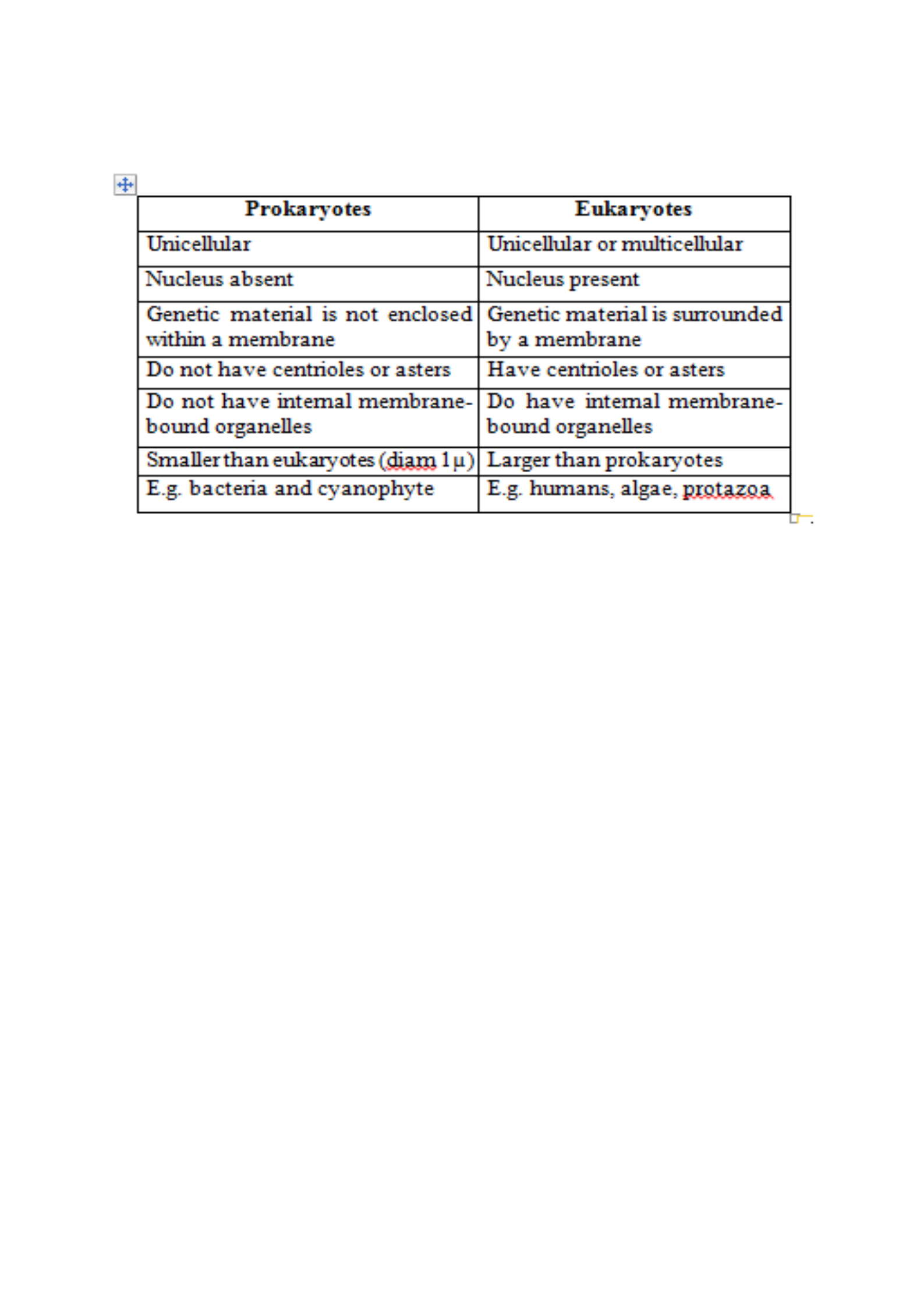
Table 1: Differences between prokaryotic and Eukaryotic cells
Eukaryotic cells are much larger and much more complex than prokaryotic cells.
These two observations are not unrelated to each other.
Eukaryotic cells have a true nucleus, bound by a double membrane. Prokaryotic cells
have no nucleus.
Eukaryotic DNA is complexed with proteins called "histones," and is organized into
chromosomes; prokaryotic DNA is "naked," meaning that it has no histones
associated with it, and it is not formed into chromosomes.
A eukaryotic cell contains a number of chromosomes; a prokaryotic cell contains only
one circular DNA molecule and a varied assortment of much smaller circlets of DNA
called "plasmids." The smaller, simpler prokaryotic cell requires far fewer genes to
operate than the eukaryotic cell.
Both cell types have many, many ribosomes, but the ribosomes of the eukaryotic cells
are larger and more complex than those of the prokaryotic cell. A eukaryotic ribosome
is composed of five kinds of rRNA and about eighty kinds of proteins. Prokaryotic
ribosomes are composed of only three kinds of rRNA and about fifty kinds of protein.
The cytoplasm of eukaryotic cells is filled with a large, complex collection of
organelles, many of them enclosed in their own membranes; the prokaryotic cell
contains no membrane-bound organelles which are independent of the plasma
membrane.
CELL ORGANELLES
An organelle is a membrane bound structure found within a cell. Organelles are found in the
cytoplasm, a viscous liquid found within the cell membrane that houses the organelles and is
the location of most of the action happening in a cell.
Advantages of Membrane Bound Organelles
Many metabolic processes involve membrane embedded enzymes

Peris Moraa Mokua
7
Enzymes of a particular pathway are within organelles thus products are within
proximity
Potentially harmful reactants and products are in isolation hence won‘t damage the
rest of the cell.
Main components of Eukaryotic cell (plant/animal cell)
1. Cytoplasm- The jelly-like substance composed of mainly water and found between
the cell membrane and nucleus. Makes up most of the "body" of a cell and is
constantly streaming. Organelles are found here and substances like salts may be
dissolved in the cytoplasm.
2. Nucleus- Contains genetic information (DNA) on special strands called
chromosomes. The nucleus is the "control center" of the cell, for cell metabolism
and reproduction.
3. Plasma membrane/ cell membrane- Separates the cell from its external environment,
selectively permeable. Protects the cell and provides stability.
NUCLEUS
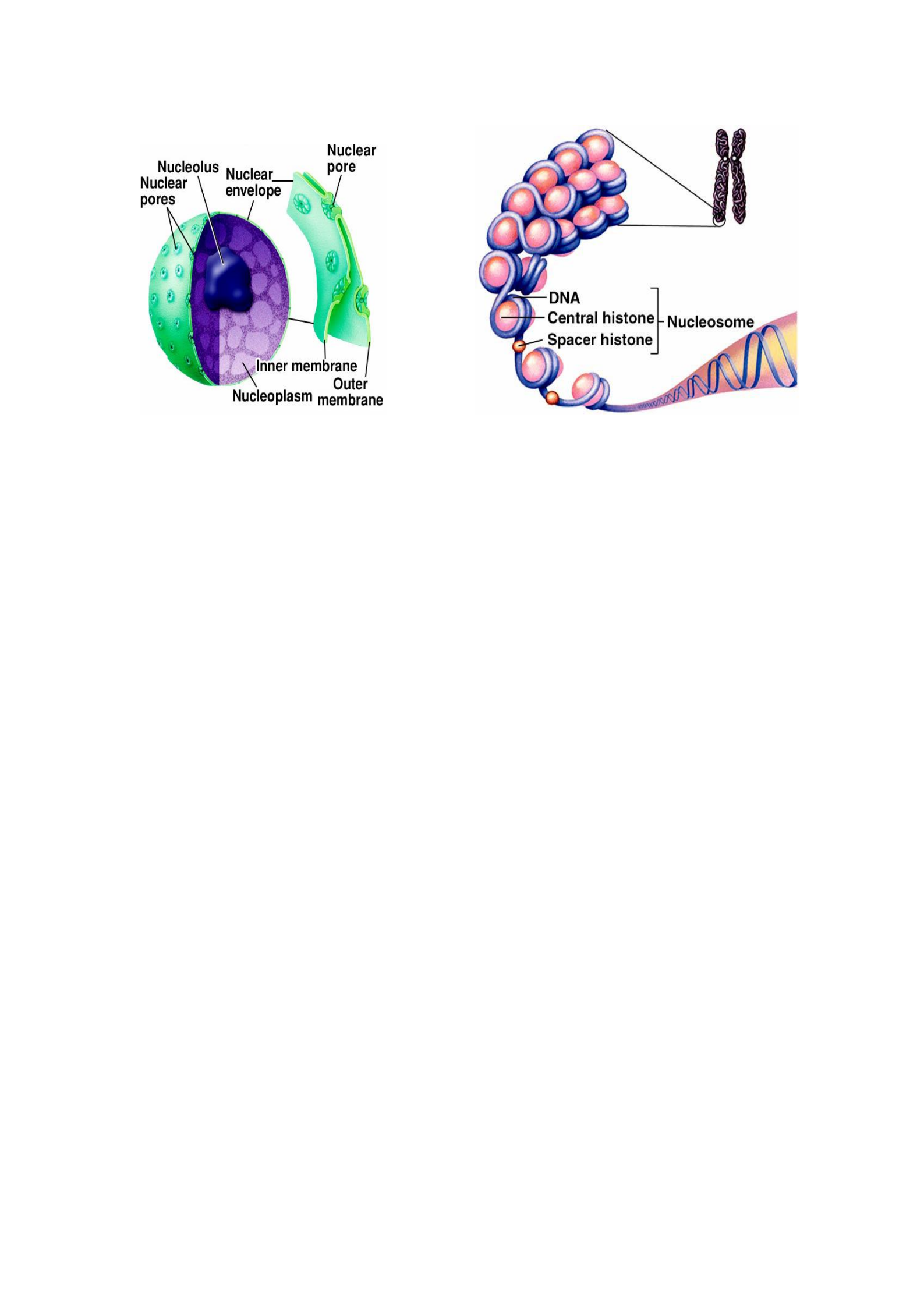
Is the largest organelle in the cell- controls cell activities. It is dark and round, and is
surrounded by a double membrane called the nuclear envelope/membrane. The
nuclear envelop creates a room within the cell to both protect the genetic information
and to house all the molecules that are involved in processing and protecting that info.
It also keeps molecules responsible for DNA transcription and repair close to the
DNA itself
In spots the nuclear envelope fuses to form nuclear pores which are selectively
permeable- control entry and exit of materials.
The nucleus contains the nucleolus, a darkened region where formation of ribosomes
and ribosomal RNA synthesis takes place.
It also contains genetic information (DNA) on special strands called chromosomes
(tightly packed strands of DNA containing all our blueprints). Chromosomes are
found in the nucleoplasm (fluid that fills nucleus) which is similar in structure and
function to cytoplasm. They consist of both DNA and protein which are seen as
chromosomes when highly condensed in preparation for cell division. At other times
they are threadlike and called chromatins. Histones are the most common proteins.
DNA is coiled around histones in a regular pattern which produce structures called
nucleosomes.
Functions
The nucleus is the "control center" of the cell, for cell metabolism and reproduction.
Separate DNA from the rest of the cell.
Transcription (making a complementary strand of RNA from DNA)
Peris Moraa Mokua
8
Fig. 3: Structure of nucleus
CYTOPLASM
Cytoplasm is the internal volume bounded by the plasma membrane. Is also a clear fluid in
the cytosol where particles are suspended. Translation (protein synthesis from RNA) takes
place in the cytoplasm. It contains:
Dissolved proteins
Electrolytes
Glucose
Organelles e.g Endoplasmic reticulum (ER), golgi apparatus, mitochondria,
lysosomes, peroxisomes, ribosomes, chloroplasts.
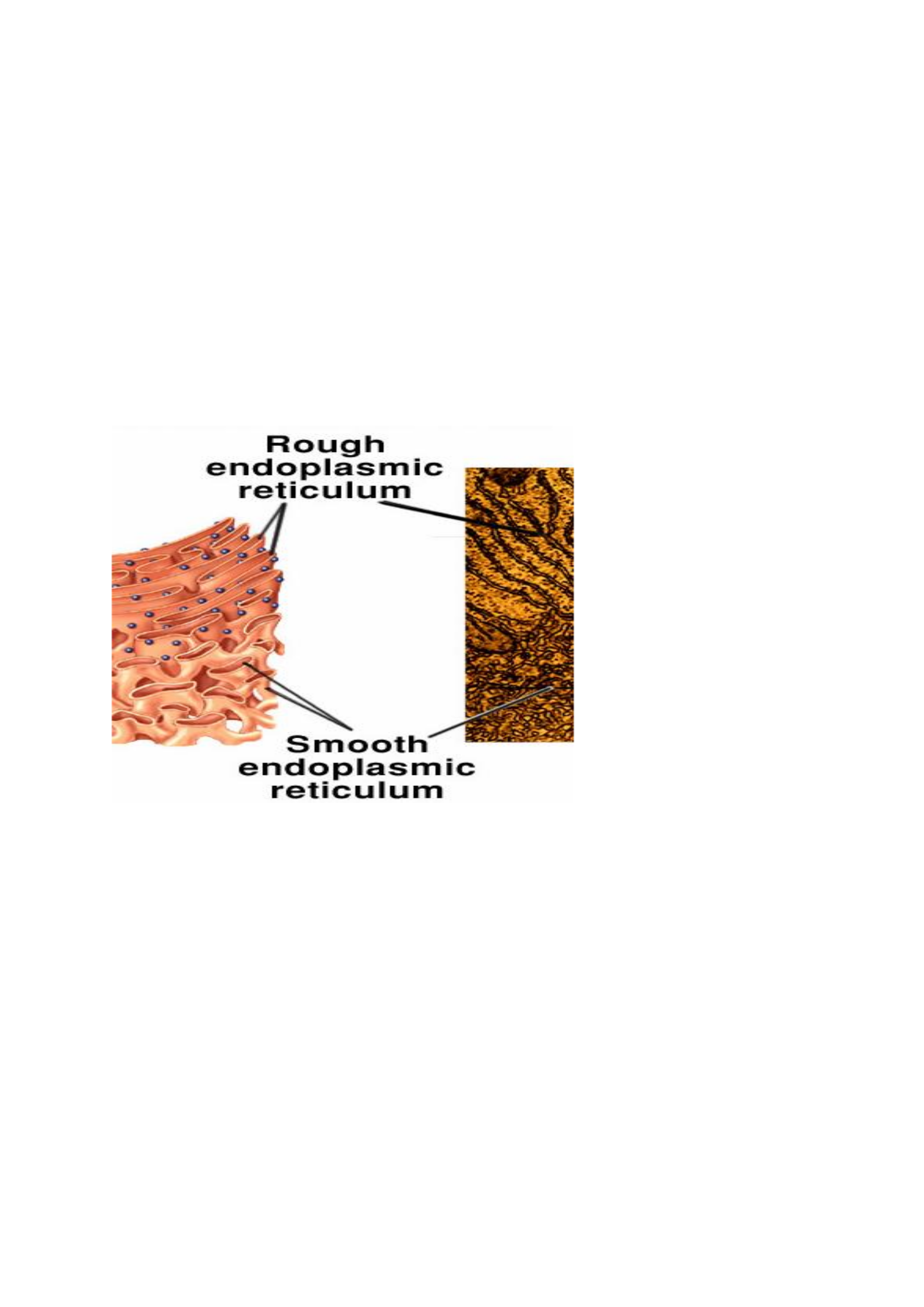
Endoplasmic Reticulum (E.R)
Endoplasmic means inside (endo) the cytoplasm (plasm). Reticulum comes from the Latin
word for net. Basically, an endoplasmic reticulum is a plasma membrane found inside the cell
that folds in on itself to create an internal space known as the lumen. The ER is a network of
membranous canals within the cytoplasm. They look like flattened sheets, sacs, tubes. They
create many membrane enclosed spaces spread across the cytoplasm. ER has connections
with the outer membrane of the nucleus and the plasma membrane. It has an interior space
called cisternae filled with fluid called endoplasmic matrix.
Types of ER
There are two types of ER: Rough ER and Smooth ER.
a) Rough ER: Is studded (lined) with ribosomes (the molecules in charge of protein
production) and is rough in appearance. The rough ER is continuous with the nuclear
envelope, and looks like a series of canals near the nucleus.
Function:
Site of synthesis of many proteins- Proteins made in the rough ER are destined to
either be a part of a membrane, or to be secreted from the cell membrane out of the
cell. Without a rough ER, it would be a lot harder to distinguish between proteins that
should leave the cell, and proteins that should remain.
Helps cells specialize and allows for greater complexity in the organism.
Peris Moraa Mokua
9
b) Smooth ER- Contains no ribosomes and is smooth in appearance. It is more tubular than
the rough ER, and is not necessarily continuous with the nuclear envelope. Every cell has a
smooth ER, but the amount will vary with cell function. For example, the liver, which is
responsible for most of the body’s detoxification, has a larger amount of smooth ER.
Fuctions:
Site for synthesis of steroids and other lipids- These are fat-based molecules that are
important in energy storage, membrane structure, and communication (steroids can
act as hormones).
Detoxifying the cell of drugs, toxins, alcohol (especially the liver)
Ca++ storage in muscles
The highly convoluted surface provides a large surface area for enzymatic activities.
Many enzymes are imbedded in the membranes.
Fig. 4: Structure of ER
Ribosomes
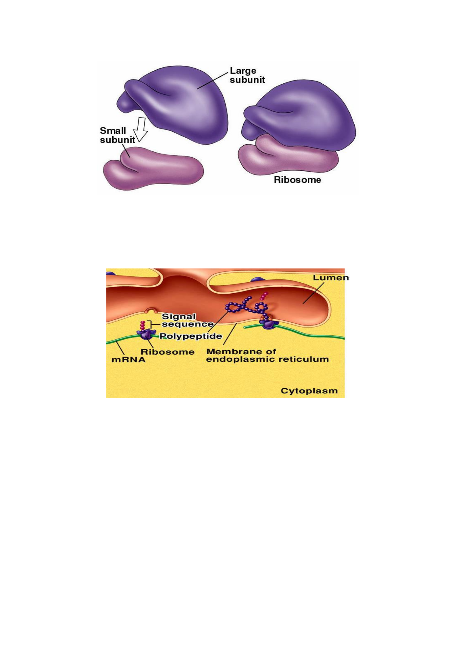
Ribosomes are small particles which are found individually in the cytoplasm and also line the
membranes of the rough endoplasmic reticulum.
Ribosomes are protein synthesis machinery i.e they produce proteins.
They are composed of two subunits, large and small, each made of protein and
ribosomal RNA (rRNA).The subunits associate when they are synthesizing proteins.
Peris Moraa Mokua
10
Fig. 5: Structure of the ribosome
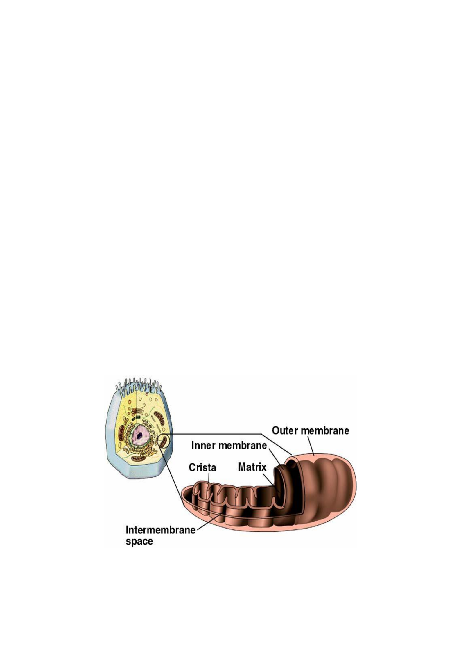
Protein synthesis occurs on ribosomes that are free-floating in the cytoplasm and on
ribosomes attached to ER.
rRNA is synthesized in the nucleolus ( a small subspace within the nucleus).
The figure below shows ribosomes attached to membrane of ER.
Fig. 6: Ribosomes attached to the membrane of ER
Golgi apparatus
Are also called Golgi bodies or Golgi. They are stacks of flattened membranous stacks. They
are present in all cells except the RBCs.
Functions: Is functionally associated with the ER.
Packing proteins from the rough ER into membrane-bound vesicles (tiny
compartments of lipid bilayer that store molecules) which then translocate to the cell
membrane
Temporarily stores protein which can then leave the cell via vesicles pinching off
from the Golgi.
Different molecules have different fates upon entering the Golgi. This determination is done
by tagging the proteins with special sugar molecules that act as a shipping label for the
protein. The “shipping department” identifies the molecule and sets it on one of 4 paths:
Peris Moraa Mokua
11
1. Cytosol: the proteins that enter the Golgi by mistake are sent back into the cytosol).
2. Cell membrane: proteins destined for the cell membrane are processed continuously.
Once the vesicle is made, it moves to the cell membrane and fuses with it. Molecules
in this pathway are often protein channels which allow molecules into or out of the
cell, or cell identifiers which project into the extracellular space and act like a name
tag for the cell.
3. Secretion: some proteins are meant to be secreted from the cell to act on other parts
of the body. Before these vesicles can fuse with the cell membrane, they must
accumulate in number, and require a special chemical signal to be released.
4. Lysosome: The final destination for proteins coming through the Golgi is the
lysosome. Vesicles sent to this acidic organelle contain enzymes that will hydrolyze
the lysosome’s content.
Mitochondria
The mitochondria are round "tube-like" organelles that are surrounded by a double
membrane, Inner and outer membranes. The inner membrane is highly folded into inward
projections called cristae- increases surface area for the site of reactions. Mitochondria are
often referred to as the "powerhouse" of the cell (releases food energy from food molecules to
be used by the cell in a process called respiration. There are two spaces within the
mitochondrion - the matrix and intermembrane space.
Unique features of mitochondria
Have their own DNA (almost as if they were a completely separate cell) that is similar
to prokaryotic DNA
Have their own ribosomes that are similar in construction to prokaryotic ribosomes
o Synthesize many, but not all, of their own proteins
Mitochondria are self-replicating i.e replicate by binary fission - similar to prokaryotic
cell division
Fig. 7: Structure of Mitochondria

Peris Moraa Mokua
12
Lysosomes
The lysosome is the cell’s recycling center. They are vescular organelles formed from golgi
and dispersed throughout the cytoplasm. They are also called suicide sacs.
They contain digestive and hydrolytic enzymes that digest food (carbohydrates, proteins,
lipids), nucleic acids and part of the cell-worn-out organelles.
Lysosomal proteins only being active in an acidic environment acts as safety mechanism for
the rest of the cell - if the lysosome were to somehow leak or burst, the degradative enzymes
would inactivate before they chopped up proteins the cell still needed.
They are also covered by a thick membrane-prevents enclosed hydrolytic enzymes from
coming in contact with other substances in the cell thus preventing their digestive actions.
Functions:
Destruct foreign materials-bacteria
Removal of excessive secretory products in cells of the glands
Removal of unwanted cells in embryo
Peroxisome
The peroxisome is a spherical organelle responsible for destroying its contents.
Functions
Unlike the lysosome, which mostly degrades proteins, the peroxisome is the site of
fatty acid breakdown.
It also protects the cell from reactive oxygen species (ROS) molecules which could
seriously damage the cell. ROSs are molecules like oxygen ions or peroxides that are
created as a by-product of normal cellular metabolism, but also by radiation, tobacco,
and drugs. They cause what is known as oxidative stress in the cell by reacting with
and damaging DNA and lipid-based molecules like cell membranes. These ROSs are
the reason we need antioxidants in our diet.
Vacuoles
Vacuoles are fluid filled organelles enclosed by a membrane. They can store materials such
as food, water, sugar, minerals and waste products.
Animal cell organelles not found in plant cells:
Cilia and flagella
Both cilia and flagella are hair-like organelles which extend from the surface of many animal
cells. The structure is identical in both, except that flagella are longer and whiplike and cilia
are shorter. There are usually only a few flagella on a cell, while cilia may cover the entire
surface of a cell.
Functions
Locomotion for one-celled organisms
Move substances over cell surfaces in multi-celled organisms.
Organelles and other features found only in plant cells:

Peris Moraa Mokua
13
1. Cell wall
The cell wall is a rigid organelle composed of cellulose and lying just outside the cell
membrane. The cell wall gives the plant cell its box-like shape. It also protects the cell. The
cell wall contains pores which allow materials to pass to and from the cell membrane.
2. Plastids
Plastids are double membrane bound organelles. It is in plastids that plants make and store
food. Plastids are found in the cytoplasm and there are two main types: Pigmented and non-
pigmented plastids:
a) Leucoplasts - colorless organelles which store starch or other plant nutrients.
(Example - starch stored in a potato)
b) Chromoplasts - contain different colored pigments. The most important type of
chromoplast is the chloroplast, which contains the green pigment chlorophyll. This
is important in the process of photosynthesis.
Chloroplasts
Are sites of photosynthesis - in nearly all plants and some protists.
Functions
They trap light energy and convert it into chemical energy to create food by
photosynthesis. Synthesize and store starch in roots and tubers.
They have pigments and give fruits ripened color.
Characteristics of Chloroplasts
They have a double membrane structure with an inner space called the stroma.
Have their own DNA, similar to prokaryotic DNA.
They can synthesize many of their own proteins using prokaryote-like ribosomes.
Replicate through division similar to prokaryotic cell division
PLASMA MEMBRANE STRUCTURE AND FUNCTION
Introduction
Cell membrane-also known as plasma membrane (PM) is a thin biological membrane that
surrounds the cytoplasm of the cell. It also separates the interior of all cells from the outside
environment and divides the interior of the cell into compartments:
Fluid outside the cell, Extracellular fluid (ECF)
Fluid inside the cell, Intacellular Fluid (ICF).
The PM is selectively permeable to ions and organic molecules and controls the movement of
subastances in and out of cells. The basic function of the cell membrane is to protect the cell
from its surroundings.
Functions of the Plasma Membrane
Protect the integrity of the interior of the cell by allowing certain substances into the
cell, while keeping other substances out.
The outer plasma membrane forms a boundary between a living cell and its
surroundings; Exhibits selective permeability, Controls traffic of molecules in and
out.

Peris Moraa Mokua
14
Internal membranes provide structural order for metabolism. Form the cell's
organelles. Compartmentalize chemical reactions, organizes the chemical activities of
cells.
It serves as a base of attachment for the cytoskeleton in some organisms and the cell
wall in others.
It helps support the cell and help maintain its shape and size.
Regulate cell growth through the balance of endocytosis and exocytosis. In
endocytosis, lipids and proteins are removed from the cell membrane as substances
are internalized. In exocytosis, vesicles containing lipids and proteins fuse with the
cell membrane increasing cell size.
Encased internal organelles.
Communication
Recognition
Cell membranes are involved in a variety of cellular processes such as cell adhesion,
ion conductivity and cell signalling.
Structure of the Plasma Membrane
Fluid Mosaic Model
The currently accepted model for the structure of the plasma membrane, called the fluid
mosaic model, was first proposed in 1972. This model has evolved over time, but it still
provides a good basic description of the structure and behavior of membranes in many cells.
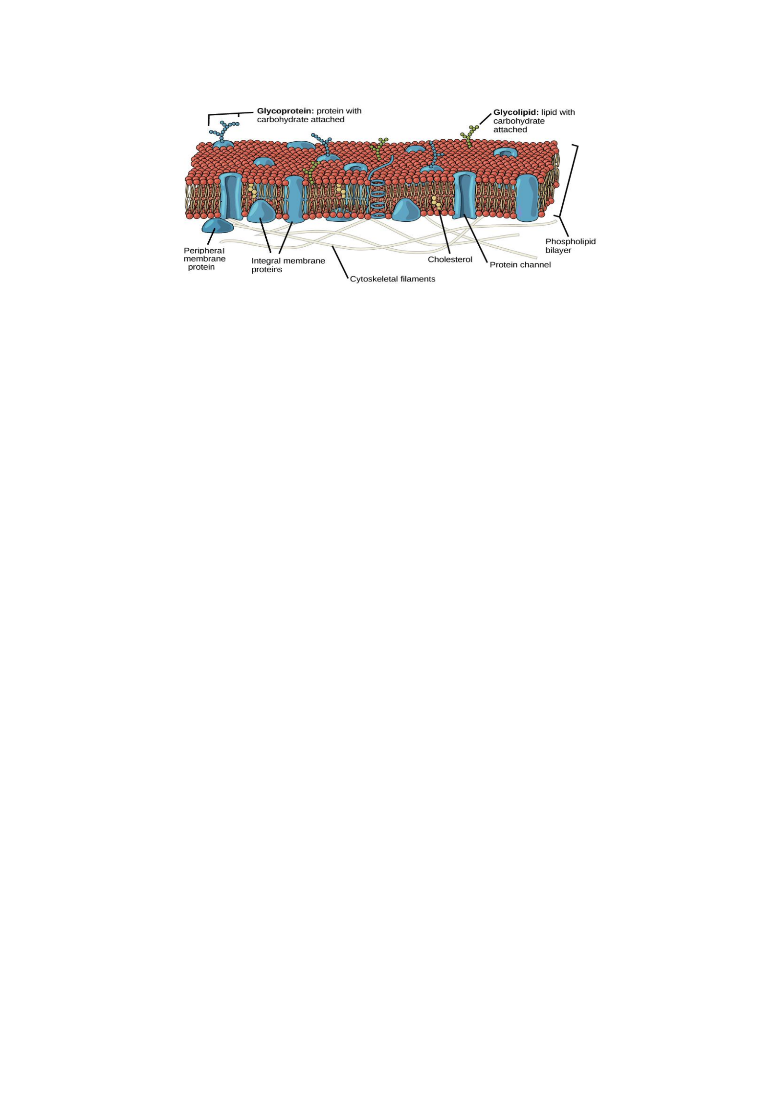
According to the fluid mosaic model, the plasma membrane is a mosaic of components—
primarily, phospholipids, cholesterol, and proteins—that move freely and fluidly in the plane
of the membrane. Proteins and other molecules are embedded in a framework of
phospholipids. Most proteins and phospholipid molecules can move laterally. The fluid
nature of the PM is important for normal functioning.
Chemical Composition
The principal components of the plasma membrane are lipids (phospholipids and cholesterol),
proteins, and carbohydrate groups that are attached to some of the lipids and proteins.
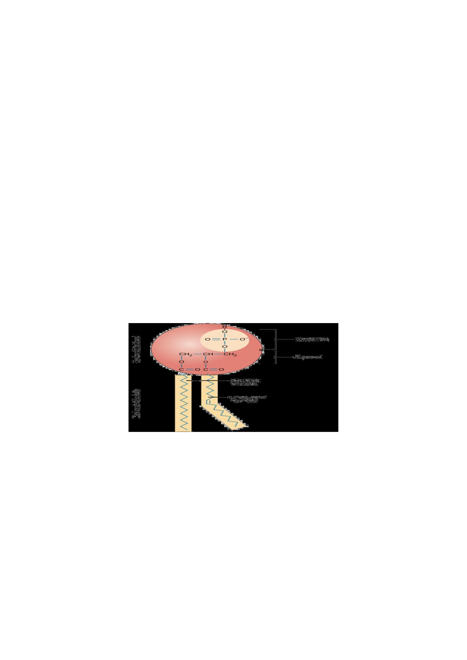
A phospholipid is a lipid made of glycerol, two fatty acid tails, and a phosphate-
linked head group. Biological membranes usually involve two layers of phospholipids
with their tails pointing inward, an arrangement called a phospholipid bilayer.
Cholesterol, another lipid composed of four fused carbon rings, is found alongside
phospholipids in the core of the membrane.
Membrane proteins may extend partway into the plasma membrane, cross the
membrane entirely, or be loosely attached to its inside or outside face.
Carbohydrate groups are present only on the outer surface of the plasma membrane
and are attached to proteins, forming glycoproteins, or lipids, forming glycolipids.
1. Membrane Lipids
They are organized into lipid bilayer. Major classes of membrane lipids include:
Phospholipids
Glycolipids
Cholesterol: stability to the membrane
Peris Moraa Mokua
15
Phospholipids: Phospholipids, arranged in a bilayer, make up the basic fabric of the plasma
membrane. They are well-suited for this role because they are amphipathic, meaning that
they have both hydrophilic and hydrophobic regions.
The hydrophilic, or “water-loving,” portion of a phospholipid is its head, which contains a
negatively charged phosphate group as well as an additional small group which may also or
be charged or polar. The hydrophilic heads of phospholipids in a membrane bilayer face
outward, contacting the aqueous (watery) fluid both inside and outside the cell. Since water is
a polar molecule, it readily forms electrostatic (charge-based) interactions with the
phospholipid heads.
The hydrophobic, or “water-fearing,” part of a phospholipid consists of its long, nonpolar
fatty acid tails. The fatty acid tails can easily interact with other nonpolar molecules, but they
interact poorly with water. Because of this, it’s more energetically favourable for the
phospholipids to tuck their fatty acid tails away in the interior of the membrane, where they
are shielded from the surrounding water. The phospholipid bilayer formed by these
interactions makes a good barrier between the interior and exterior of the cell, because water
and other polar or charged substances cannot easily cross the hydrophobic core of the
membrane.
Functions
Provide membrane with form
Selective barrier
Fig. 8: Phospholipid structure
2. Membrane Proteins
Proteins are the second major component of plasma membranes. There are two main
categories of membrane proteins: Integral and Peripheral.
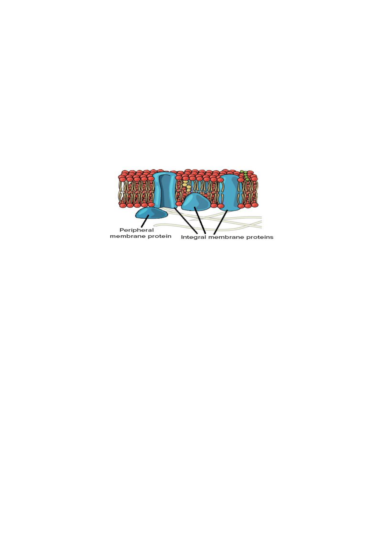
a) Integral/Intrinsic membrane proteins: Are partially or fully immersed/integrated into
the lipid bilayer. They have at least one hydrophobic region that anchors them to the
hydrophobic core of the phospholipid bilayer. Some stick only partway into the membrane,
while others stretch from one side of the membrane to the other and are exposed on either
side. Proteins that extend all the way across the membrane are called transmembrane
proteins.
Peris Moraa Mokua
16
The portions of an integral membrane protein found inside the membrane are hydrophobic,
while those that are exposed to the cytoplasm or extracellular fluid tend to be hydrophilic.
Transmembrane proteins may cross the membrane just once, or may have as many as twelve
different membrane-spanning sections. Some integral membrane proteins form a channel that
allows ions or other small molecules to pass.
Function as: Channels, Carrier proteins, Enzymes, Receptors and Support.
b) Peripheral/Extrinsic membrane proteins: Are found on the outside and inside surfaces
of membranes, attached either to integral proteins or to phospholipids by hydrogen or
electrostatic bonds. Unlike integral membrane proteins, peripheral membrane proteins do not
stick into the hydrophobic core of the membrane, and they tend to be more loosely attached.
Functions: Enzymes, Receptors
Fig. 9: Lipid bilayer with membrane proteins
3. Membrane Carbohydrates
Are the third major component of plasma membrane. They are found on the outside surface
of cells and are bound either to proteins (forming glycoproteins) or to lipids (forming
glycolipids). These carbohydrate chains may consist of 2-60 monosaccharide units and can be
either straight or branched.
Functions
Along with membrane proteins, these carbohydrates form distinctive cellular markers
that allow cells to recognize each other. These markers are very important in the
immune system, allowing immune cells to differentiate between body cells, which
they shouldn’t attack, and foreign cells or tissues, which they should.
Carbohydrates negatively charged, Membrane negative charge; Repel negatively
charged molecules
Act as receptor site
Attachment to other cells
Peris Moraa Mokua
17
Fig. 10: Plasma Membrane Structure
Movement of Substances Across the Plasma Membrane
The PM is differentially permeable: some substances readily pass through, others do not. It is
most permeable to small molecules and lipid-soluble substances. Water(!) and other small
molecules like CO2 and O2 can pass through easily. There are molecules that do not pass
through easily: amino acids, sugars, ions. Passage of some molecules across the membrane is
assisted with special channels to allow or speed up the passage. The specific selectivity can
vary depending on the membrane.
There are two different types of exchange of molecules between the outside and inside of the
cell.
1. Passive transport- Movement of substances is due to gradient differences in
concentration, pressure and charge. There are 3 forms:
-Diffusion
-Faciliatetd transport; Uniport transport
-Osmosis
2. Active transport; Movement of substances through the cell mebrane that require use of
energy (ATP).
-Sodium-Potassium pump
-Endocytosis
-Exocytosis
-Symport transport systems
-Antiport transport systems
1. Passive Transport
a) Diffusion
Is the random movement of molecules from the area of higher concentration to the area of
lower concentration (net movement), until they are equally distributed. Examples: Sugar in
tea (We stir the tea to speed up the diffusion). The odour of food cooking, perfumes etc. CO2
entering the stomata of leave. Oxygen diffusing out of the stomata and lenticels of leaves

Peris Moraa Mokua
18
Factors Affecting the Rate of Diffusion
Concentration gradient difference
Distance over which diffusion takes place
The area over which diffusion takes place-surface area
The nature of any structure across which diffusion occurs e.g number of pores
The size and nature of the diffusing molecules
o Small in size and Fat soluble diffuse faster
b). Osmosis
Movement of water molecules from a region of higher concentration to a region of low
concentration through a semi-permeable membrane. Movement of substances from more
dilute solution to a more concentrated solution (solvent particles are moving from higher to
lower solvent concentration across the membrane, they are moving from lower to higher
solute concentration). Example; Absorption of water by plant roots.
The movement of liquids in and out cells is dependent on the concentration of the solution
surrounding it. There are 3 types of situations in which this could vary:
1. Isotonic: External solution concentration and the internal concentration of the organism
are the same i.e. there is the same concentration of non-diffusible solutes and water on both
sides of the plasma membrane.
2. Hypotonic: External solution concentration is less than the concentration of the organism
i.e. concentration of solutes is lower outside the cells In this case water will rush into the
organism.
3. Hypertonic: External solution concentration is greater than the concentration of the
organism i.e. the concentration of solutes is higher outside the cells In this case the water will
rush out of the organism.
Peris Moraa Mokua
19
Table 2: Differences between diffusion and osmosis
Diffusion
Osmosis
Movement of all types of substances from the
area of their higher free energy to the area of
their lower free energy
Movement of only solvent or water from its
higher concentration to lower concentration
across a membrane
Can operate in any medium
Operates only in a liquid medium
Applicable to all types of substances; solid,
liquid, gases
Applicable to only solvent part of the
solution
Does not require any semi-permeable
membrane
Requires a semi-permeable membrane
Turgor pressure does not normally operate in
diffusion
Is opposed by turgor pressure of the system
Not influenced by solute potential
Dependent upon the solute potential
Diffusion of a substance mostly dependent of
the presence of other substances
Osmosis is dependent upon the number of
particles of the other substances dissolved in
the liquid
c) Facilitated Transport
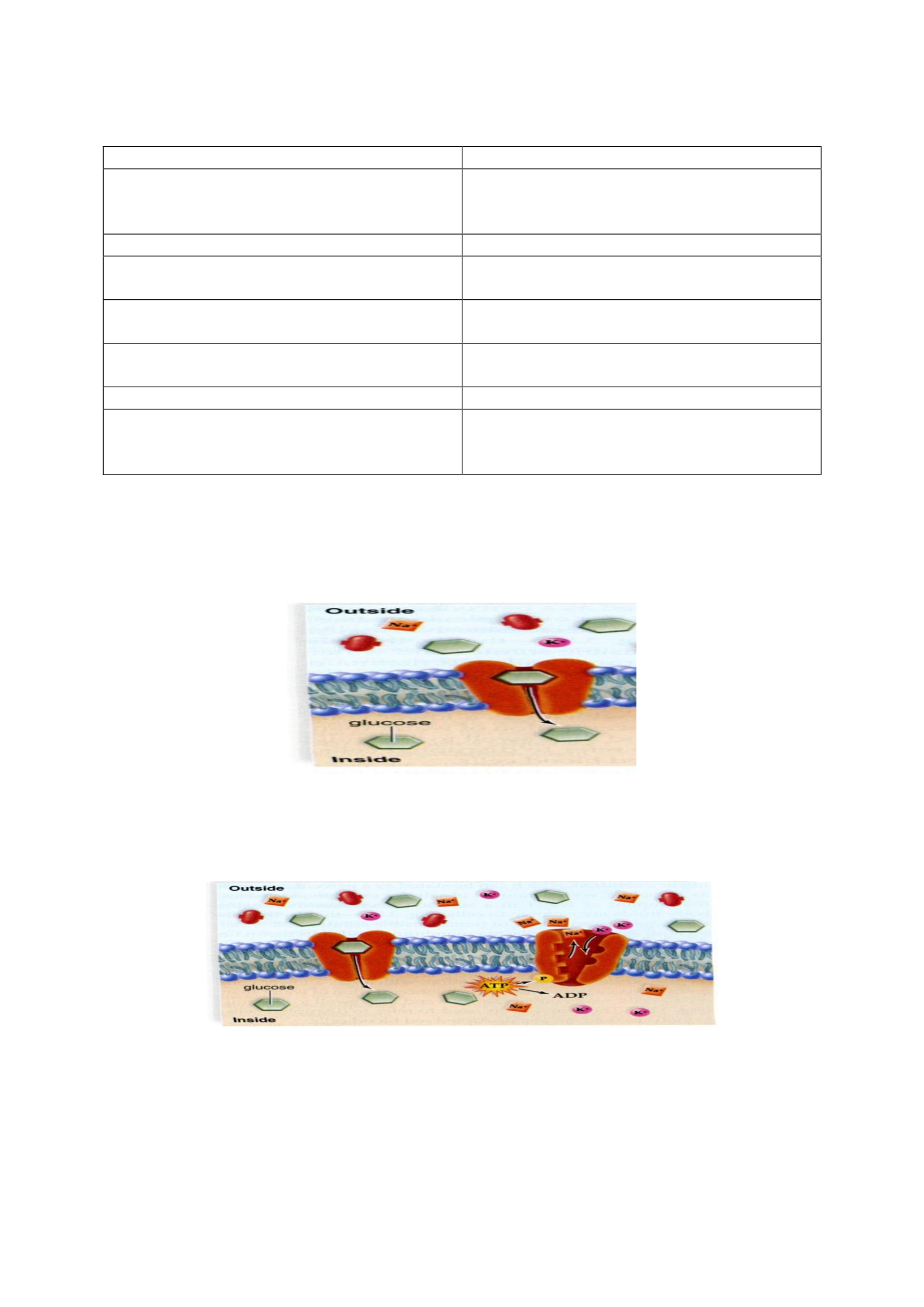
Non-diffusible solutes such as sugars or amino acids can be transported by means of protein
carriers in the plasma membrane at a rate higher than usual across the plasma membrane from
the side of higher concentration to the side of lower concentrations. Each protein carrier is
sometimes called ‘transporter’. Energy – not necessary.
Fig. 11: Facilitated diffusion
2. Active Transport
A molecule moves from lower concentration to a higher concentration by means of ‘pump’.
Thus, the solute can be concentrated in either side. Energy in form of ATP is necessary.
Fig. 12: Active transport
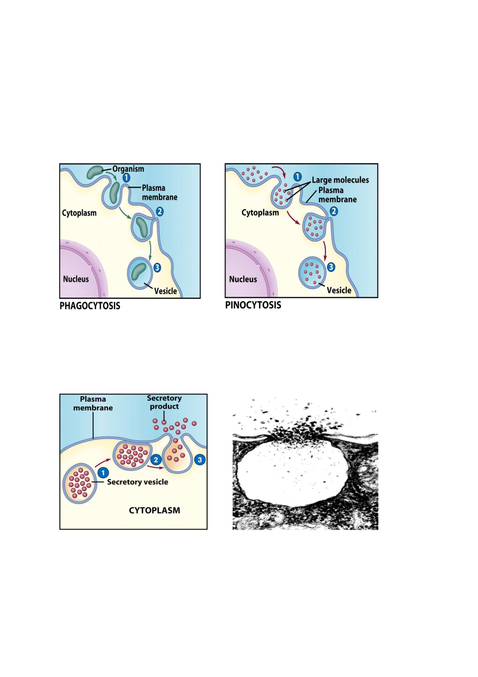
a) Endocytosis
Is the movement of large material examples particles, organisms and other large molecules
into the cells. There are 2 types of endocytosis:
Bulk-phase (nonspecific)
Receptor-mediated (specific)
Peris Moraa Mokua
20
Process of Endocytosis
o The Plasma membrane surrounds material
o Edges of membrane meet
o Membranes fuse to form vesicle
Forms of endocytosis: Phagocytosis – cell eating
Pinocytosis – cell drinking
Fig. 13: Process of endocytosis
b) Exocytosis
Is the reverse of endocytosis whereby the cell discharges the material.
Vesicle moves to cell surface
Membrane of vesicle fuses
Materials expelled
Fig. 14: Process of exocytosis
Peris Moraa Mokua
21
CELL DIVISION
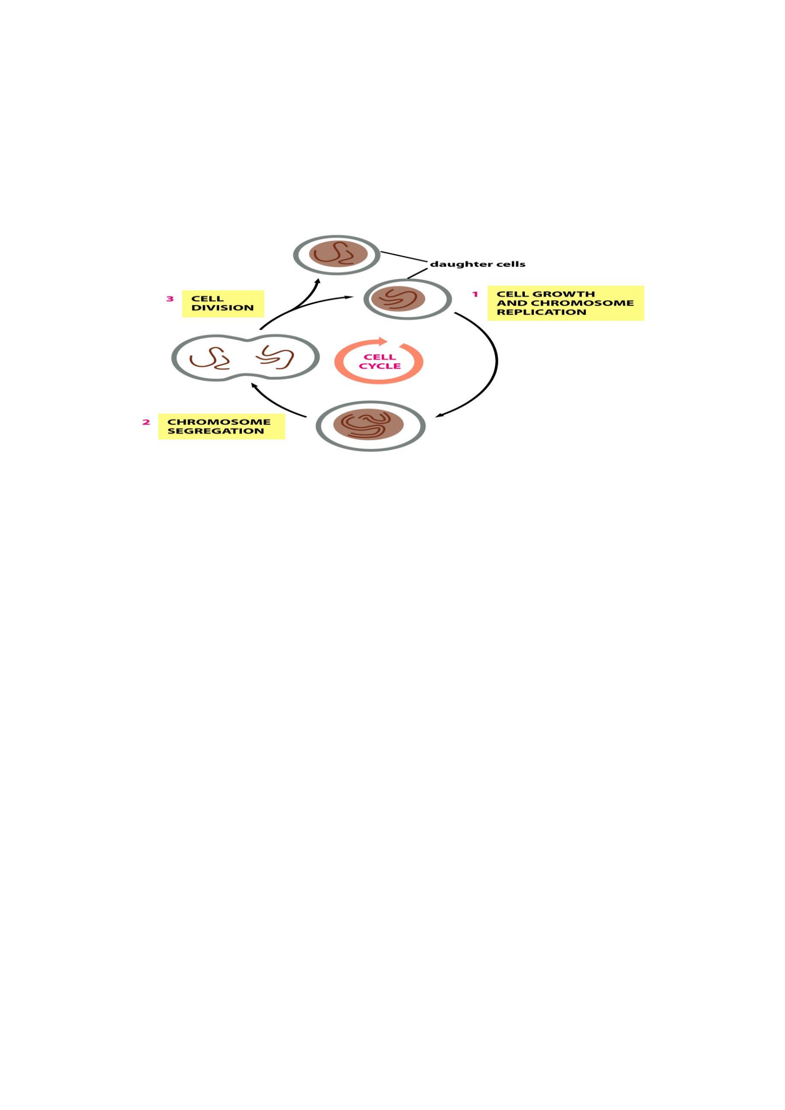
Is the process by which a parent cell divides into two or more daughter cells. It usually occurs
as part of a larger cell cycle (Sequence of events that occurs between one cell division and the
next). The cell cycle has 3 stages:
1. Interphase: Cells grow and replicate their DNA
2. Nuclear division: Separation of chromatids
3. Cytokinesis: Division of cytoplasm and organelles
Fig. 15: Stages of cell cycle
Cell division differs in prokaryotes and eukaryotes. Prokaryotes such as bacteria undergo a
vegetative cell division known as binary fission, where their genetic material is segregated
equally into two identical daughter cells; a single chromosome makes a copy of itself and the
cell wall forms between the chromosomes dividing the cell. All cell divisions, regardless of
organism, are preceded by a single round of DNA replication. For simple unicellular
organisms such as the amoeba, one cell division is equivalent to reproduction – an entire new
organism is created.
In eukaryotes, cell division starts with division of the nucleus which occurs in two distinct
types of cell division: a vegetative division, whereby each daughter cell is genetically
identical to the parent cell (mitosis) and a reproductive cell division, whereby the number of
chromosomes in the daughter cells is reduced by half to produce haploid gametes (meiosis).
Meiosis results in four haploid daughter cells by undergoing one round of DNA replication
followed by two divisions. Replication is the process of duplicating a chromosome.
Homologous chromosomes are separated in the first division, and sister chromatids are
separated in the second division. Both of these cell division cycles are used in the process of
sexual reproduction at some point in their life cycle.
Importance of Cell division
Maintenance of the original cell's genome. Before division can occur, the genomic
information that is stored in chromosomes must be replicated, and the duplicated
genome must be separated cleanly between cells.
Growth
Repair: worn-out or damaged cells
Basis of reproduction
Peris Moraa Mokua
22
Phases of cell division
1. Interphase
Interphase is the process a cell must go through before mitosis, meiosis, and cytokinesis.
Interphase consists of four main stages: G1 (growth 1), S (synthesis phase), G2 (growth 2
phase), and M (Mitotic/meiotic phase). G1 is a time of growth for the cell. If the cell does not
progress through G1, the cell then enters a stage called G0 (resting phase). In G0, cells are
still living but they are put on hold. The cells may later be called back into interphase if
needed at a later time. There are checkpoints during interphase that allow the cell to be either
progressed or denied further development. In S phase, the chromosomes are replicated in
order for the genetic content to be maintained. During G2, the cell undergoes the final stages
of growth before it enters the M phase. The M phase, can be either mitosis or meiosis
depending on the type of cell. Germ cells undergo meiosis, while somatic cells will undergo
mitosis. After the cell proceeds through successfully through the M phase, it may then
undergo cell division through cytokinesis. The control of each checkpoint is controlled by
cyclin and cyclin dependent kinases. The progression of interphase is the result of the
increased amount of cyclin. As the amount of cyclin increases, more and more cyclin
dependent kinases attach to cyclin signaling the cell further into interphase. The peak of the
cyclin attached to the cyclin dependent kinases this system pushes the cell out of interphase
and into the M phase, where mitosis, meiosis, and cytokinesis occur.
Fig. 16: Interphase stage
2. Prophase
Prophase is the first stage of division. The nuclear envelope is broken down, long strands of
chromatin condense to form shorter more visible strands called chromosomes, the nucleolus
disappears, and microtubules attach to the chromosomes at the kinetochores present in the
centromere. Microtubules associated with the alignment and separation of chromosomes are
referred to as the spindle and spindle fibers. Chromosomes will also be visible under a
microscope and will be connected at the centromere. During this condensation and alignment

Peris Moraa Mokua
23
period, homologous chromosomes may swap portions of their DNA in a process known as
crossing over.
3. Metaphase
Chromosomes line up in the middle of the cell. The chromosomes are still condensing and are
currently at one step away from being the most coiled and condensed they will be. Spindle
and spindle fibers have already connected to the kinetochores. At this point, the
chromosomes are ready to split into opposite poles of the cell towards the spindle to which
they are connected.
4. Anaphase
Anaphase is a very short stage of the cell cycle and occurs after the chromosomes align at the
mitotic plate. After the chromosomes line up in the middle of the cell, the spindle fibers will
pull them apart. The chromosomes are split apart as the sister chromatids move to opposite
sides of the cell.
5. Telophase
Telophase is the last stage of the cell cycle. Two cells form around the chromatin at the two
poles of the cell. Two nuclear membranes begin to reform and the chromatin begin to
unwind.
Mitotic Cell Division
Mitosis is how somatic (non-reproductive) cells divide. Somatic cells make up most of your
body's tissues and organs, including skin, muscles, lungs, gut, and hair cells. In mitosis, the
important thing to remember is that the daughter cells each have the same chromosomes and
DNA as the parent cell. The daughter cells from mitosis are called diploid cells. Diploid cells
have two complete sets of chromosomes. Since the daughter cells have exact copies of their
parent cell's DNA, no genetic diversity is created through mitosis in normal healthy cells.
Fig. 17: Mitotic cell division
In mitotic cell division, the nucleus divides once creates two genetically identical daughter
diploid cells. The major steps of mitosis are shown here.
The Mitosis Cell Cycle
Before a cell starts dividing, it is in the "Interphase." Cells must be constantly dividing but
each cell spends most of its time in the interphase. Interphase is the period when a cell is
getting ready to divide and start the cell cycle. During this time, cells are gathering nutrients
Peris Moraa Mokua
24
and energy. The parent cell is also making a copy of its DNA to share equally between the
two daughter cells.
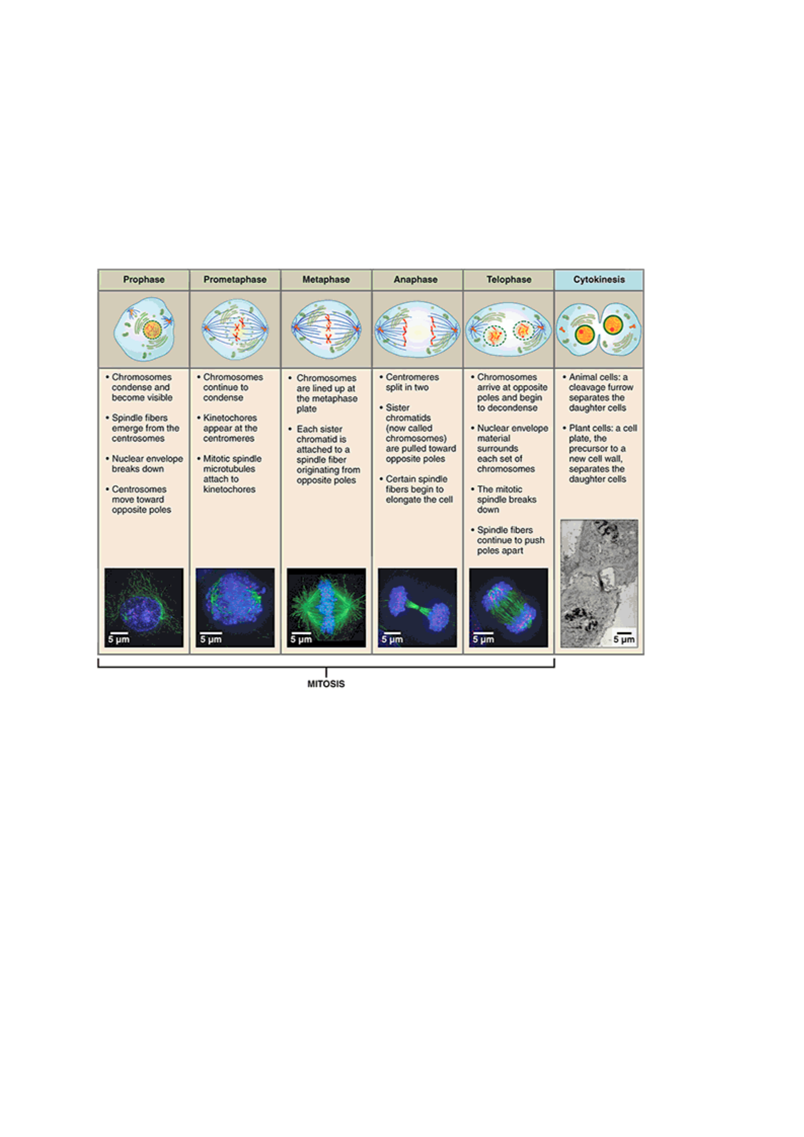
The mitosis division process has several steps or phases of the cell cycle—interphase,
prophase, prometaphase, metaphase, anaphase, telophase, and cytokinesis—to successfully
make the new diploid cells.
Fig. 18: Mitotic stages
When a cell divides during mitosis, some organelles are divided between the two daughter
cells. For example, mitochondria are capable of growing and dividing during the interphase,
so the daughter cells each have enough mitochondria. The Golgi apparatus, however, breaks
down before mitosis and reassembles in each of the new daughter cells.
Significance of mitosis
Formation of new cells for growth
Asexual reproduction
Repair of worn-out or damaged cells
Meiosis Cell Division
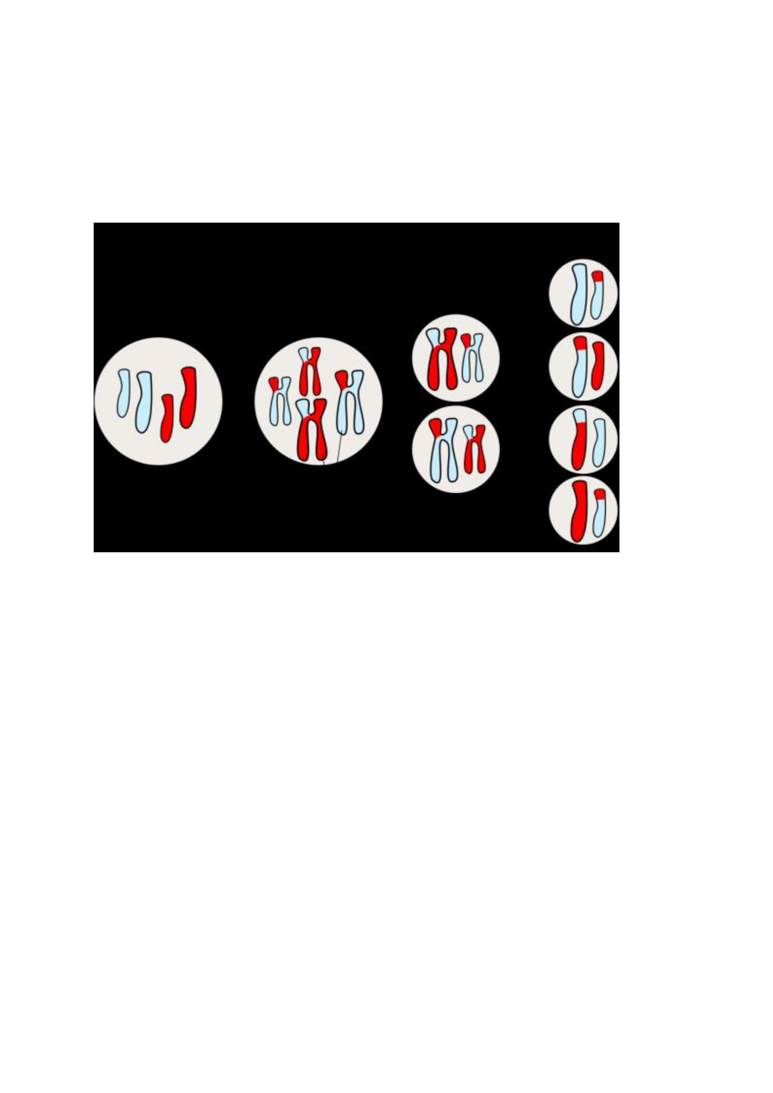
Meiosis is cell division that creates sex cells, like female egg cells or male sperm cells. In
meiosis, each new cell contains a unique set of genetic information. After meiosis, the sperm
and egg cells can join to create a new organism. Meiosis is why we have genetic diversity
in all sexually reproducing organisms. During meiosis, a small portion of each chromosome
breaks off and reattaches to another chromosome. This process is called "crossing over" or
Peris Moraa Mokua
25
"genetic recombination." Genetic recombination is the reason full siblings made from egg
and sperm cells from the same two parents can look very different from one another.
The meiotic stage has 2 main stages of division: Meiosis I and II. The end product is four
haploid daughter cells each having different genetic information from each other and the
parent cell
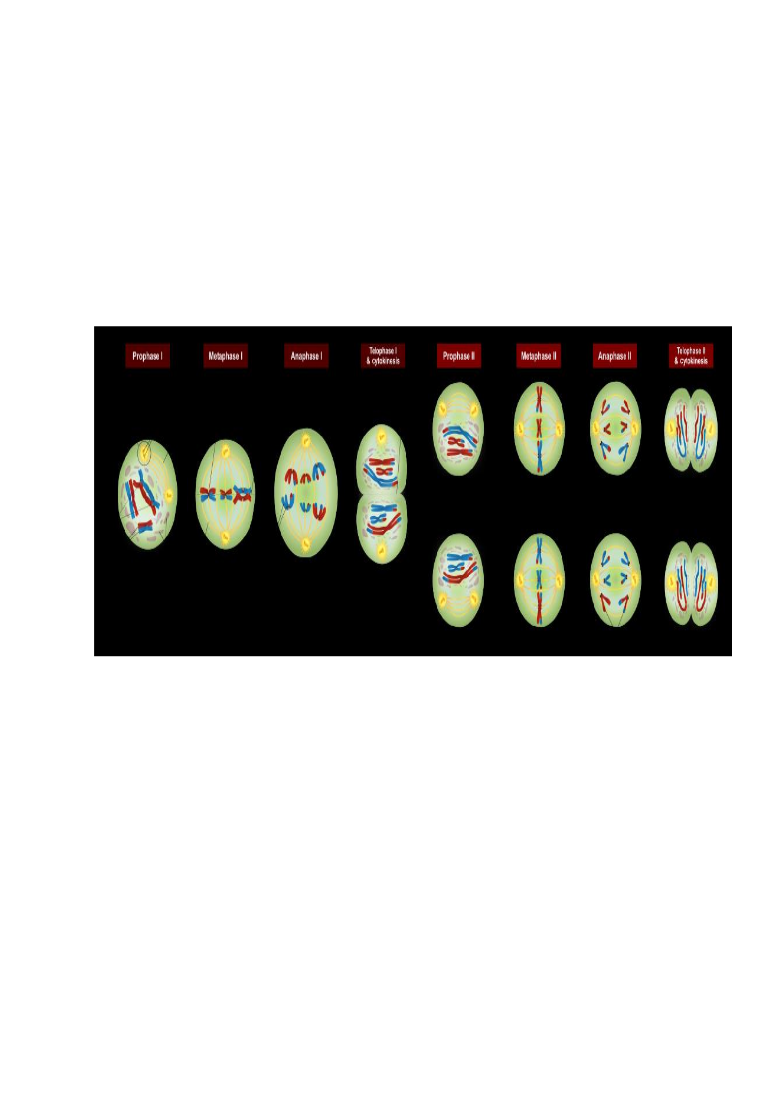
Fig. 19: Meiotic cell division
The Meiosis Cell Cycle
Meiosis has two cycles of cell division, called Meiosis I and Meiosis II. Meiosis I halves the
number of chromosomes and is also when crossing over happens. Meiosis II halves the
amount of genetic information in each chromosome of each cell. The end result is four
daughter cells called haploid cells. Haploid cells only have one set of chromosomes - half the
number of chromosomes as the parent cell.
Before meiosis I starts, the cell goes through interphase. Just like in mitosis, the parent cell
uses this time to prepare for cell division by gathering nutrients and energy and making a
copy of its DNA. During the next stages of meiosis, this DNA will be switched around during
genetic recombination Interphase is divided into three phases:
Growth 1 (G1) phase: Is a very active phase, the cell synthesizes its vast array of
proteins, including the enzymes and structural proteins it will need for growth. In G1,
each of the chromosomes consists of a single linear molecule of DNA.
Synthesis (S) phase: The genetic material is replicated; each of the cell's
chromosomes duplicates to become two identical sister chromatids attached at a
centromere. This replication does not change the ploidy of the cell since the
centromere number remains the same. The identical sister chromatids have not yet
condensed into the densely packaged chromosomes visible with the light microscope.
This will take place during prophase I in meiosis.
Growth 2 (G2) phase: G2 phase as seen before mitosis is not present in meiosis.
Meiotic prophase corresponds most closely to the G2 phase of the mitotic cell cycle.
Peris Moraa Mokua
26
Interphase is followed by meiosis I and then meiosis II. Meiosis I separates homologous
chromosomes, each still made up of two sister chromatids, into two daughter cells, thus
reducing the chromosome number by half. During meiosis II, sister chromatids decouple and
the resultant daughter chromosomes are segregated into four daughter cells. For diploid
organisms, the daughter cells resulting from meiosis are haploid and contain only one copy of
each chromosome. In some species, cells enter a resting phase known as interkinesis
between meiosis I and meiosis II.
Meiosis I and II are each divided into prophase, metaphase, anaphase, and telophase stages,
similar in purpose to their analogous sub phases in the mitotic cell cycle.
Phases
Fig. 20: Phases of meiosis cell division
Meiosis I
Meiosis I segregates homologous chromosomes, which are joined as tetrads (2n, 4c),
producing two haploid cells (n chromosomes, 23 in humans) which each contain chromatid
pairs (1n, 2c). Because the ploidy is reduced from diploid to haploid, meiosis I is referred to
as a reductional division.
Prophase I
Is typically the longest phase of meiosis. During prophase I, homologous chromosomes pair
and exchange DNA (homologous recombination). This often results in chromosomal
crossover. This process is critical for pairing between homologous chromosomes and hence
for accurate segregation of the chromosomes at the first meiosis division. The new
combinations of DNA created during crossover are a significant source of genetic variation,
and result in new combinations of alleles, which may be beneficial. The paired and replicated
chromosomes are called bivalents or tetrads, which have two chromosomes and four
chromatids, with one chromosome coming from each parent. The process of pairing the
homologous chromosomes is called synapsis. At this stage, non-sister chromatids may cross-

Peris Moraa Mokua
27
over at points called chiasmata. Prophase I is divided into a series of sub stages named
according to the appearance of chromosomes.
a) Leptotene
The first stage of prophase I, also known as leptonema (thin threads). In this stage, individual
chromosomes, each consisting of two sister chromatids, become "individualized" to form
visible strands within the nucleus. The two sister chromatids closely associate and are
visually indistinguishable from one another. During leptotene, lateral elements of the
synaptonemal complex assemble. Leptotene is of very short duration and progressive
condensation and coiling of chromosome fibers takes place.
b) Zygotene
The zygotene stage is also known as zygonema (paired threads) occurs as the chromosomes
approximately line up with each other into homologous chromosome pairs. The telomeres
cluster at one end of the nucleus. At this stage, the synapsis (pairing/coming together) of
homologous chromosomes takes place, facilitated by assembly of central element of the
synaptonemal complex. Pairing is brought about in a zipper-like fashion and may start at the
centromere (procentric), at the chromosome ends (proterminal), or at any other portion
(intermediate). Individuals of a pair are equal in length and in position of the centromere.
Thus pairing is highly specific and exact. The paired chromosomes are called bivalent or
tetrad chromosomes.
c) Pachytene
The pachytene stage, also known as pachynema (thick thread). At this point a tetrad of the
chromosomes has formed known as a bivalent. This is the stage when homologous
recombination, including chromosomal crossover (crossing over), occurs. Non-sister
chromatids of homologous chromosomes may exchange segments over regions of homology.
Sex chromosomes, however, are not wholly identical, and only exchange information over a
small region of homology. At the sites where exchange happens, chiasmata form. The
exchange of information between the non-sister chromatids results in a recombination of
information; each chromosome has the complete set of information it had before, and there
are no gaps formed as a result of the process. Because the chromosomes cannot be
distinguished in the synaptonemal complex, the actual act of crossing over is not perceivable
through the microscope, and chiasmata are not visible until the next stage.
d) Diplotene
During the diplotene stage, also known as diplonema (two threads) the synaptonemal
complex degrades and homologous chromosomes separate from one another a little. The
chromosomes themselves uncoil a bit, allowing some transcription of DNA. However, the
homologous chromosomes of each bivalent remain tightly bound at chiasmata, the regions
where crossing-over occurred. The chiasmata remain on the chromosomes until they are
severed at the transition to anaphase I.
In mammalian and human fetal oogenesis all developing oocytes develop to this stage and are
arrested before birth. This suspended state is referred to as the dictyotene stage or dictyate.
It lasts until meiosis is resumed to prepare the oocyte for ovulation, which happens at puberty
or even later.

Peris Moraa Mokua
28
e) Diakinesis
Chromosomes condense further during the diakinesis (moving through) stage. This is the first
point in meiosis where the four parts of the tetrads are actually visible. Sites of crossing over
entangle together, effectively overlapping, making chiasmata clearly visible. Other than this
observation, the rest of the stage closely resembles prometaphase of mitosis; the nucleoli
disappear, the nuclear membrane disintegrates into vesicles, and the meiotic spindles begins
to form.
Metaphase I
Homologous pairs of chromosomes move together and align along the equator of the
cell/metaphase plate: As kinetochore microtubules from both centrosomes attach to their
respective kinetochores, the paired homologous chromosomes align along an equatorial plane
that bisects the spindle, due to continuous counterbalancing forces exerted on the bivalents by
the microtubules emanating from the two kinetochores of homologous chromosomes. This
attachment is referred to as a bipolar attachment. The protein complex cohesin holds sister
chromatids together from the time of their replication until anaphase. In mitosis, the force of
kinetochore microtubules pulling in opposite directions creates tension. The cell senses this
tension and does not progress with anaphase until all the chromosomes are properly bi-
oriented. In meiosis, establishing tension requires at least one crossover per chromosome pair
in addition to cohesin between sister chromatids.
Anaphase I
Kinetochore microtubules shorten, pulling homologous chromosomes (which consist of a pair
of sister chromatids) to opposite poles. Non-kinetochore microtubules lengthen, pushing the
centrosomes farther apart. The cell elongates in preparation for division down the center.
Unlike in mitosis, only the cohesin from the chromosome arms is degraded while the cohesin
surrounding the centromere remains protected. This allows the sister chromatids to remain
attached together at their centromeres while homologs are segregated.
Telophase I
The first meiotic division effectively ends when the chromosomes arrive at the poles. Each
daughter cell now has half the number of chromosomes but each chromosome consists of a
pair of chromatids. The microtubules that make up the spindle network disappear, and a new
nuclear membrane surrounds each haploid set i.e. the nuclear envelope reassemble. The
chromosomes uncoil back into chromatin. Cytokinesis, the pinching of the cell membrane in
animal cells or the formation of the cell wall in plant cells, occurs, dividing the cell into two
thus completing the creation of two daughter cells. Sister chromatids remain attached during
telophase I.
Cells may enter a period of rest known as interkinesis or interphase II. No DNA
replication occurs during this stage.
Meiosis II
Meiosis II is an equational division analogous to mitosis, in which the sister chromatids are
segregated, creating four haploid daughter cells (1n, 1c). The four main steps of Meiosis II
are: Prophase II, Metaphase II, Anaphase II, and Telophase II.
Prophase II

Peris Moraa Mokua
29
Disappearance of the nucleoli and the nuclear envelope. Shortening and thickening of the
chromatids. Centrosomes move to the polar-regions and arrange spindle fibers for the second
meiotic division.
Metaphase II
The centromeres contain two kinetochores that attach to spindle fibers from the centrosomes
at opposite poles. Chromosomes align along equator of cell.
Anaphase II
The remaining centromeric cohesin is cleaved allowing the sister chromatids to separate. The
sister chromatids by convention are now called sister chromosomes as they move toward
opposing poles.
Telophase II
Last stage of meiotic division which is similar to telophase I, and is marked by
decondensation and lengthening of the chromosomes and the disassembly/disappearance of
the spindle. Nuclear envelopes reform/assemble and cytokinesis (cleavage or cell plate
formation) divides the cell into two eventually producing a total of four daughter cells, each
with a haploid set of chromosomes. Meiosis is now complete and ends up with four new
daughter cells and n divided between four haploid cells.
Importance of Meiosis
It is the fundamental basis of sexual reproduction; It produces gametes (eggs &
sperms)
Two haploid (1n) gametes are brought together through fertilization to form a diploid
(2n) zygote
Results of Meiosis
Gametes (egg & sperm) form
Four haploid cells with one copy of each chromosome
One allele of each gene
Different combinations of alleles for different genes along the chromosome
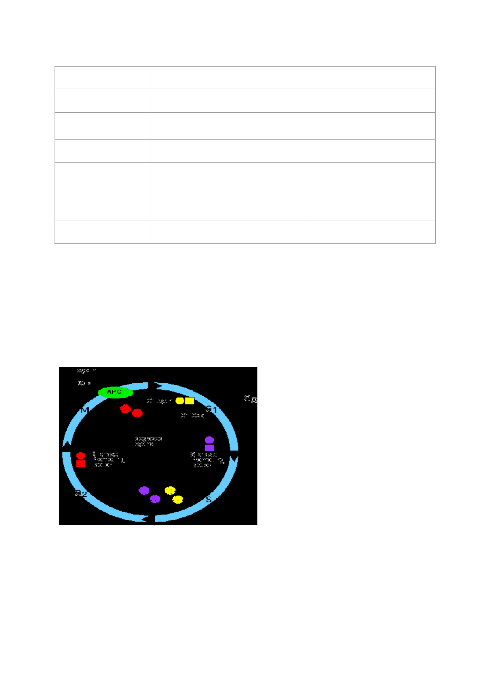
Table 3: Comparison of Meiosis and Mitosis
Meiosis
Mitosis
End result
Normally four cells, each with half
the number of chromosomes as the
parent
Two cells, having the same
number of chromosomes as
the parent
Function
Production of gametes (sex cells) in
sexually reproducing eukaryotes
Cellular reproduction, growth,
repair, asexual reproduction
Where does it
happen?
Germ cells; Reproductive cells of
almost all eukaryotes (animals,
plants, fungi, and protists)
Somatic cells; All
proliferating cells in all
eukaryotes except brain cells
Chromosome
number
Half of parent
Same as parent
Peris Moraa Mokua
30
When
At sexual maturity
Throughout life
Number of divisions
2
1
Genetically same as
parent?
No
Yes
Crossing over
happens?
Yes, normally occurs between each
pair of homologous chromosomes
Very rarely
Pairing of
homologous
chromosomes?
Yes
No
Cytokinesis
Occurs in Telophase I and Telophase
II
Occurs in Telophase
Centromeres split
Does not occur in Anaphase I, but
occurs in Anaphase II
Occurs in Anaphase
Regulation of the Cell Cycle
A eukaryotic cell cannot divide into two, the two into four, etc. unless two processes
alternate:
• Doubling of its genome (DNA) in S phase
(synthesis phase) of the cell cycle;
• Halving of that genome during mitosis (M phase).
The period between M and S is called G1; that between S and M is G2.
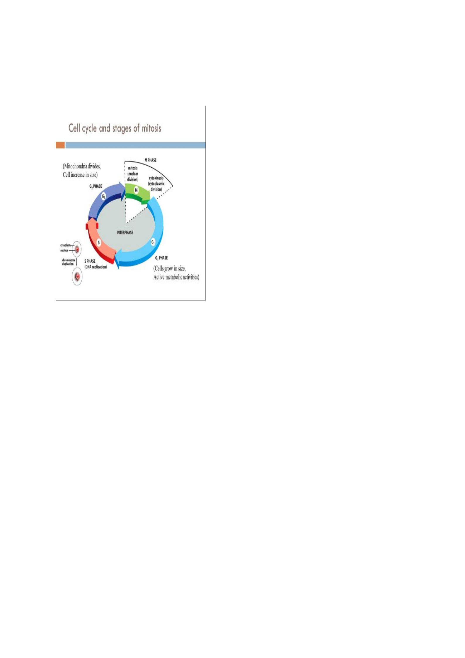
Fig. 21: The Cell Cycle
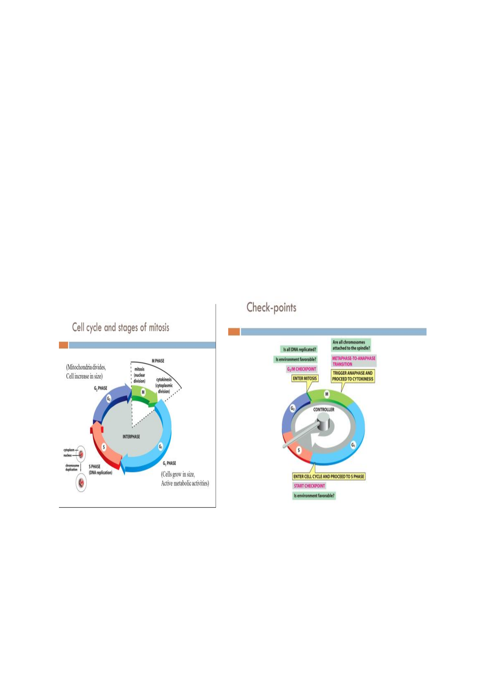
The cell cycle consists of 3 main stages:
• G1 phase- Growth and preparation of the chromosomes for replication. Cells
grow in size. There is active metabolic activities.
• S phase- Synthesis of DNA (and centrosomes) i.e. DNA replication.
Peris Moraa Mokua
31
• M phase- Mitosis (nuclear division), Cytokinesis (cytoplasmic division).
When a cell is in any phase of the cell cycle other than mitosis, it is often said to be in
interphase.
Fig. 21: Cell cycle stages
Steps in the cycle
• A rising level of G1-cyclins bind to their Cyclin dependent kinases (Cdks) and signal
the cell to prepare the chromosomes for replication.
• A rising level of S-phase promoting factor (SPF) — which includes cyclin A bound to
Cdk2 — enters the nucleus and prepares the cell to duplicate its DNA (and its
centrosomes).
• As DNA replication continues, cyclin E is destroyed, and the level of mitotic cyclins
begins to rise (in G2).
• M-phase promoting factor (the complex of mitotic cyclins with the M-phase Cdk)
initiates;
Assembly of the mitotic spindle
Breakdown of the nuclear envelope
Condensation of the chromosomes
• These events take the cell to metaphase of mitosis.
• At this point, the M-phase promoting factor activates the anaphase-promoting
complex/cyclosome (APC/C) which;
Allows the sister chromatids at the metaphase plate to separate
and move to the poles (= anaphase), completing mitosis.
Destroys cyclin B. It does this by attaching it to the protein ubiquitin which targets it
for destruction by proteasomes.
Turns on synthesis of G1 cyclin for the next turn of the cycle.
Degrades geminin, a protein that has kept the freshly-synthesized DNA in S phase
from being re-replicated before mitosis. This is only one mechanism by which the

Peris Moraa Mokua
32
cell ensures that every portion of its genome is copied once — and only once —
during S phase.
Quality control of the cell cycle
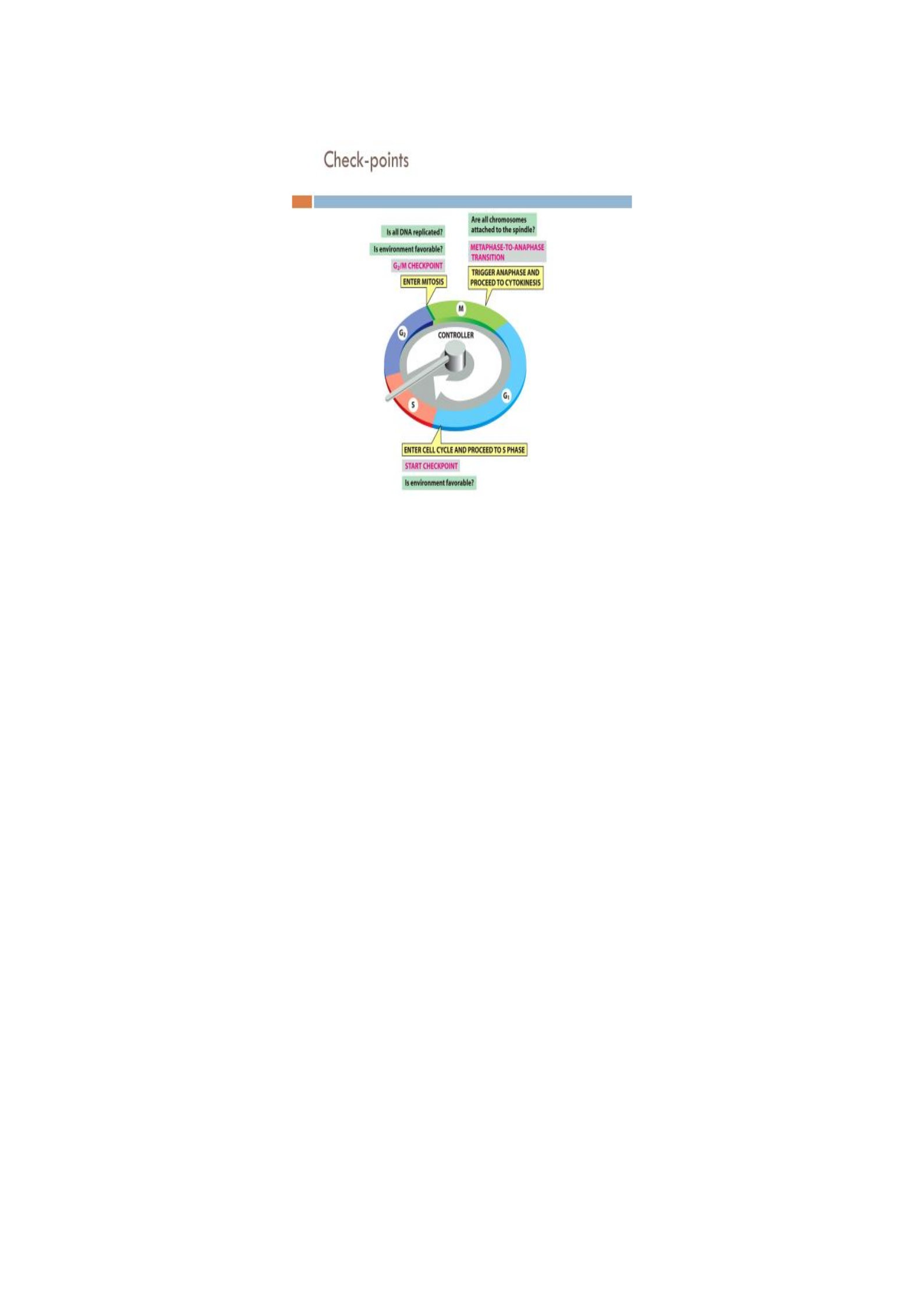
Cell cycle checkpoints
A checkpoint is a stage in the eukaryotic cell cycle at which the cell examines internal (e.g.
DNA damage) and external (e.g. molecular signals) cues and "decides" whether or not to
move forward with division. The cell has several systems (checkpoints) for interrupting the
cell cycle if something goes wrong.
1. A check on completion of S phase/G1 checkpoint. Transition of S. The cell seems to
monitor the presence of the Okazaki fragments on the lagging strand during DNA replication.
The cell is not permitted to proceed in the cell cycle until these have disappeared. Checks for
cell size, nutrients, growth factors and DNA damage.
2. DNA damage checkpoints/G2 checkpoints. Transition of M. Checks for DNA damage
and completeness of DNA replication. These sense DNA damage caused by e.g. UV radiation
from the sun. p53, a famous tumor suppressor protein called guardian of genome is the first
line of DNA damage response. It acts in the following ways;
a). Stops cell cycle at the G1 checkpoints by triggering production of Cdk inhibitor proteins-
bind to Cdk-cyclin complex and block their activity buying time for DNA repair.
b). Activate DNA repair enzymes.
c). Trigger programmed cell death so damaged DNA is not passed on. This prevents
mutations;
Before the cell enters S phase (a G1 checkpoint)
During S phase
After DNA replication (a G2 checkpoint).
3. Spindle checkpoints/M checkpoint. Transition of metaphase to anaphase. The cell
examines whether all the sister chromatids are correctly attached to the spindle microtubules.
Because the separation of the sister chromatids during anaphase is an irreversible step, the
cycle will not proceed until all the chromosomes are firmly attached to at least two spindle
fibers from opposite poles of the cell.
How this checkpoint work: Cells don't actually scan the metaphase plate to confirm that all of
the chromosomes are there. Instead, they look for "straggler" chromosomes that are in the
wrong place (e.g., floating around in the cytoplasm). If a chromosome is misplaced, the cell
will pause mitosis, allowing time for the spindle to capture the stray chromosome.
Some of these have been discovered to;
Detect any failure of spindle fibers to attach to kinetochores and arrest the cell in
metaphase (M checkpoint);
Detect improper alignment of the spindle itself and block cytokinesis;
Trigger apoptosis if the damage is irreparable.
All the checkpoints examined require the services of a complex of proteins. Mutations in the
genes encoding some of these have been associated with cancer; that is, they are oncogenes.
This should not be surprising since checkpoint failures allow the cell to continue dividing
despite damage to its integrity.
Peris Moraa Mokua
33
Fig.22: Cell cycle check-points
Control of the Cell Cycle: Cell cycle regulators
The core control system of the cell cycle are proteins in the cytoplasm which include the
Cyclins, Cyclin-dependent kinases (Cdks), Maturation-Promoting Factor (MPF) and the
Anaphase Promoting Complex/Cyclosome (APC/C). They cause key events such as DNA
replication or chromosome separation to take place. They also make sure the cell cycle events
take place in the right order and that one phase for example G1 triggers the onset of the next
phase such as the S phase.
a). Cyclins
Are the most important core cell cycle regulator. They are a group of related proteins. Are of
various types;
G1 cyclin (cyclin D)
G1/S cyclins
S-phase cyclins (cyclins E and A)
Mitotic (M) cyclins (cyclins B and A)
Each cyclin is associated with a particular phase, transition or sets of phases in the cell cycle
and helps drive the events of that phase or period. For example, M cyclin promotes events of
M phase such as nuclear envelope breakdown and chromosome condensation. The levels of
the different cyclins vary considerably (rise and fall) across the cell cycle. A typical cyclin is
present at low levels for most of the cycle but increases strongly at the stage where it is
needed. G1 cyclins are unusual in that they are needed for much of the cell cycle.
b). Cyclin-dependent kinases (Cdks)
These are enzymes which phosphorylate (attach phosphate groups to) specific target proteins.
They work together with the cyclins. Alone Cdks are inactive but the binding of a cyclin
activates it making it a functional enzyme and allowing it to modify target proteins. The
attached phopsphate group acts like a switch making the target protein more or less active.
There are three types of Cdks:

Peris Moraa Mokua
34
G1 Cdk (Cdk4)
S-phase Cdk (Cdk2)
M-phase Cdk (Cdk1)
Their levels in the cell remain fairly stable/constant across the cell cyle but Cdk activity and
target proteins change as levels of their various cyclins rise and fall., but each must bind the
appropriate cyclin (whose levels fluctuate) in order to be activated. They add phosphate
groups to a variety of protein substrates that control processes in the cell cycle.
When a cyclin attaches to a Cdk, it has 2 effects;
It activates the Cdk as a kinase,
It directs the Cdk to a specific set of target proteins, ones appropriate to the cell cycle
period controlled by the cyclin. For example, G1/S cyclins sends Ckds to s phase targets
(e. g promoting DNA replication) while the M cyclins send C.dks to M phase targets (e. g
making the nuclear membrane breakdown).
c). Maturation-promoting factor (MPF)
Is a Cdk bound to its M cyclin partner. Like a typical cyclin, M cyclin stays at low levels for
much of the cell cycle but builds up as the cell approaches the G2/M transition. As M cyclin
accumulates it binds to Cdks already present in the cell forming complexes posed to trigger
M phase. Once these complexes receive an additional signal (cell’s DNA is intact) they
become active and set the events of M phase in motion. The MPF complexes add phosphate
tags to several different proteins in the nuclear envelope resulting in its breakdown (key event
of early M phase) and also activates targets that promote chromosome condensation and other
M phase events.
d). The Anaphase-Promoting Complex/Cyclosome (APC/C)
The APC is also called the cyclosome, and the protein complex is often designated as the
APC/C. Is activated by the MPF. Causes M cyclins to be destroyed starting in anaphase. This
pushes the cell out of mitosis allowing the daughter cells to enter G1. It also causes
destruction of the proteins that hold sister chromatids together allowing them to separate in
anaphase and move to opposite poles of the cell.
The APC/C adds a small protein tag called ubiquitin (Ub).When a target is tagged with Ub it
is sent to proteasome (recycle bin of the cell) and destroyed. E.g. the APC attaches a UB tag
to M cyclins causing them to be chopped up by the proteasome and allowing the newly
forming daughter cells to enter G1 phase. It also uses Ub tagging to trigger the separation of
sister chromatids during mitosis. If it gets the rights signals at metaphase, it sets off a chain fo
events that destroys cohesion (a protein holding sster chromatids together); The APC first
adds a Ub tag to a protein called securing sending it for recycling. Securing normally binds to
and inactivates a protein called separase. When securing is sent for recycling, separase
becomes active, it chops up the cohesin that holds sister chromatids together allowing them to
separate.
Triggers the events leading to destruction of the cohesins thus allowing the sister
chromatids to separate;
Degrades the mitotic cyclin B.

