


oy,
oy
bo
oat
s
4
a5
pecc
some
i
a
com>
Na”
dosed
meee
er
earn
oe
Te
ae
randeam
‘
cH,
oT
He
oy
pain
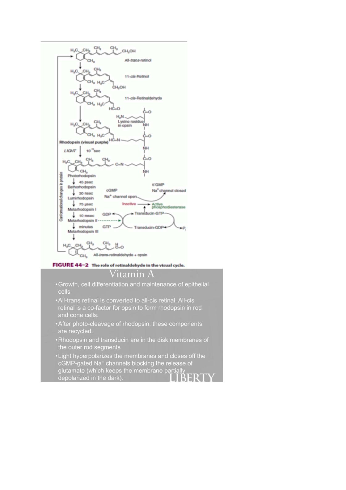
FIGURE
44-2
The
role
of
retinaldehyde
in
the
visual
cycle.
Vitamin
A
*
Growth,
cell
differentiation
and
maintenance
of
epithelial
cells
*All-trans
retinal
is
converted
to
all-cis
retinal.
All-cis
retinal
is
a
co-factor
for
opsin
to
form
rhodopsin
in
rod
and
cone
cells.
«After
photo-cleavage
of
rhodopsin,
these
components
are
recycled.
«Rhodopsin
and
transducin
are
in
the
disk
membranes
of
the
outer
rod
segments
*Light
hyperpolarizes
the
membranes
and
closes
off
the
cGMP-gated
Na*
channels
blocking
the
release
of
glutamate
(which
keeps
the
membrane
partiall
depolarized
in
the
dark).
.

Vitamin
A
Contd.
*
Glutamate
inhibits
bipolar
neurons
of
the
optic
nerve
from
being
stimulated,
therefore
light
relieves
that
inhibition
leading
to
impulse
transmission.
Vitamin
A
deficiency
leads
to
night
blindness,
metaplasia
of
the
corneal
epithelium,
xerophthalmia,
Bitot
spots,
pneumonia
and
follicular
hyperkeratosis
8-carotene
is
an
orange
pigment
and
is
found
in
yams,
carrots,
potatoes
and
yellow
squash
Thema
Somenzaton
ies
—
7-Dehydrocholesteral
FIGURE
44-3
The
synthesis
of
vitamin
D
in
the
skin.
{calciol;vitamin
D,)
HO.
FIGURE
44-4
Metabolism
of
vitamin
D.

TABLE
44-6
Classification
of
Minerals
According
to
Their
Function
Involved
in
membrane
function
Function
as
prosthetic
:
aa
"
Regulatory
role
or
role
in
hormone
action
Have
effects
in
the
body,
but
essentiality
is
not
established
known
be
toxic
inexcess
Occurs
in
all
tissues,
making
proteins
like
Hb,
Myoglobin,
Cytochromes,
catalase,
peroxidase
and
guanylate
cyclase.
In
the
liver,
the
rate
limiting
step
enzyme
is
6-
aminolevulinate
synthase
(ALA
synthase)
and
its
expression
is
repressed
by
heme.
ALA
synthase
uses
pyridoxine
(Vit
B6)
as
a
co-
factor.
ALA
dehydrase
is
inhibited
by
lead,
as
in
lead
poisoning
Oto
eh
UNIVERSITY
TABLE
31-1
Examples
of
Some
Important
Human
Hemoproteins
and
Animal
Storage
of
oxygen
in
muscle
Involvement
inelectron
transport
chain
Hydroxylation
of
xenobiotics
Degradation
of
hydrogen
peroxide
Oxidation
of
tryptophan
"The
functions
of
the
above
proteins
are
described
in
various
chapters
of
this
text.

Fer".
Protoporphiyrin
Ill
|.
|.
ai
Hychrosyerettyitxtane
to
in
cohamn
1
of
Table
31-2.
Enzymes
1,6,
7,
and
8
are
located
Porphotinagen
ger
ding
y
cause
lin
anemia.
Mutations
in
the
genes
encoding
enzymes
2-8
cause
drtbae
ee
iL
adh
yot
enzyme
ke
k
=
A
ds
ALA
synthesis
occurs
at
ALA
synthase
(ALAS!)
by
a
repression-
Seems,
(EA
|...
by
Enzyme
3
is
also
=
SUCCINYLCok
«
GPycites
deaminase
ot
hydroxymethylbilane
synthaie,
FIGURE
31-11
siochemical
causes
of
the
major
signs
and
symptoms
of
the
porphyrias.

TABLE
31-2
Summary
of
Major
Findings
in
the
Porphyrias'
4
Uroporptynnogen
erythrepowtic
Protosensitrty
Urinary,
red
cell
(erytheopowetic!
(OMIM
263700)
uroporphynin|increased
6
Coproporphyrinagen
axidase
ca
Photosensitivity,
abdominal
Urinary
ALA,
PRG,
(OMIM
121300)
coproporphynin
fecal
coproporphynin
increased
I
8.
Ferrochetatase
maa
Photosansitivity
_condimons
Condisons
1,5,
and
#
are
generally
te
most
prevalent
poephytas.
Condon
2s
rae
wa
kL
=.
a
‘.
Acute
Intermittent
Porphyria
Autosomal
dominant
disease
with
late
onset
due
to
uroporphyrinogen-|
synthase
deficiency
leads
to
ALA
and
PBG
accumulation
Episodic
expression
of
symptoms
Anxiety,
paranoia,
confusion,
recurrent
acute
abdominal
pain
No
photosensitivity
Port-wine
urine
coloration
Worsened
by
barbiturates
because
they
stimulate
cytochrome
P-450
synthesis
and
depletes
heme.
The
decreased
heme
levels
lets
go
of
th
featasd
of
ALA
synthase
which
increases
the
PaaS
Sra
and
PBG
and
makes
the
symptoms
worse.”
'
“°'
'°
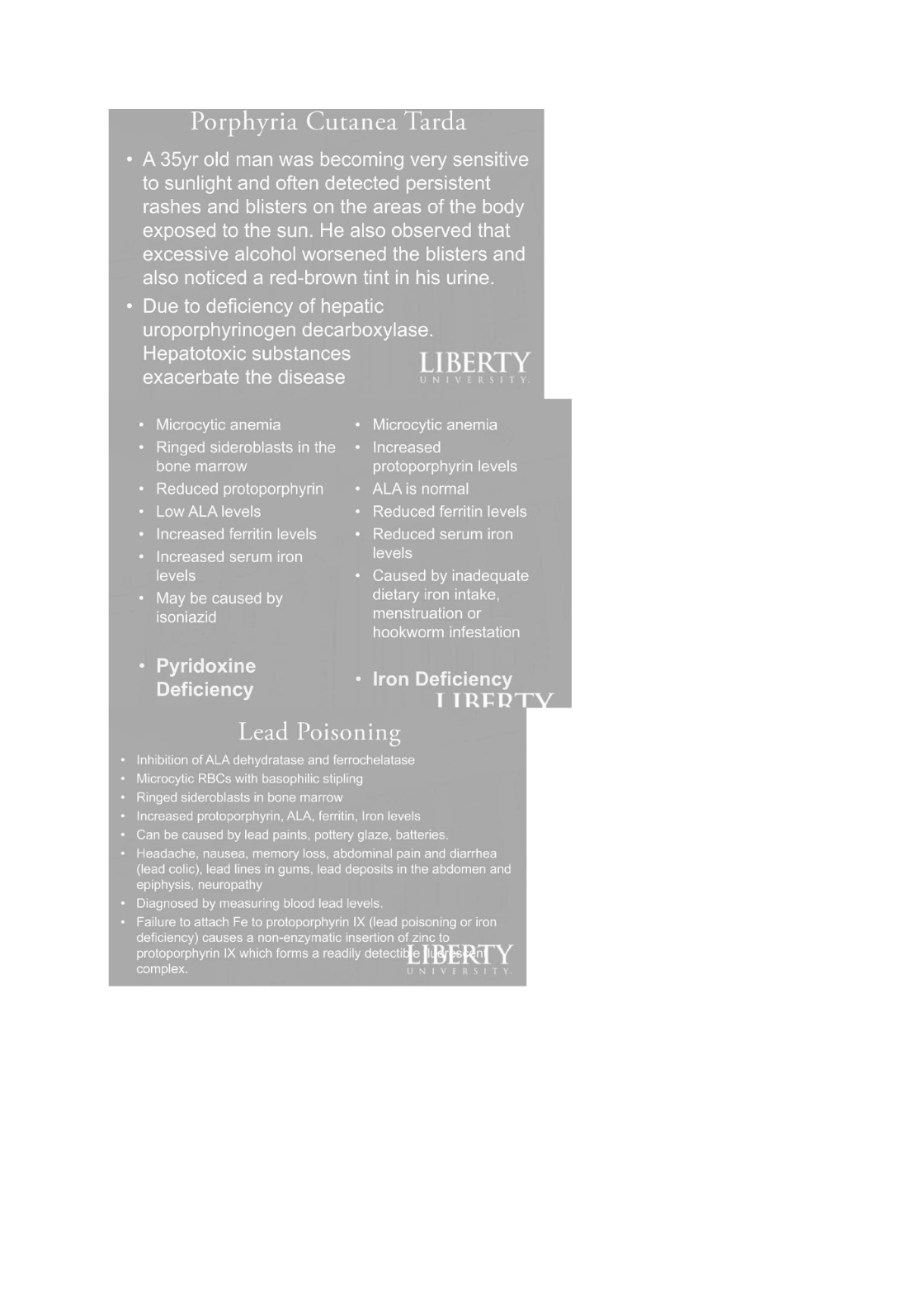
Porphyria
Cutanea
Tarda
¢
A
35yr
old
man
was
becoming
very
sensitive
to
sunlight
and
often
detected
persistent
rashes
and
blisters
on
the
areas
of
the
body
exposed
to
the sun.
He
also
observed
that
excessive
alcohol
worsened
the
blisters
and
also
noticed
a
red-brown
tint
in
his urine.
Due
to
deficiency
of
hepatic
uroporphyrinogen
decarboxylase.
Hepatotoxic
substances
LIBERTY
exacerbate
the
disease
rns
wwe
act
Microcytic
anemia
Microcytic
anemia
Ringed
sideroblasts
in
the
Increased
bone
marrow
protoporphyrin
levels
Reduced
protoporphyrin
ALA
is
normal
Mo)
A
Wee
iol)
Reduced
ferritin
levels
Increased
ferritin
levels
Reduced
serum
iron
Increased
serum
iron
NeAel
levels
Caused
by
inadequate
May
be
caused
by
dietary
iron
intake,
isoniazid
menstruation
or
hookworm
infestation
Pyridoxine
al.
Byvi
tel
etalea
lron
Deficiency
IT
tTRERT
Lead
Poisoning
+
Inhibition
of
ALA
dehydratase
and
ferrochelatase
*
Microcytic
RBCs
with
basophilic
stipling
«
Ringed
sideroblasts
in
bone
marrow
Increased
protoporphyrin,
ALA,
ferritin,
Iron
levels
*
Can
be
caused
by
lead
paints,
pottery
glaze,
batteries.
Headache,
nausea,
memory
loss,
abdaminal
pain
and
diarrhea
(lead
colic),
lead
lines
in
gums,
lead
deposits
in
the
abdomen
and
epiphysis,
neuropathy
»
Diagnosed
by
measuring
blood
lead
levels.
+
Failure
to
attach
Fe
to
protoporphyrin
IX
(lead
poisoning
or
iron
deficiency)
causes
a
non-enzymatic
insertion
of
zinc
to
eee)
(e
ToT
AN
AMPA
TCA
RCM
Rc
Tle
detectitfe
fifa
Ps
fan]
Y
complex.
UNIVERSITY

Iron
Transport
and
Storage
lron
is
needed
to
form
Hb
and
when
Hb
is
broken
down,
Fe**
is
bound
to
ferritin
and
transported
in
the
blood
by
transferrin
for
delivery
to
tissues
for
heme
Aa
Ueto
oe
Ceruloplasmin
(a
Cu2*
protein)
oxidizes
Fe2*
to
Fe**
for
transport
and
storage
in
the
liver.
tcl
AdIt
are
liso
Meo
(e)g>t-m
ae)
geet=]
(e072)
ome)
ml
fe]a
Mam
ct10
[oom
The
body does
not
have
a
means
of
excreting
excess
iron
so
its
levels
are
controlled
by
regulating
intestinal
absorption.
Hemosiderin
binds
excess
Fe**
to
preye
free
iron into
the
blood
where
it
is
haba
holt
UDP-Glucose
UDP-Glucuronic
acid
2NAD*
2NADH
+
2H*
°
Biinbin
+
a
Bilirubin
monoglucuronide
ube
FIGURE
31-14
Conjugation
of
bilirubin
with
glucuronic
acid.
The
glucuronate
donor,
UDP-glucuronic
acid,
is
formed
from
UDP-
glucose
as
depicted.
The
UDP-glucuronosyltransferase
is
also
called
bilirubsn-UGT.
re
ae
+
UDP

Porphyrins
&
Bile
Pigments
FIGURE
31-15
btagrammatic
representation
of
the
three
process
affected
in
a
number
of
conditions
causing
jaundice
is
also
TABLE
31-3
Some
Causes
of
Unconjugated
and
Conjugated
Hyperbilirubinemia
Hemolytic
anemias
Obstruction
of
the
biliary
tree
Neonatal
“physiological
Dubin-Johnson
syndrome
jaundice"
Crigler-Najjar
syndromes
types
Rotor
syndrome
land
Gilbert
syndrome
Liver
diseases
such
as
the
various
types
of
hepatitis
‘Toxic
hyperbilirubinemia
These
causes
are
discussed
briefly in
the
text.
Common
causes
of
obstruction
of
the
biliary
tree
are
a
stone
in
the
common
bile
duct
and
cancer
of
the
head
of
the
pancreas.
Various
g,
the
types
of
hep
q
e
saan
Causes
-—
7
yP

i
FIGURE
31-16
Cisgrammetk
representation
of
some
major
causes
of
jaundice.
Prehepatic
indk
event
in
the
blood
stream,
the
major
Cause
would
be
various
forms
of
hemolytic
anemia
(see
Chapter
$2).
Mepatic
agente:
event
in
the
Iver,
such
as
the
various
types
of
hepatitis
or
other
forms
of
liver
dixease
(eg,
cancer).
A
Te
are obstruction
of
the
common
bile
duct
by
a
oes
calculus)
of
by
cancer of
the
head
of
the
pancreas.
TABLE
31-4
Laboratory
Results
in
Normal
Patients
and
Patients
with
Three
Different
Causes
of
Jaundice
and
Decmased
micro-
Present
if
micro-
obstruction
present
(betraction
cows
most
common
of
obstructive
(pouthepaic)
jundhce
cancer
of
the
head
pancreas
snd
gallstone
im
common
duct
presence
of
Reinabe
the
urine
4
scemetemes
rederned
therefore
hepubies
Obetracton
of
common
bie
Quct
Cate
CMC
jaurchce
whereas
the
jaurccice
of
ihemeiytc
anema
6
eferend
to
achotunc.
The
Wbortory
results
patents
with
hepats
are
varuble,
dep
extent
of
micr>-ctrtractor
to
tie
ductules
Serum
level
of
alanine
aminotransierase
anc
aspartate
amimotranstorase
\AST)
uncaty
murtechy
erated
©
hegutts
wheres:
levels
of
alkane
phosphatase
are
chrepted
obtrctoe
leet
deawe

