



Vitamin A Deficiency in Children
1. What are the nutritional sources of Vitamin A and what are the functions of Vitamin A? The sources of vitamin A
include green and yellow veggies and fruits (sources of provitamin A); cream, butter, liver, egg yolk, kidney (vitamin A).
Retinol, Retinal, and Retinoic Acid are the active forms of vitamin A. Retinol and retinal are important for reproduction
and vision. Retinoic Acid is important for gene expression, maintenance of epithelial tissues by supporting the barrier-
based defense against pathogens and mucus secretion, and promotion of growth. Retinoic acid is also important for
normal cell proliferation and differentiation of WBCs, and intestinal epithelium.
2. What are the different forms of Vitamin A and why are children most susceptible to its deficiency? How does the
body maintain adequate levels of Vitamin A? The different forms of vitamin A are retinol, retinal, retinoic acid, and B-
carotene. Vitamin A is stored in the liver, so there are adequate levels of vitamin A in the body. The amount of retinol +
RBP in the body is a diagnostic tool for vitamin A deficiency because if there isn’t any RBP being released, then there
isn’t any retinol to bind with in the blood, so there is insufficient vitamin storage. Children are susceptible to vitamin A
deficiencies because it is not readily found in their diet at first and their vitamin stores are low at birth and if their
mothers had vitamin A deficiency then the child is more vulnerable to having the deficiency.
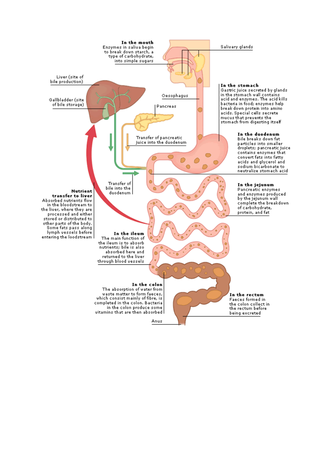
3. What is the role of bile in Vitamin A absorption? Bile breaks down lipid in the duodenum Since bile increases the .
absorption of fats, it is an important part of the absorption of the fat-soluble substances, such as the vitamins A, D, E,
and K. Besides its digestive function, bile serves also as the route of excretion for bilirubin, a byproduct of red blood
cells recycled by the liver.
4. What are the pathologies that characterize an excess of Vitamin A? Hypervitaminosis A is due to excessive intake
(>7.5mg/day) of vitamin A. both the size and numbers of lipid droplets increase as response to vitamin A deficiency.
Hypervitaminosis A promotes cheilitis (dryness and cracking of lips) as well as dryness of nasal mucosa, eyes, skin; hair
loss, and nail fragility because of decreased keratin synthesis; later indicators are bone pain, hepatomegaly, nausea, a
rise of intracranial pressure that may mimic a brain tumor and vomiting…maybe even irreversible liver damage.
Teratogenesis can occur in pregnant women.
5. Describe the role of Vitamin A in vision, gene regulation. Retinoic acid is involved in gene regulation: all trans RA
and 9-cis RA are ligands for three RARs ( ) and three retinoid X receptors (RXRs) ( ), respectively. The RXRs can
form heterodimers with RARs, TRs, VDRs, and PPAR. So, vitamin A had a broad effect on many hormonally and
nutrient-responsive genes. The RARs and RXRs bind to RAREs and RXREs in the promoter region of genes, then vitamin
A nuclear receptors are able to interact with coactivators and corepressors in the nucleus. 11-cis retinol is required for
vision. It binds to opsin making rhodopsin that when hit my light, changes to all trans retinal.
6. Describe the various classes of xerophthalmia and their features.
Xerophthalmia Classification Symptoms
XN: night blindness Cannot see properly in dim light
X1A: conjunctival xerosis Dryish, sand-like change in conjunctiva
X1B: Bitot’s spot Foamy, cheese like patches in whites of eyes
X2: corneal xerosis Drying on cornea, scaly appearance
X3A: corneal
ulceration/keratomalacia
Formation of holes on the cornea (involving
less than a third of corneal surface)

X3B: corneal ulceration
keratomalacia
Cornea becomes cloudy and soft (involving
more than a third of corneal surface)
XS: Corneal Scar Scar on cornea
XF: Xerophthalmia fundus Structural damage to rods in cornea,
followed by degeneration of rods and cones
7. Why did the patient in Case 1 have pneumonia? Vitamin A is needed for normal cell differentiation including
immune system function. Vitamin A deficiency is associated with impaired humoral and cellular immune function,
keratinization of the respiratory epithelium, and decreased mucus secretion, which weaken barriers to infection.
Calcium-Deficiency Ricketts
1. What are the major components (cellular and inorganic) of bone and what are the functions of bone in humans? The
major components of bone are calcium and phosphate as hydroxyapatite and type 1 collagen.
2. Why is calcium physiologically important and how is it distributed in the body compartments? Calcium is the most
abundant mineral in the body. 98% if calcium is found in bones. The remainder is involved in signaling, muscle
contraction, and blood clotting.
3. What are the sources of calcium, how and where is it absorbed and stored? Be detailed! Look at notes
4. Describe the sources and biological significance of Vitamin D and how it is synthesized and activated in the body.
Look at notes
5. What are the levels of Vitamin D in breastmilk and how does bile affect plasma? Vitamin D levels? 1% of breastmilk
is vitamin D. Bile breaks down lipid in the duodenum. Since bile increases the absorption of fats, it is an important part
of the absorption of the fat-soluble substances, such as the vitamins A, D, E, and K. Besides its digestive function, bile
serves also as the route of excretion for bilirubin, a byproduct of red blood cells recycled by the liver.
6. What happens when there is an excess of Vitamin D in the blood? Vitamin D is toxic. This can cause loss of appetite
nauseam thirst, and weakness. Also, hypercalcemia that can cause metastatic calcification.
7./8. Describe the roles of calcitriol, PTH and calcitonin in serum calcium regulation. What are the effects of PTH and
Vitamin D on serum phosphate levels? Calcitriol and PTH increase serum calcium levels by increase bone resorption,
kidney reabsorption, and calcium absorption in gut (calcitriol only) Calcitonin is a hormone that is produced in .
humans by the parafollicular cells (commonly known as C-cells) of the thyroid gland. Calcitonin is involved in helping
to regulate levels of calcium and phosphate in the blood, opposing the action of parathyroid hormone. Calcitonin acts
to reduce calcium levels, inhibits activity of osteoclasts, and decreases resorption of calcium in the kidneys. Calcitonin
increases phosphate levels. PTH decreases phosphate levels.
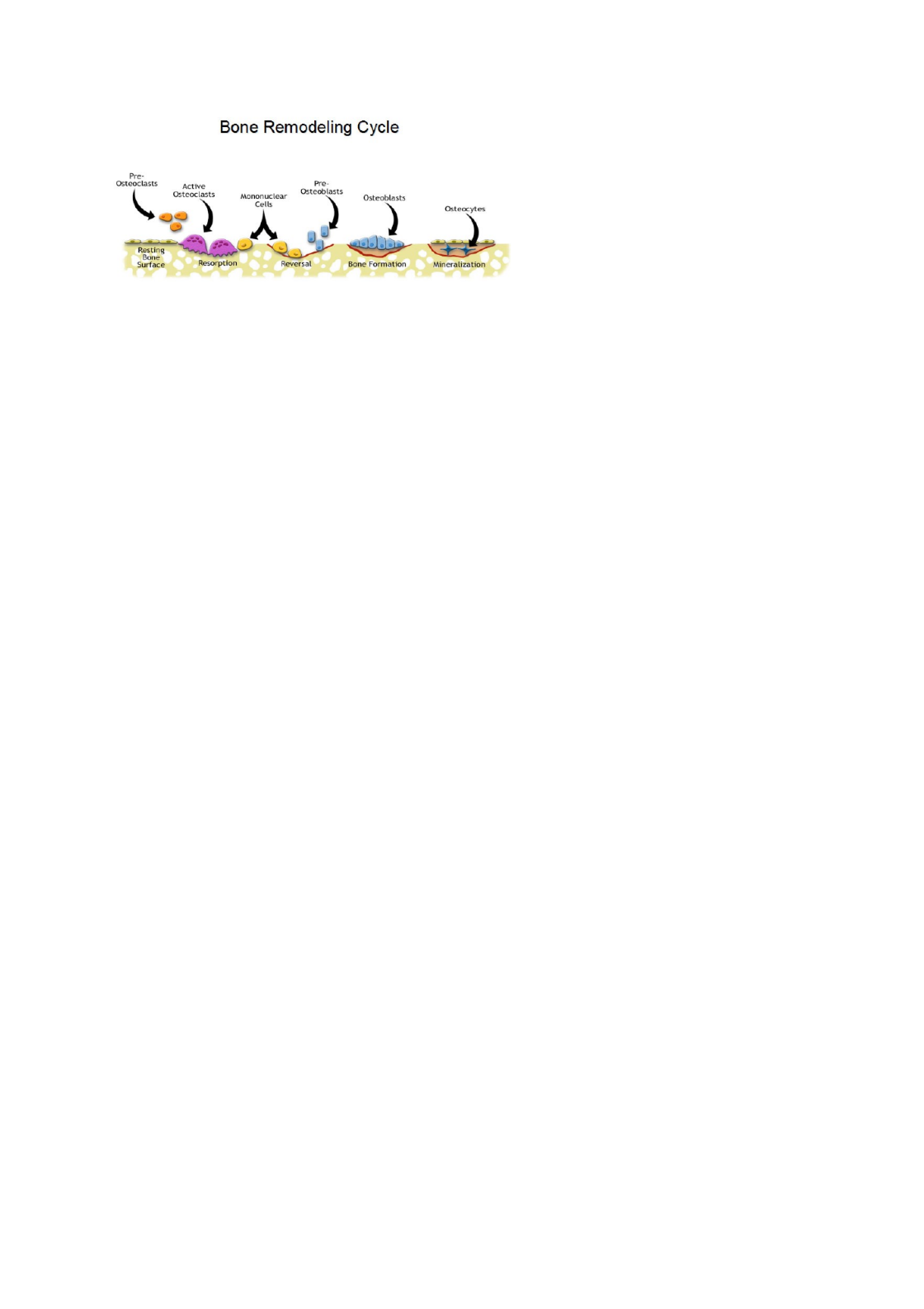
9. Describe the process of bone remodeling and the roles of RANKL, RANK and OPG. How can these be employed in the
treatment of osteoporosis?
10. What are examples of markers of bone turnover, resorption and bone synthesis? Markers of bone turnover
(remodeling) include alkaline phosphatase and osteocalcin. Markers of resorption include deoxypyrolidone, NTx (N-
teleopeptide), and CTx (C-teleopeptide). NTx and CTx are increased in individuals with metabolic bone diseases
associated with increase bone resorption, primary hyperparathyroidism, and are decreased in hypoparathyroidism.
Markers of bone synthesis include alkaline phosphatase.
11. Differentiate the pathophysiology of osteomalacia and Rickets. Rickets occurs in children, osteomalacia occurs in
adults. Rickets is characterized by an increase in osteoid protein; cupping and fraying of the metaphysis and
longitudinal widening of growth plates are hallmarks of rickets. In adults the growth plate has closed and decreased
mineralization of osteoid is terms osteomalacia. 12. What are the
signs and symptoms of Rickets? Explain the reasons for the low calcium, phosphorus, PTH, urinary calcium in this
patient? The ratio of demineralization and mineralization is increased in Rickets (but not in osteoporosis). Rickets
shows bowed legs. The type of deformity depends on the age of the patient: in infants, deformities are in the forearms
and tibias, craniotabes, hypotonia, and tetany. The patient has low calcium levels and low urinary calcium levels
because of the lack of access to milk. The patient had high PTH levels because PTH causes calcium resorption in bone,
reabsorption of kidneys, and decrease phosphorus levels in the kidney.
13. What are other causes of Rickets? Rickets can be caused by low calcium levels, low phosphate levels (x-linked or
autosomal dominant), or low V levels (type 1 is a defect in 1 hydroxylase and type 2 is a mutation in a V
DD receptor).
Hereditary Hemochromatosis (HH)
1. What are the possible causes of hemochromatosis? What are the complications of hemochromatosis (Iron overload)?
HH causes the body to absorb too much iron. Normally humans extract needed iron from food via the intestines. When
there is an adequate amount of iron, the body reduces its absorption to avoid excessive accumulations. In a person
with HH, the mechanism for regulating iron absorption is faulty and the body absorbs too much iron. Remember
people are not able to excrete absorbed iron! An absorption problem (not likely) or a feedback problem in which the
HFE gene is unable to recognize the replenishment of adequate storage.
2. What are the signs and symptoms of HH? Arthritis from iron in joints, hypogonadism, cirrhosis of liver, fibrosis of
liver, chronic tiredness, bronze discoloration (iron deposits in skin), hair loss, diabetes mellitus (iron deposits in
pancreas), cognitive problems, hemosiderosis (accumulation of hemosiderin when ferritin is used).
3./4. What are possible laboratory indicators of iron overload and how can you establish a diagnosis of HH? What are
the most common genetic abnormalities that cause HH? What is the pattern of inheritance?
transferrin levels, ferritin or hemosiderin level (unreliable because other diseases can also cause those high levels),
genetic testing of HFE gene in chromosome 6. Homozygotes of HH have most a C282Y change (cysteine to tyrosine
change in AA 282); heterozygotes have both a C282Y change and a H63D (aspartate to histamine in AA 63). HH is
autosomal recessive.
5./6. Describe how the body gets iron and how iron is used by the body. Where is iron normally stored in the body?
What are the roles of ferritin, hemosiderin, transferrin, ferric reductase, DMT-1, ferroportin, hephaestin, TfR1, heme
oxygenase in iron homeostasis? The body gets iron from diet or from heme. In the intestinal tract, usually the
duodenum and jejunum, heme is digested by heme carrier proteins (HCP) (apical side only) and is oxidized by heme
oxygenase to ferrous iron (soluble). Iron from the diet changed from insoluble ferric iron to soluble ferrous iron by
duodenal ferric reductase and is absorbed by divalent metal ion transporter 1 (DMT-1) in the apical (villus) side or
basolateral side (crypt). Intestinal mucins also help with absorptions in the apical side. From absorption, iron can
either be stored in ferritin or hemosiderin or can be transported outside of the cells into circulation. If transported, it
must first pass through ferroportin-1 that is regulated by hepcidin (secreted by the liver). Then ferrous iron is reduced
to ferric iron by hephaestin, a ferroxidase. Ferric iron then attaches to transferrin in circulation. If two ferric irons

attach to transferrin, it is termed diferric transferrin. Transferrin then attaches to transferrin receptor 1 in RBCs and is
taken in. Transferrin and its receptor are recycled. 7. Describe the iron stores regulator hypothesis. Also describe the
HFE gene and gene product and its role in iron homeostasis. The iron stored regulator hypothesis states that the body
slowly accumulates dietary iron which prevents iron overload after iron stored are seem adequate. This involves TF
bound iron, serum ferritin, and serum TF. The stored regulator hypothesis is thought to be impaired in HH, so that
implies that there is abnormal feedback from the store’s regulator. HFE is the iron store regulator. When the HFE
protein is attached to transferrin receptor 1, the receptor cannot bind to a protein called transferrin. When transferrin
receptor 1 is bound to transferrin, iron enters liver cells. So, it is likely that the HFE protein regulates iron levels in liver
cells by preventing transferrin from binding to transferrin receptor 1.
The HFE protein regulates the production of a protein called hepcidin. Hepcidin is produced by the liver, and it
determines how much iron is absorbed from the diet and released from storage sites in the body. When the HFE protein
is not bound to transferrin receptor 1, it binds to a group of other proteins that includes hepcidin. The formation of this
protein complex triggers the production of hepcidin. So, when the HFE protein is bound to transferrin receptor 1,
hepcidin production is turned off and when the HFE protein is not bound to transferrin receptor 1, hepcidin production
is turned on. SO IN HRE MUTATION, HFE BINDs TO THE RECEPTOR SO IRON DOESN’T FLOW IN. HFE is expressed in
crypt cells, basolateral side. Mutated HFE, may lead to decreased uptake of plasma iron by crypt cells, thus diminishing
iron pools in the cell. This might in turn result in increased expression of mRNA for DMT-1 and ferroportin 1 in villus
cells that mature from crypt cells. This can cause overabsorption of iron from villus cells. Crypt cells are stem cells that
can either mature and migrate upwards to the apical layer and become villi. Hepcidin mRNA is lower in patients with
HH.
8. Describe the biochemical dangers of iron overload and toxicity. Iron overload cause excess free iron that increases
ROS production that attacks polyunsaturated FA, protein, and nucleic acids. Increase iron also increases ECM
components and leads to fibrosis of the liver.
9. How will you manage a case of HH? Why is phlebotomy not a long-term solution in HH? Either through phlebotomy
or Fe chelates.
Liver
(site
of
bile
production)
Gallbladder
(site
of
bile
storage)
Nutrient
transfer
to
liver
Absorbed
nutrients
flow
in
the
bloodstream
to
the
liver,
where
they
are
processed
and
either
stored
or
distributed
to
other
parts
of
the
body,
Some
fats
pass
along
lymph
vessels
before
entering
the
loodstream
In
the
mouth
Enzymes
in
saliva
begin
to
break
down
starch,
a
type
of
carbohydrate,
into
simple
sugars
Oesophagus
Pancreas
Transfer
of
pancreatic
juice
into
the
duodenum
Transfer
of
bile
into
the
duodenum
In
the
ileum
The
main
function
of
the
ileum
is
to
absorb
nutrients;
bile
is
also
absorbed
here
and
returned
to
the
liver
through
blood
vessels
In
the
colon
The
absorption
of
water
from
waste
matter
to
form
faeces,
which
consist
mainly
of
fibre,
is
completed
in
the
colon,
Bacteria
in
the
colon
produce
some
vitamins
that
are
then
absorbed
Anus
Salivary
glands
In
the
stomach
Gastric
juice
secreted by
glands
in
the
stomach
wall
contains
acid
and
enzymes,
The
acid
kills
bacteria
in
food;
enzymes
help
break
down
protein
into
amino
acids,
Special
cells
secrete
mucus
that prevents
the
stomach
fram
digesting
itself
In
the
duodenum
Bile
breaks
down
fat
particles
into
smaller
droplets;
pancreatic
juice
contains
enzymes
that
convert
fats
into
fatty
acids
and
glycerol
and
sodium
bicarbonate
to
neutralize
stomach
acid
In
the
jejunum
Pancreatic
enzymes
and
enzymes
produced
by
the
jejunum
wall
complete
the
breakdown
of
carbohydrate,
protein,
and
fat
In
the
rectum
Faeces
formed
in
the
colon
collect
in
the
rectum
before
being
excreted

