


1
EFFECTS OF EXERCISE-INDUCED MUSCLE DAMAGE ON HUMAN PERFORMANCE IN
DYNAMIC ACTIVITIES
CHAPTER ONE
INTRODUCTIO
N

2
1.1 Introduction
Exercise-Induced Muscle Damage (EIMD) is a common phenomenon that occurs due to
unusual physical activity, especially those involving high eccentric components. Eccentric
contractions occur when muscles are actively elongated, and this type of contraction is
often encountered in everyday activities such as descending stairs. In addition, many
athletic activities also involve eccentric muscle action, specifically during the landing or
impact phases when running, jumping, or spinning.
Compared to concentric contractions (shortening of muscles) and isometric (muscles in a
static state), eccentric muscle action is mechanically more efficient. However, this type of
contraction uses a unique activation strategy, so the muscles are more susceptible to
damage (Enoka, 1996; McHugh et al., 2000).
1.2 Mechanisms of muscle damage
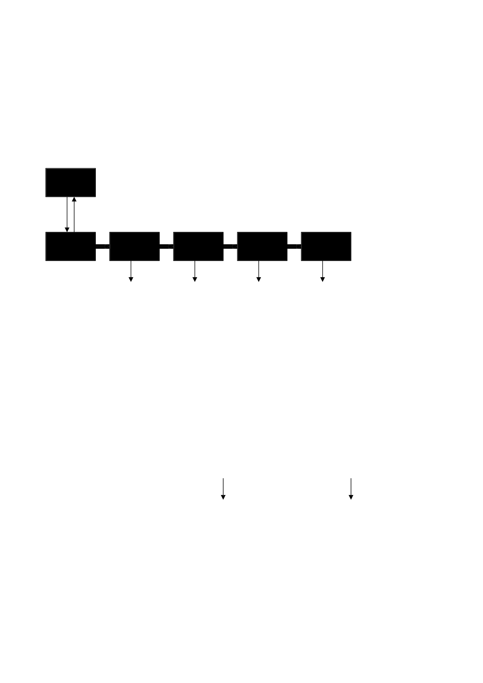
Proske and Morgan (2001) put forward a series of early events to explain how muscle
damage is caused by eccentric exercises (Figure 1.1). This model suggests that during
eccentric contractions, weaker sarcomers will be stretched beyond the overlap of
myofilamylates. This process occurs as the muscle passes through a long tension curve in
the leg descends, where the length inconsistency of the sarcomer begins to develop (see
Figures 1.2 & 1.3) (Gordon et al., 1966). As a result, the sarcomere is disturbed, which is
then followed by membrane damage and subsequent dysfunction in the excitation-
contraction (EC) mechanism. Muscle biopsy data show evidence of disorders in sarcomers,
3
Impaired
sarcomer
Excessive
sarcomer Membrane
Damage Dead
Fiber
Local
contracture
Sarkomer
Normal
including Z-line streaming (Newham et al., 1983a; Fridén, 1984), as well as damage to the
t-tubules, sarcoplasmic reticulum, and sarcolemma (Lieber et al., 1994, 1996; Fridén &
Lieber, 1998).
Eccentric
Contractions
Optimal
Length
Shift
EC Pair
Dysfuncti
on
Increasing
Passive
Tension
Delayed tension
(Irreversible)
Falling in
Tension
(Reversibe
)
Swelling
and Pain

4
Figure 1.1 A series of postulated events that cause muscle damage from eccentric
exercises (Proske & Morgan, 2001, p. 334).
These disturbances are reported to occur mainly in type II fibers, suggesting that they are
exceptionally damaged (Fridén et al., 1983; Fridén & Lieber, 1992). More recently,
intravital microscopic observations have also revealed substantial microvascular
dysfunction including an increase in red blood cell-flowing capillaries and an enlarged
capillary diameter (Kano et al., 2005). This ultrastructural disruption initiates the entry of
Ca2+ into the sarcoplasm activating proteolytic pathways involved in the breakdown and
repair of muscle fibers (Peake et al., 2005, Tidball, 2005). These events in turn produce
symptoms associated with muscle damage, including an increase in intramuscular proteins
in the bloodstream (Hortobágyi & Denahan, 1989), prolonged loss of muscle strength,
decreased range of motion and increased muscle pain (Clarkson et al., 1992; Cleak &
Eston, 1992).
5
Descending limbs
Sarcomer length (μ)
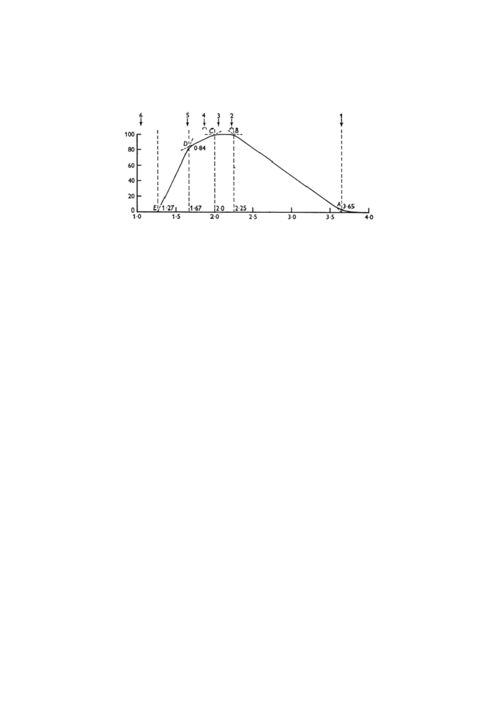
Figure 1.2 The relationship between length and tension in skeletal muscles. Adapted from
Gordon et al. (1966), p.185.
% of maximum
tension
6
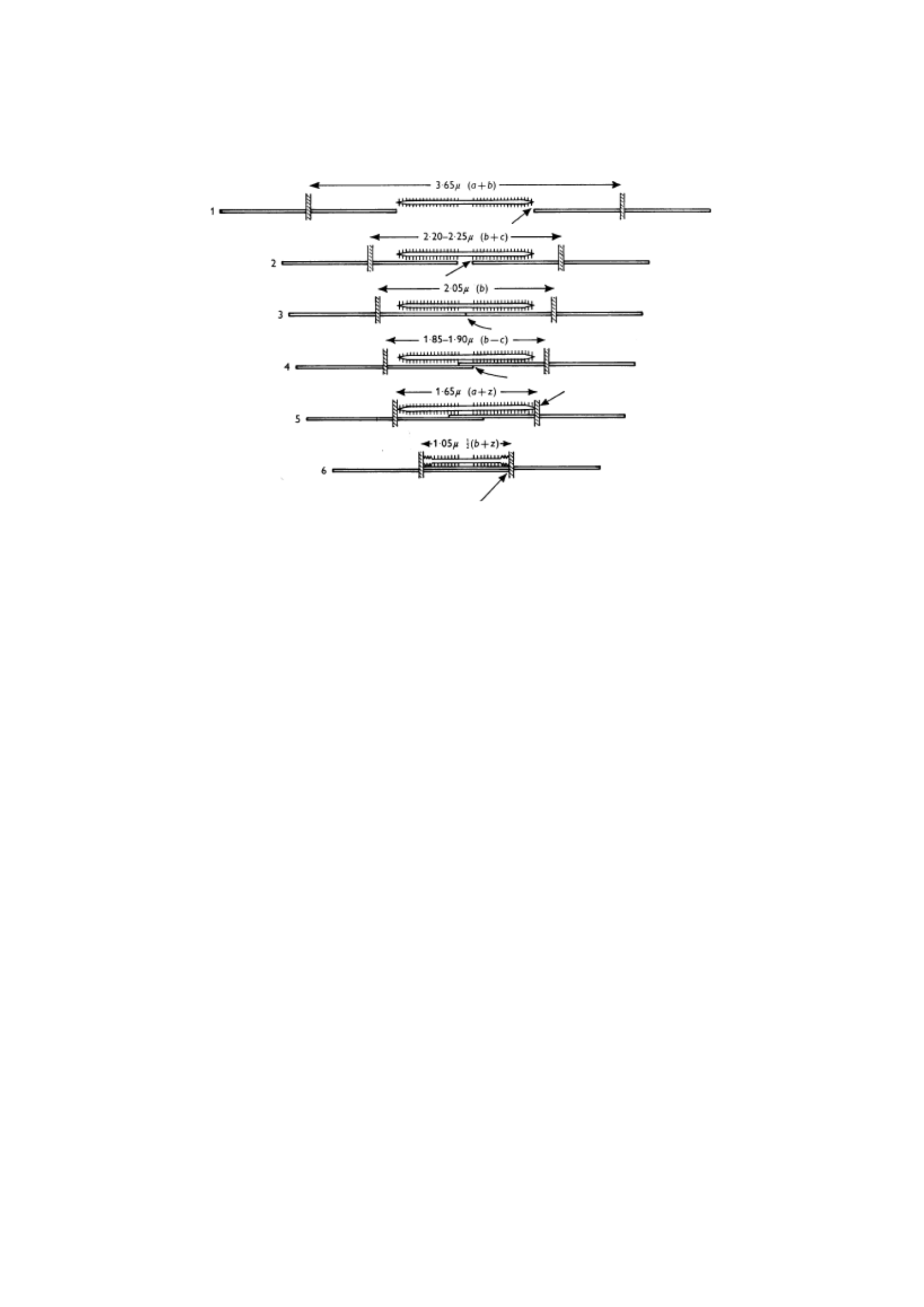
Figure 1.3 The critical stage in the increase in myofilament overlap corresponding to the
key points (1–6) labeled on the length-voltage curve in figure 2.1. Adapted from Gordon
et al. (1966), p.186.

7
1.3 Muscle function after eccentric exercise
Although there are some symptoms associated with EIMD, it has been suggested that
muscle function measurements provide the best way to assess the magnitude and duration
of muscle injury (Warren et al., 1999). In addition, the immediate and prolonged loss of
power-generating capacity resulting from eccentric exercise may be the most important
symptom when considering the effect of EIMD on human responses to dynamic exercise
(Byrne et al., 2004).
Isometric strength measured at a fixed joint angle is the most commonly used assessment
of muscle function after eccentric exercise (Warren et al., 1999), with the largest decrease
in maximal voluntary contraction (MVC) occurring immediately after eccentric exercise,
followed by linear recovery (Byrne et al., 2001). Evaluation of dynamic muscle function
after EIMD can be achieved using an isokinetic dynamometer. While it is not possible to
replicate the sport-specific patterns and speed of movement, insights into dynamic muscle
function have been gained using this technique. The magnitude of strength loss and the
degree of recovery between isometric, concentric and eccentric muscle action appear to be
similar after EIMD (Byrne & Eston, 2002a; Michaut et al., 2002). However, conflicting
findings have been reported in relation to changes in peak torque at different angular
speeds. Observation that the greatest decrease in peak torque is at higher angular velocity
(Fridén et al., 1983; Eston et al., 1996) supports the idea that type II fibers can be
selectively damaged during eccentric exercise (Fridén et al., 1983; Fridén & Lieber, 1992);
but it is countered by conflicting evidence that peak torque is affected to a greater degree

8
at slower angular speeds rather than faster (Gibala et al., 1995; Deschenes et al., 2000;
Michaut et al., 2002).
1.4 Dynamic training
Athletic performance that relies on the ability of muscles to generate strength quickly is
negatively affected after exercise-induced muscle damage (EIMD). The loss of force
generation capacity results in impaired sprint performance due to reduced peak power and
increased time to peak power. Direct and prolonged reductions have been observed in peak
power output at cycle ergometers (Sargeant & Dolan, 1987; Byrne & Eston, 2002b), in
time to peak power (Twist & Eston, 2007) and in cycle sprint performance (Twist & Eston,
2005). However, recovery was not immediate and further declines at 24 and 48 hours were
observed, suggesting that delayed-onset muscle pain (DOMS) may affect dynamic
response.
The effect of EIMD on durability performance is still debated. Some studies have reported
no change in sub-maximal running performance after a previous attack from exercise that
damaged eccentric muscles (Hamill et al., 1991; Scott et al., 2003; Paschalis et al., 2005;
Marcora & Bosio, 2007). Other investigations have observed that EIMD has a negative
effect on performance (Braun & Dutto, 2003; Chen et al., 2007b). Summary
The purpose of this thesis is to investigate the effect of exercise-induced muscle damage
(EIMD) on response to dynamic exercise in human subjects. This topic is rarely
investigated and the findings are vague. Therefore, the main goal is to provide empirical
evidence to advance scientific knowledge and understanding of the EIMD phenomenon;

9
E
especially by investigating physiological responses, perceived exertion, and metabolism
to dynamic exercise performance with EIMD. This thesis consists of 4 studies, as outlined
below. Studies 1 and 4 used a total sample of 13 participants, 7 of which were common to
both studies. Similarly, studies 2 and 3 used a total sample of 16 participants, 7 of which
were common to both studies.
Study 1 Effect of exercise-induced muscle damage on ventilation response
and perceived exertion on moderate and vigorous intensity cycle exercise
This study examined the effects of exercise-induced muscle damage (EIMD) on the
ventilation and exertion responses felt to cycle exercise. Ten healthy, physically active
men cycled for six minutes at moderate intensity and fatigue at a strenuous intensity before
and 48 hours after eccentric exercise (100 squats with a load corresponding to 70% body
mass). Changes in ventilation and perceived exertion ratings (RPE) are calculated for each
individual and expressed against time (moderate and vigorous exercise) and as a
percentage of fatigue time (vigorous exercise). Ventilation increases during moderate
exercise at 48 hours ( V ; 34.5 ± 5.0 to 36.3 ± 3.8 L.min-1, P<0.05) but the RPE increase is
not
important. During strenuous training at 48 hours, the time of fatigue (TTE) is
reduced and
V
E

10
(87.1 ± 14.1 to 93.8 ± 11.7 L.min-1) and RPE (15.5 ± 1.3 to 16.1 ± 1.4) increased (P<0.05).
When expressed as a percentage of TTE, the difference between the vent value and the
RPE disappears. The findings suggest that the enhanced ventilation response to cycle
exercise after EIMD may be an important cue in informing the perception of effort during
high-intensity exercise but not during moderate-intensity exercise. No changes were
observed in the blood lactate response, although the ventilation response was enhanced,
suggesting that other ventilation stimuli may play an important role after EIMD. Potential
dissociation of ventilation response and blood lactate during the incremental ramp cycle
after EIMD was investigated in study 2.
This study has been the basis of the following publications:
Davies, R. C., Rowlands, A.V., & Eston, R. G. (2009). Effect of exercise-induced muscle
damage on breathing and perceived exertion response to moderate and vigorous cycle
exercise. European Journal of Applied Physiology, 107(1), 11-19.
Study 2 Effect of eccentric exercise-induced muscle damage on gas exchange
threshold
Previous attacks of exercise damaging eccentric muscles improve the ventilation response
to constant weight cycle exercise without altering the blood lactate profile. However, the
effect of exercise-induced muscle damage (EIMD) on the performance of ramp
supplemental exercise has received sparse attention. This study tests the hypothesis that
EIMD will add to the ventilation response to subsequent ramp incremental cycle exercises

11
2
leading to a reduction in the gas exchange threshold (GET). In the absence of altered blood
lactate profiles, this would indicate a dissociation of GET from the lactate threshold. Ten
healthy and physically active male subjects (age, 25 ± 7 years; mass, 80.1 ±9.9 kg; height,
1.80 ± 0.08 m) performed the maximum incremental cycle exercise test before (before)
and 48 hours after (after) completing 100 squats, with a load corresponding to 70% body
mass. After the eccentric training GET occurs early (pre GET V O
2 : 1.58 ± 0.26; post-GET
V O : 1.41 ± 0.14 l.min-1) while the blood lactate response did not change (P > 0.05). The
dissociation between GET and blood lactate response during cycling with EIMD suggests
that the two phenomena are not causally related. We propose that 13%
the resulting increase in ventilation and reduction in GET is mainly caused by increased
activation of stimulated group III and IV afferents through mechanical disruption of local
muscle fibers and microvessels due to eccentric exercise.
The observed decrease in endurance capacity may be due to a shift in the metabolic profile
of muscles to an increased dependence on non-oxidative metabolism. Therefore, the
purpose of study 3 was to investigate changes in muscle metabolism during gradual
exercise to fatigue.
The study is currently under review for publication:
Davies RC, Rowlands AV, Poole DC, Jones AM and Eston RG Exercise-induced muscle
damage separates the exchange threshold of Lactate and Gas. It is currently under review.
Study 3 The 31P-MRS METABOLIC RESPONS TO GRADUAL EXERCISE AFTER
MUSCLE-DAMAGING ECCENTRIC EXERCISE

12
Performance degradation associated with EIMD includes a reduction in maximum force
generation capacity (Clarkson et al., 1992) and a shorter time to fatigue (Asp et al., 1998;
Carmichael et al., 2005, 2006). Asp et al. (1998) have proposed that EIMD results in a
shift towards more glycolytic energy production that can contribute to the acceleration of
fatigue progression. This study investigated the effect of EIMD on changes in muscle
metabolism during incremental exercise using 31phosphor magnetic resonance spectroscopy (31P-MRS).
Before and 48 hours after performing 100 squats, the 31P-MRS was used to measure dynamic
changes in [PCr], [Pi], [ADP] and pH during the incremental test of knee extensors to
fatigue. The resting ratio [Pi]:[PCr] increased 48 hours after eccentric exercise (pre: 0.12
± 0.02; post: 0.18 ± 0.05). During gradual exercise, the changes in pH, [PCr] and [Pi]:[PCr]
are similar but do not continue for that long. Time to fatigue and associated peak work rate
values are significantly reduced after
eccentric exercises (before: 519 ± 56; post: 459 ± 63 seconds) and (before: 29 ± 4; posts: 25 ± 4
W),

13
each. The pH and [PCr] values of the end of exercise were significantly higher (pre: 6.75
± 0.12; post: 6.83 ± 0.15) and (pre: 20 ± 13; post: 33 ± 15% of the baseline score),
respectively. These findings suggest that the acceleration of fatigue development after
eccentric exercise does not result from changes in muscle phosphate metabolism.
While phosphate metabolism does not appear to have a detrimental influence on dynamic
exercise performance, the substantial microvascular dysfunction observed after EIMD
(Kano et al., 2005) may contribute to impaired performance due to impaired delivery and
distribution of O2 within the capillary base of active muscle.
Thus study 4 was designed to investigate the effect of EIMD on O2 matching delivery to O2
utilization. The study is currently under review for publication: Davies RC, Eston RG, Fulford
J, Rowlands AV and Jones AM. Muscle damage alters metabolic responses to dynamic exercise
in humans: a 31P-MRS study. It is currently under review.
Study 4 Effect of eccentric exercise-induced muscle damage on the
dynamics of muscle oxygenation and pulmonary oxygen uptak
Unusual eccentric exercises have a major impact on muscle structure and function.
However, it is not known whether related microvascular dysfunction interferes matching
the delivery ofO2 ( Q2O2 ) with the utilization of O2 ( V2 ). Near infrared spectroscopy (NIRS)
was used to test the hypothesis that muscle damage caused by eccentric exercise will
increase the ratio of Q O2 : V O2 muscle during temporary heavy intensity training maintains the speed of the V O2 kinetics at
the beginning of the exercise. Nine men are physically active Complete a 'step' test for
heavy intensity training of a baseline unpacked on a cycle

14
ergometer before and 48 hours after eccentric exercise (100 squats with weights corresponding
to 70% of body mass). NIRS and lung breath by breath V O
2 Measured continuously during the
exercise test and then modeled using standard non-linear regression techniques. No change in
the kinetics
of phase II pulmonary V O 2 after the start of the exercise (time constant, pre: 25 ± 4; post:
24 ± 2 seconds; amplitude, pre: 2.36 ± 0.23; post: 2.37 ± 0.23 L/min; all P>0.05). However, the
primary (pre: 14 ± 3; post: 19 ± 3 sec) and overall (pre: 16 ± 4; post: 21 ± 4 sec) mean
response times of [HHb] responses were significantly slower after eccentric exercise (P<0.05).
Slower kinetics [HHb] observed after eccentric exercise consistent with an increase in
the ratio of
Q O2 : V O2 during the transition to heavy-intensity training. We propose that the primary phase
of the unchanged
Ki associated with an increase in the ratio of Q O2 : V O2 which conserves blood- fluks miosit O2. This research has
been the basis of the following publications/presentations: Publications:
Davies, R. C., Eston, R. G., Poole, D.C., Rowlands, A. V., Dimenna, F., Wilkerson, D. P.,
et al. (2008). Effect of eccentric exercise-induced muscle damage on the dynamics of
muscle oxygenation and pulmonary oxygen uptake. Journal of Applied Physiology,
105(5), 1387-1388.
Because the reviewers thought the paper could have a major impact on the field, Dr. Mark
Burnley was invited to write a Commentary to accompany the publication of the article:
Burnley, M. (2008). Found in translation: dependence of oxygen uptake kinetics on O2
delivery and O2 utilization. Journal of Applied Physiology, 105(5), 1387-1388

15
Presentation
Davies R, Eston R, Poole D, Rowlands A, Dimenna F, Wilkerson D, Twist C, and Jones
A. (2008). Effect of exercise-induced muscle damage on pulmonary oxygen uptake and
muscle deoxygenation kinetics during high-intensity exercise. In J. Cabri, F. Alves, D.
Araujo, J. Barreiros and A. Veloso (Eds), 13th Annual Congress of the European Congress
of Sport Science, Abstract Book (p. 630). Estoril.

16
CHAPTER TWO
LITERATURE REVIEW

17
2.1 Introduction
Temporary damage to skeletal muscles resulting from unusual exercise has been the
subject of investigation by exercise physiologists for more than 100 years. In 1902 Hough
described the delayed but temporary pain experienced when an untrained muscle made a
series of contractions against a strong spring, suggesting that it was caused by the rupture
of the muscle Since then, several hundred published investigations have attempted to
elucidate the mechanisms underlying this phenomenon and document its symptoms. Direct
histological analysis of human and animal muscle tissue has provided evidence to support
Hough's (1902) original argument that changes in skeletal muscle morphology are a
symptom of unusual exercise.
2.2 Changes in skeletal muscle morphology
Human muscle biopsy data have provided direct evidence of considerable disturbances in
the ultrastructure of skeletal muscles after unusual eccentric exercises (Fridén et al., 1981,
1983; Fridén, 1984; Newham et al., 1983a; Jones et al., 1986; Gibala et al., 1995).
Interference originating from Z-band myofibrilar is seen as streaming, dilation or total
interference. Z-line disorders are the most frequently reported ultrastructural abnormalities
and thus can represent weak links in myofibril contractile mechanisms (Newham et al.,
1983a; Fridén 1984). Reports of greater disturbances of type II fibers have led to the
understanding that these fibers are exceptionally damaged during eccentric exercise
(Fridén et al., 1983; Fridén, 1984; Jones et al., 1986 Lieber et al., 1991; MacPherson et al.,

18
1996). Eccentric contractions are understood to produce higher levels of mechanical stress
than concentric or isometric contractions due to reduced motor unit activation (Enoka,
1996; Armstrong et al., 1991; McHugh et al., 2000). This causes

19
speculation that type II motor units are recruited selectively during eccentric contractions
(Enoka, 1996; McHugh et al., 2000, 2002; Nardone & Schieppati, 1988; Nardone et al.,
1989; Howell et al., 1995) and that excessive pressure on a small amount of active fibers
causes them to become damaged (McHugh et al., 2000).
Figure 2.1 Longitudinal section of fast-twitch (FT) fibers in A) sedentary control rat
brachii tricep muscle, and B) rat brachii triceps muscle 1 day after downhill running
exercise (DH). Scale rod, 1μm. Adapted from Takekura et al. (2001). Watch for Z-line
smudges and focus disturbances from the A-band region after running downhill.
In animal models used to study EIMD, damage to the contractile and cytoskeletal
components of predominantly type II fibers has been revealed using histological staining
B

20
(Fridén and Lieber, 1992; Takekura et al., 2001) (Figure 2.1). The lack of staining for
cytoskeletal proteins has provided evidence of disorders in sarcolemma and cytoskeleton
(Lieber et al., 1994, 1996; Fridén & Lieber, 1998; Komulainen et al., 1998, 1999, 2000).
Morphological changes in skeletal muscle also include ultrastructural changes in the
arrangement of the T tubules that are believed to occur when adjacent myofilamymen
exhibit different levels of stretching (Takekura et al., 2001; Yueng et al., 2002), and the
existence of several centers

21
core (Kano et al., 2004). Furthermore, disturbances in capillary geometry including
enlarged capillary diameters have been observed using intravital microscopy, possibly due
to myocyte swelling (Kano et al., 2004).
In human studies, changes to the intermediate filament system have been interpreted as
evidence of myofibrillar and cytoskeletal damage (Fridén et al., 1984). However, this
interpretation has been challenged by Yu and colleagues (2002) who have reported an
increase rather than a decrease in cytoskeletal protein staining. These authors suggest that
their findings provide evidence of muscle repair and remodeling rather than muscle
damage from eccentric contractions (Yu et al., 2002, Yu & Thornell, 2002). While animal
models may not always accurately reflect changes in their human counterparts, they have
provided valuable insights that are not so easily gained in human models. Indeed, caution
must be taken when interpreting the results of studies using human muscle biopsies
because there is evidence to suggest that the biopsy procedure itself can produce some
changes that are erroneously associated with EIMD (Malm et al., 2000; Roth et al., 2000).
Nonetheless, direct analysis of muscle tissue has revealed substantial impairment in both
human and animal EIMD models.
2.3 Indirect marker of muscle damage
Due to the invasive nature of muscle biopsy procedures in human investigations, research
scientists are increasingly choosing to use indirect markers of muscle damage in an effort

22
to better understand the underlying mechanisms of EIMD and its symptoms.

23
2.3.1 Muscle protein flow
The structural and functional status of muscle tissue can be determined by the level of
skeletal muscle enzymes in the blood. If sarcolemmal integrity is impaired as a result of
eccentric exercise (as reported in 2.2), efflux of muscle proteins into the bloodstream will
occur (Hortobágyi & Planahan, 1989). Muscle proteins such as creatine kinase (CK) and
lactate dehydrogenase (LDH) are commonly used as indirect markers of muscle damage.
In fact, Warren et al. (1999) reported that, of the human studies reviewed, more than 50%
used changes in myofibre protein levels in the blood as evidence of EIMD with CK being
the most commonly reported. However, the time course of CK activity in the blood seems
to depend on the breakdown protocol used. After muscle-damaging exercises such as
downhill running, weightlifting or plyometric exercise, plasma CK levels peak at 24-48
hours (Paul et al., 1989; Eston et al., 1996; Horita et al., 1999; Byrne and Eston, 2002a;
Twist & Eston, 2005; Chen et al., 2007b). In contrast, high-intensity eccentric exercise
using an isokinetic dynamometer induced a delayed response with CK levels peaking on
days 4-5 (Clarkson et al., 1992; Nosaka & Clarkson, 1992, Chen et al., 2003, Zainuddin et
al., 2005). The interpretation of CK's response to eccentric exercise is further complicated
by high inter-subject variability despite similar declines in contractile function (Clarkson
and Ebbeling, 1988; Hortobágyi and Denahan, 1989). Although it is interesting to
hypothesize a link between loss of sarcolemmal integrity, increased plasma CK activity
and loss of muscle function, there is no evidence that plasma CK levels accurately reflect
the extent of caused myofibre damage (Nosaka & Clarkson, 1992).

24
2.3.2 Calcium homeostasis
Myophilic damage in EIMD animal models has been associated with loss of calcium
(Ca2+) homeostasis due to myocyte membrane disruption (Armstrong, 1990).
Sarcoplasmic reticulum (SR) disorder (Byrd, 1992; Fridén & Lieber, 1996) increases
membrane permeability and is understood to be responsible for increased intracellular
Ca2+ concentrations (Armstrong, 1984). An increase in Ca2+ can then contribute to
further degradation of muscle tissue by stimulating the release of calcium-activated neutral
proteases such as calpain that have been shown to damage Z-line-associated proteins
(Busch et al., 1972; Belcastro, 1993; Belcastro et al., 1998). However, direct investigation
of calcium SR regulation in human EIMD is very limited and has yielded conflicting
results. Nielson et al. (2005) observed no change in SR function after muscle-damaging
eccentric exercises. In contrast, Enns and colleagues (1999) reported no immediate change
in the absorption of SR Ca2+ but a prolonged change in SR function over a recovery period
of 2-14 days. Administration of calcium channel blockers (CCBs) in animal models of
EIMD has been reported to reduce or prevent intracellular Ca2+ elevation and subsequent
injury (Soza et al., 1986; Duan et al., 1990; Duarte et al., 1992; Armstrong et al., 1993).
Similarly, in humans, damage to some sarcomeric proteins is attenuated or delayed by
administration of CCB after eccentric exercise (Beaton et al., 2002). However,
administration of CCB unexpectedly increased the infiltration of inflammatory cells
including neutrophils and macrophages into muscle tissue, possibly due to the effect of
CCB on vascular and smooth muscle tone (Beaton et al., 2002).

25
2.3.3 Inflammatory response
After muscle-damaging eccentric exercises, inflammatory cells such as neutrophils and
macrophages are understood to infiltrate the muscles to eliminate necrotic tissue and
initiate the process of muscle repair and regeneration (MacIntyre et al., 1995; Peake et al.,
2005; Tidball, 2005). The infiltration of these inflammatory cells is also involved in
producing secondary cytoskeletal disorders in eccentrily trained muscles (Pizza et al.,
2001, 2005)
The first inflammatory cells to accumulate are neutrophils (Fielding et al., 1993; Malm et
al., 2000). These cells eliminate necrotic tissue by phagocytosis and release cytokines to
attract additional inflammatory cells. Neutrophils can penetrate human skeletal muscle
within an hour of eccentric exercise and remain present for up to 5 days (Fielding et al.,
1993) although significant increases are more often reported lasting up to 24 hours
(MacIntyre et al., 1996, 2000, 2001, Beaton et al., 2002). Neutrophil accumulation is
understood to activate population macrophages and attract further macrophage invasions.
Macrophages are not only active phagocytes but can also promote repair and regeneration
through the release of cytokines known to cause myobular proliferation in vitro (Hawke &
Garry, 2001). However, a recent review revealed that only 55% of human studies,
compared to 85% of animal studies, have detected neutrophil infiltration in exercise-
damaged muscles (Schneider & Tiidus, 2007). Animal models of EIMD are most
commonly used to study the accumulation of inflammatory cells due to the difficulties
inherent in the double biopsy procedure in human subjects (Pizza et al., 2008). Thus the

26
inflammatory response to eccentric exercise in humans remains controversial, not least
because it seems to depend on a variety of factors including mode, intensity, and duration

27
exercise, muscle groups examined and detection methods (directly using muscle biopsies
or indirectly through blood analysis) (Peake et al., 2005; Tidball, 2005; Schneider &
Tiidus, 2007).
2.3.4 Metabolic disorders
A number of studies have shown that intramuscular glycogen stores are depleted after
muscle-damaging eccentric exercises. O'Reilly et al. (1987) showed a prolonged depletion
of muscle glycogen content after 45 minutes of eccentric cycle. Muscle biopsy samples
showed that the muscle glycogen content had dropped to 61% of the baseline value
immediately after eccentric exercise and further depleted to 44% of the baseline value 10
days after exercise exercise. Subsequent studies have corroborated these findings
suggesting that EIMD can interfere with muscle glycogen resination (Asp et al., 1995; Asp
et al., 1998; Costill et al., 1990).
Resting muscle glycogen absorption decreases after eccentric exercise due to decreased
insulin sensitivity. Transient insulin resistance was reported after eccentric exercise
(Kirwan et al., 1992; del Aguila et al., 2000; Asp et al., 1996) has been associated with a
decrease in the main glucose transport protein, GLUT-4 (Asp et al., 1995; Asp et al., 1996).
Translocation of GLUT-4 to cell membranes when additional glucose is needed begins
with insulin binding to membrane-bound insulin receptors (IRS-1) (Tee et al., 2007). The
physiological stress associated with EIMD appears to interfere with insulin stimulation of

28
IRS-1 and subsequent activation of GLUT-4, leading to decreased insulin-mediated
glucose uptake (del Aguila et al., 2000).

29
While type I muscle fibers are mostly oxidative, type II fibers, which are selectively
recruited (Enoka, 1996; McHugh et al., 2000, 2002; Nardone & Schieppati, 1988; Nardone
et al., 1989; Howell et al., 1995) and are exceptionally damaged (Fridén et al., 1983;
Fridén, 1984; Jones et al., 1986 Lieber et al., 1991; MacPherson et al., 1996) during
eccentric contractions, mostly glycolytic. Asp et al. (1998) have reported that the resting
glycogen content of type II fibers is more severely depleted than type I fibers after eccentric
exercise (Asp et al., 1998). These observations have led to speculation that increased
glycogenolysis may result from EIMD and may also be responsible for the higher resting
blood lactate concentrations [La] reported with EIMD (Asp et al., 1996, Asp et al., 1998).
An increase in [La] has also been reported during exercises with EIMD and is also
associated with a shift to more glycolithic energy production (Braun & Dutto, 2003; Chen
di al., 2007b, 2008; Gleeson et al., 1995, 1998). It has been suggested that damage to type
II fibers will require greater recruitment of these fibers during exercise with EIMD to
maintain the required strength production (Gleeson et al., 1998). However, a higher [La]
does not appear to be a mandatory consequence of the EIMD. Some studies have reported
no change in [La] response to exercise after previous eccentric exercise (Hamill et al.,
1991; Scott et al., 2003; Marcora & Bosio, 2007; Moysi et
al., 2005). A recent study by Schneider et al. (2007) reported unchanged phase II V−O2
Kinetics that indicate that EIMD does not interfere with oxidative function. These authors
suggest that the observed increase in [La] should be due to an increase in lactate efflux
from active muscle due to increased membrane permeability after muscle-damaging
exercise (Schneider et al., 2007).

30
2.3.5 Delayed onset muscle pain
Delayed-onset muscle pain (DOMS) was first described by Hough (1902) and is a
characteristic manifestation most commonly associated with EIMD. In a review of the
measurement tools used to assess EIMD, Warren et al. (1999) reported that subjective and
objective assessments of DOMS were reported on 63% and 12% of the human studies
reviewed, respectively; The most commonly observed measurement of muscle damage is
used (Warren et al., 1999). However, there is a poor temporal relationship between DOMS
and changes in muscle morphology (Newham et al., 1983a; Jones et al., 1986) and changes
in muscle function (Newham et al., 1983b; Howell et al., 1993; Rodenburg et al., 1993;
Saxton et al., 1995; Prasartwuth et al., 2005). The 'delay' in pain experience seems to vary
among individuals but peak pain generally develops between 24 and 48 hours after muscle-
damaging exercise (Newham et al., 1983b, 1988; Jones et al., 1987, 1989; Clarkson et al.,
1992; Cleak & Eston, 1992) and gradually subside, usually disappearing within 96 hours
(Jones et al., 1987; Cleak & Eston 1992). In contrast, changes in muscle morphology were
found immediately after completion of the 20-minute step test with the severity of damage
increasing in biopsy samples taken at 24 and 48 hours (Newham et al., 1983a). In a
previous investigation, Fridén and colleagues (1981) showed that ultrastructural
disturbances were 3 times greater in biopsy samples taken 2 compared to 7 days after an
eccentric exercise fight although no biopsies were taken immediately after a breakdown
protocol involving repeated downwards (Fridén et al., 1981). Functional disorders,
specifically changes in strength also follow different time courses for the development of
DOMS (Rodenburg et al., 1993; Newham et al., 1983b: Nosaka et al., 2002). The greatest
drop in strength immediately after eccentric exercise and shows a linear recovery back to

31
baseline measurements (Clarkson et al., 1992;

32
Sayers & Clarkson, 2001; Howell et al., 1993; Byrne & Eston, 2002a) usually within 5-7
days (Armstrong, 1984; Clarkson & Tremblay 1988) although in some individuals,
recovery may take up to several weeks (Clarkson et al., 1992; Howell et al., 1993). Thus
DOMS is a poor indicator of functional impairment and the magnitude of morphological
muscle damage.
Several theories have been proposed to explain the mechanisms responsible for the
sensation of DOMS, including the theory of muscle damage originally presented by Hough
more than 100 years ago (Hough, 1902). Mechanical disturbances of structural elements,
(for more details see section 2.2) are believed to contribute to the stimulation of pain
receptors (nociceptors). These are mainly group III and IV thin fiber afferent neurons,
which are located in muscles, connective tissue, musculostotendinous junctions, arterioles
and capillaries and their stimulation is understood to cause pain sensations (Cheung et al.,
2003). Several studies have shown an association between increased muscle soreness and
impaired dynamic muscle function (Horita et al., 1999; Proske et al., 2003; Weerokkody
et al., 2003b). However, as explained above, the difference in temporal relationships
between DOMS and mechanical disorders suggests that the theory of muscle damage can
only partially explain the development of DOMS. The harmful stimulus of lactic acid has
been involved in producing DOMS sensation (Armstrong, 1984). Increased blood lactate
[La] concentrations have been reported in several studies during exercise after EIMD
(Gleeson et al., 1995, 1998; Braun & Dutto, 2003). However, the lactic acid theory has
been largely rejected because there appears to be no association between pain ratings and
blood levels [La] after a downhill running fight designed to

33
induces EIMD (Schwane et al., 1983) and higher lactate levels have been shown not to
cause pain in concentric training (Armstrong, 1984; Schwane et al., 1983).
Inflammation and swelling triggered by damage are currently the most widely accepted
mechanisms for DOMS (Smith, 1991, Proske & Morgan, 2001; Cheung et al., 2003).
Eccentric exercise pain or pain manifests itself as a dull pain stimulated by palpation or
contractions and is absent at rest (Cleak & Eston, 1992; Avela et al., 1999; Komi, 2000).
Several studies have proposed that swelling after EIMD may be mechanically involved in
the development of DOMS due to increased local tissue pressure (Howell et al., 1985;
Bobbert et al., 1986; Fridén et al., 1986). Smith (1991) has hypothesized that an increase
in intramuscular pressure during contraction or palpation will provide sufficient
mechanical stimulus for the sensitization of mechanical nociceptors (Smith, 1991). These
thin fiber afferents are also known to be sensitive to various inflammatory mediators
including bradykinin, prostaglandins and histamines that are released during the
proteolytic breakdown and repair process (Clarkson & Hubal, 2002). It has been suggested
that delayed inflammatory response can lead to delayed DOMS symptoms (Smith, 1991).
However, Taguchi and colleagues (2005) have shown that chemical stimuli including pH
5.5, adenosine triphosphate, and bradykinin, do not affect the afferent response of group
III and IV muscles to eccentric exercise (Taguchi et al., 2005).
2.3.6 Changes in power

34
The immediate and prolonged loss of strength that occurs after eccentric exercise is one of
the most frequently used markers of EIMD. In the review of the measurement tools used
in

35
The EIMD study, Warren et al. (1999) reported that 50% of human studies measured
maximum voluntary contraction torque (MVC), a measure that the authors say provides
the most accurate and reliable indirect marker of muscle damage in human studies (Warren
et al., 1999). Loss of direct force generation capacity can also be observed after non-
destructive concentric exercise but recovery to baseline strength occurs within a few hours
(Newham et al., 1983b; Jones et al., 1989). The greatest loss of strength and the longest
recovery time have been attributed to high-force eccentric protocols such as those used by
Newham and colleagues (1987) which involve maximum eccentric contraction of the
elbow flexors. These authors and others have reported a decrease in strength of more than
50% compared to pre-workout values, with linear recovery to baseline typically lasting 1
– 2 weeks (Newham et al., 1987; Saxton et al., 1995; Nosaka et al., 1991) although in some
individuals, recovery may take up to several weeks (Clarkson et al., 1992; Howell et al.,
1993). Instead, downhill running (Eston et al., 1996, Eston et al., 2000) and bar-bell squat
protocols (Byrne & Eston 2002a, 2002b; Moysi et al., 2005) designed to trigger muscle
breakdown, typically results in a simpler loss in force-generating capacity with a decrease
in strength between 10 and 30% and recovery to the baseline occurring within 4-7 days.
2.4 Muscle function after muscle damage caused by exercise
Morgan and Allen (1999) proposed that the earliest events in muscle-damaging eccentric
exercise involve randomly distributed sarcomere overstretching, although the initial
decline in strength may be associated with metabolic fatigue, damaged muscles or a
combination of both (Morgan & Allen 1999). If the sarcomere is stretched to a minimal

36
point of overlap between actin and myosin filaments, the formation of a cross bridge will
be

37
and the ability to produce force will be impaired (Clarkson et al., 1992). To support this
hypothesis, it has been reported that strength loss is greater when destructive exercises are
performed with muscles at longer lengths rather than shorter (Newham et al., 1988; Child
et al., 1998).
2.4.1 Optimal muscle length change
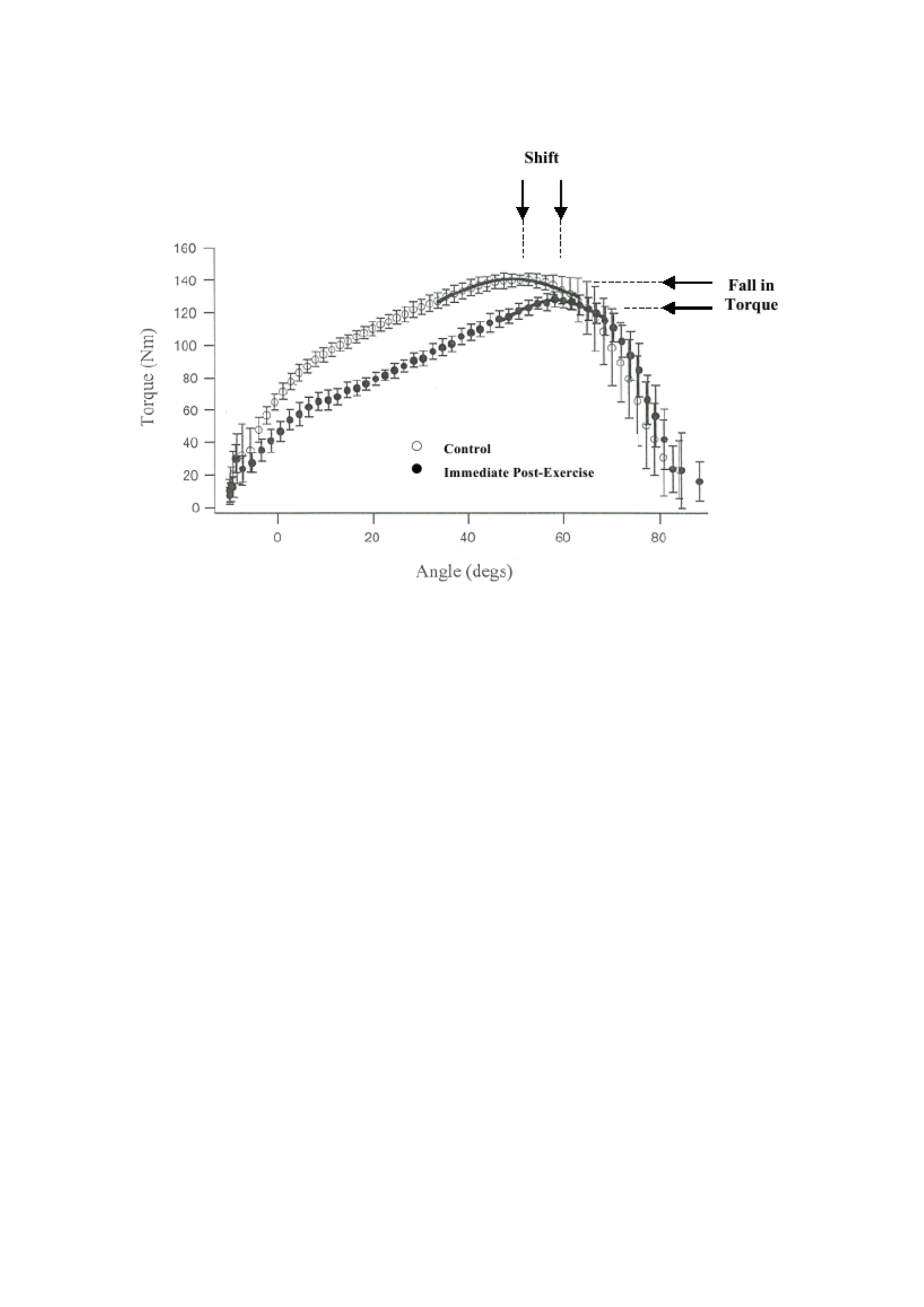
The shift in the length-tension relationship for a particular force to a longer muscle length
after eccentric exercise was first observed by Katz (1939), who proposed that excessive
muscle stretching rapidly beyond its optimal length tends to damage contractile elements.
Katz's (1939) original findings based on frog and tortoise muscles have been confirmed by
more recent investigations using human models (Jones et al., 1997; Whitehead et al., 1998;
Brockett et al., 2001; Philippou et al., 2003) (Figure 2.2).
These findings provide evidence to support the thesis that longer muscle lengths are
required to achieve the same myofilar overlap after eccentric exercise due to increased
series adherence resulting from excessive sarcomer stretching (Morgan & Allen, 1999;
Proske & Morgan, 2001). In further support of this theory, greater strength loss is reported
in the short term compared to longer or optimal muscle lengths after eccentric exercises
that show a shift towards longer muscle lengths to produce maximum strength (Saxton &
Donnelly, 1996; Anak et al., 1998; Byrne et al., 2001; Sayers & Clarkson, 2001; Byrne &
Eston, 2002b).
38
Figure 2.2 Angle curve-torque of the hamstrings before eccentric exercise (Control) (O)
and immediately after exercise (●). The Gaussian curve has been fixed to the top 10% of
each curve. Adapted from Brockett et al., (2001). Notice the right shift directly from the
angular-torque relationship after eccentric exercise.
2.4.2 Low-frequency fatigue
An alternative hypothesis to explain the loss of force-generating capacity and the shift in
the long-voltage relationship proposes that changes in calcium homeostasis may be

39
responsible. Decreased release of calcium ions (Ca2+) from the sarcoplasmic reticulum
(Warren et al., 1993; Westerblad et al., 1993) and an increase in intracellular Ca2+ (Chin
& Allen, 1996) have been shown to cause failure of the excitation-contraction (EC)
coupling process in rat muscle preparation. In human biopsy samples, Hill et al. (2001)
have reported that the release and absorption of the sacchaplasmic reticulum Ca2+ is
significantly depressed after exercise that triggers a decrease in the MVC of the knee
extensor. This and

40
Other authors have also observed a significant correlation between decreased torque
production at low electrical stimulation frequencies and Ca2+ release after exercise (Hill
et al., 2001, Neilsen et al., 2005).
The disproportionate loss of force at low frequencies (20Hz) compared to high frequencies
(100Hz) of electrical stimulation is known as low-frequency fatigue (LFF) and is likely
caused by disruption of the excitation-contraction coupling process (Edwards et al, 1977).
This is known to be caused by strenuous training (Edwards et al., 1977; Newham et al.,
1983b; Jones et al., 1989), with the most profound effects triggered by eccentric exercises
(Newham et al., 1983b; Jones et al., 1989). Thus there is evidence from animal and human
studies that suggests that reduced release of the sarcoplasmic reticulum Ca2+ is the main
cause of LFF. However, there is evidence that, in addition to reduced Ca2+ release, LFF
following eccentric training may be caused by changes in muscle morphology and
subsequent remodeling (Jones et al., 1996; Westerblad et al., 2000). The length
redistribution of sarcomer after eccentric exercise may explain the length-dependent
effects of strength loss associated with EIMD, especially given the fact that, like the
decrease in MVC, LFF is more pronounced at short muscle length than length. (Jones et
al., 1989; Byrne et al., 2001)
2.4.3 Changes in nerve control
Muscle strength generation is not only a product of the contractile function of muscles and

41
EC coupling but also of nerve impulses. Therefore, the inability to generate maximum
strength in muscles damaged by eccentric exercise could theoretically be the result of
changes in nerve impulses, the estimates of which can be obtained using electromyography
(EMG).

42
Deschenes et al. (2000) have provided evidence to show that neuromuscular efficiency,
the ratio of the resulting torque to integrated EMG activity (iEMG), decreases after EIMD.
These authors reported an increase in iEMG activity and a decrease in the torsion:iEMG
ratio during maximum isometric contractions lasting for 10 days. Other symptoms of
EIMD include plasma CK activity, perceived pain and peak torque all recovered within 7
days (Deschenes et al., 2000). Subsequent investigations have reported similar
disturbances in the torque:iEMG ratio with the greatest effect seen at low forces; arguably
where the precision of stylistic production is most functionally relevant (Weerakkody et
al., 2003a; Lavender & Nosaka, 2006; Semmler et al., 2007). An increase in isometric
force fluctuations has also been observed after muscle-damaging eccentric exercises but
does not appear to be an artifact of muscle damage as the action returns to baseline levels
within 24 hours (Lavender & Nosaka, 2006; Semmler et al., 2007).
Changes have also been reported in the perception of force production and joint position
after eccentric exercises. Saxton et al. (1995) showed that after forearm flexor eccentric
exercises, participants consistently overestimated the amount of strength they could
generate. Instead of matching the target strength of 35% MVC generated in the arm
(control) that was not damaged, participants weakened their target strength during the 5
days of the study after the initial insult (Saxton et al., 1995). However, when the force
applied by the damaged arm and control is expressed as the MVC proportion for that arm
at the time, the error in the estimation is reduced or absent (Saxton et al., 1995). In addition
to disturbances in the matching of the sense of strength, disturbances in the reproduction
of limb or joint positions have been reported after eccentric exercise (Saxton et al., 1995;

43
Brockett et al., 1997; Walsh et al., 2004). However, conflicting results were noted.
Brockett et

44
al. (1997) reported that participants produced a larger joint angle after eccentric exercises
whereas Saxton et al. (1995) and Walsh et al. (2004) reported that participants produced
smaller joint angles after eccentric exercises. It has been proposed that changes in the
perception of strength and position of the limbs after eccentric exercise may be caused by
damaged sensory receptors within the muscles (Saxton et al., 1995; Brockett et al., 1997;
Carson et al., 2002; Proske et al., 2003). It is believed that the sense of force originates
from peripheral receptors in the muscles and tendon organs of the Golgi and that the
muscular spindle provides signals for the sense of position (McCloskey, 1978; Gandevia,
1996). Twist et al. (2008) reported impaired unilateral balance performance 24 hours after
plyometry exercise, damaging the muscles that the authors associate with changes in
proprioceptive control. However recent work by Gregory and his colleagues using an
anesthetized cat model has shown that the responsiveness of tendon organs and muscle
spindles is not altered by eccentric exercise (Gregory et al., 2002, 2004). Thus the effects
of eccentric and muscle-damaging exercises on proprioceptive function have not been
described.
It has been proposed that an increase in iEMG associated with changes in proprioception
after EIMD may be an indication of changes in motor unit activation (Proske et al., 2004;
Weerokkody et al., 2003b; Prasartwuth et al., 2005; Semmler et al., 2007). Proske et al.
(2003) have suggested that DOMS may be involved in neural control changes after
eccentric exercise. They propose that pain from DOMS leads to reduced motor cortical
stimulation, which may serve to protect the muscle during further damage during the repair
process (Proske et al., 2003). However, the mechanism or mechanisms by which EIMD

45
affects changes in motor unit recruitment remains to be determined.

46
2.5 Repeated Combat Effects
Protective adaptations to a single eccentric exercise attack have been called the 'repetitive
fight effect' (Nosaka & Clarkson, 1995). Whenever an unusual eccentric exercise is
repeated, over a period of time, the magnitude of the characteristic symptoms of EIMD
decreases. Changes in muscle morphology, muscle protein efflux, inflammation, loss of
strength and other symptoms are weakened by the repetition of the same exercise but the
damaging effects are not prevented (Nosaka & Clarkson, 1995; McHugh et al., 1999;
McHugh, 2003). The effect of repeated fights can be given as early as 2 days after the
initial practice fight (Paddon-Jones et al., 2000; Nosaka & Newton, 2002) and for most
EIMD symptoms, it lasts at least 6 months but disappears between 9 and 12 months
(Nosaka et al., 2001a). Very little prior practice is required to take effect. At least 2
maximal eccentric contractions have been shown to provide protection against EIMD
symptoms when the same elbow flexor muscle performs 24 maximal contractions 2 weeks
later (Nosaka et al., 2001b). Similarly, Lavender and Nosaka (2008) showed that light
eccentric exercise, 30 (6 x 5) contractions of only 10% MVC were effective in weakening
muscle damage against subsequent eccentric exercise attacks of 40% MVC performed 48
hours later.
The mechanisms underlying the effects of repeated fights are not fully understood although
some potential mechanisms have been proposed. It has been suggested that protection is
provided as a result of neurological, cellular and mechanical adaptations that can work
independently of each other or simultaneously (McHugh et al., 1999; McHugh 2003).

47
Neural adaptation theory proposes that during subsequent eccentric exercises, the
recruitment of motor units is altered to redistribute the workload and thus increase the

48
motor unit efficiency (Nosaka & Clarkson, 1995). Warren et al. (2000) observed a 30%
decrease in the average frequency of EMG in the anterior tibial muscle in the second of
two attacks
The 50 eccentric MVCs are separated by one week. These authors concluded that the data
showed that an increase in slow motor unit activation and a concomitant decrease in fast
unit activation occurred in repeated combat (Warren et al., 2000). The findings of Chen
(2003) provide further support for the thesis of reducing the activation of rapid twitch
motor units during the second eccentric fight. However, McHugh et al. (2001) were unable
to find evidence of neural adaptation after performing repetitive relatively low-intensity
eccentric exercises.
The theory of cellular adaptation involves the potential adaptation of contractile
machinery. Proske and Morgan (2001) have suggested that after eccentric exercise the
optimal muscle length increases due to the addition of sarcomer in series. The shift of the
optimal angle towards longer muscle length after EIMD has been confirmed in several
studies (see section 2.4.3). However, in a recent study, Chen and colleagues (2007a)
revealed that while the shift to the right in the optimal angle showed a relationship with
the level of muscle damage associated with the initial fight, it did not appear to be directly
related to the mechanism responsible for the effects of repeated fights (Chen et al., 2007a).
Several investigations have used blood markers to show that there is a reduction in the
inflammatory response associated with EIMD after repeated attacks of eccentric exercise
(Pizza et al., 1996, 2001; Hirose et al., 2004; Smith et al., 2007). However, a more recent
study using muscle biopsies, showed that some inflammatory genes were transcriptionally

49
regulated (not attenuated) after repeated attacks of eccentric foot exercises (Hubal et al.,
2008).

50
Mechanical adaptation to eccentric exercises may involve renovating the intermediate
filament system to provide mechanical reinforcement against subsequent attacks. Yu and
Thornell (2002) showed that a single attack on the lower floor that runs increases actin and
desmin staining, suggesting that this reflects an increase in protein synthesis as part of the
adaptation process. Recently Lehti and colleagues (2007) have used mouse biopsy data to
show that previous eccentric exercises produce adaptive responses that protect
sarcolemma, intermediate filaments, and sarcomer proteins against subsequent
disturbances. While there may be some mechanisms underlying the effects of repetitive
fights that may work separately or to complement each other, the unified theory for
explaining the mechanisms of protective adaptation remains elusive.
The presence of cross-over or contralateral adaptations that have recently been revealed to
eccentric exercises has shown that the effects of repeated fights likely involve complex
interactions of all three proposed mechanisms (Howatson & van Someren, 2007). After
the second fight of 45 eccentric MVCs of elbow flexors, the symptoms of EIMD were
reduced when the second fight was performed with the opposite or contralateral limb
although the magnitude of the change was not as deep as when the same arm was trained
twice. The authors have suggested that contralateral adaptation is most likely mediated by
neural mechanisms because there is no direct stimulus for cellular mechanical changes in
the contralateral arm (Howatson & van Someren, 2007). Thus deeper adaptations in the
same lateral arm are likely to result from neurological, cellular and mechanical
mechanisms working together.

51
2.6 Dynamic muscle function
Of all the symptoms of EIMD, immediate and prolonged impaired muscle function has the
potential to be the most debilitating when considering the human response to dynamic
exercise after eccentric exercise. However, studies of dynamic muscle function during
athletic performance have received only limited attention. The first study that directly
assessed the effect of muscle-damaging eccentric exercises on dynamic muscle function
was conducted by Sargeant and Dolan (1987). These authors used a thorough downhill
walk intervention (-25%) to induce damage followed by an assessment of the maximum
short-term power output using an isokinetic cycle ergometer. Cycling at 110 rev.min-1 for
20 seconds, peak power was reduced by 23% 24 hours after the eccentric training protocol
and was still 8% lower than baseline at 96 hours (Sargeant & Dolan, 1987). The decline in
MVC has a magnitude greater than peak power, with a loss of 45% of the pre-eccentric
training value at 24 hours, recovering to a loss of 30% at 72 hours (Sargeant & Dolan,
1987).
2.6.1 Wingate 30-second cycle test
These findings have been supported by Byrne and Eston (2002b) using the 30-second
Wingate test to investigate changes in power generation capabilities after EIMD. These
authors reported a decrease in peak power output and isometric MVC following the
performance of a 100 bar-bell squat with a load corresponding to 80% concentric one
maximum repetition. There is a striking difference in the magnitude of strength and loss

52
of power and the recovery pattern. The isometric MVC drops by 35% immediately after
the squatting protocol and then follows a linear recovery; 26% at 24 hours and 19% at 48
hours. In comparison, Wingate's peak power was reduced by 13% immediately after the
eccentric intervention but

53
fell further, by 18% at 24 hours and 16% at 48 before recovery (Byrne & Eston, 2002b).
In a more recent study, Nottle and Nosaka (2007) used a 40-minute downhill run damage
protocol (-7%) to investigate changes in Wingate's peak power. In accordance with
Sargeant and Dolan (1987) and Byrne and Eston, 2002b), a greater decrease in strength
than peak power was observed immediately after the downhill run (17% and 5%,
respectively, peak strength and power), but neither strength nor power loss persisted
beyond these initial post-eccentric exercise measurements. In contrast to the previous two
studies (Sargeant & Dolan, 1987; Byrne & Eston, 2002b), rapid recovery and peak power
were unexpectedly 5% higher than baseline at 120 hours (Nottle & Nosaka, 2007). A
previous investigation, Malm et al. (1999) reported no change in Wingate's peak strength
after a stepping protocol designed to induce muscle damage. However, the modest increase
in pain reported by participants and unchanged CK responses suggest that the stepping
protocol used may not be intense enough to alter muscle function.
2.6.2 Performa sprint
Malm and colleagues (1999) also reported changes in the performance of intermittent cycle
sprint tests (10 x 10 seconds of all-out cycling interspersed with 50-second rest periods)
which unexpectedly increased by 8% at 48 hours. No change in the performance of the 30
m sprint was observed 48 hours after completing 70 (7 x 10) jump falls designed to induce
muscle damage (Semark et al., 1999). However, participants in the study were trained
rugby union and field hockey players who, although they reported moderate pain, showed
no increase in plasma CK levels and may have been protected from changes in muscle

54
function from jumping falls through the effects of repeated fights (see section 2.5).
Effective

55
Muscle-damaging protocols such as the 100 plyometric jump used by Highton et al. (2009)
have been shown to result in a decrease in isokinetic peak torque and an increase in 5 and
10 m sprint running times at 24 hours and 48 hours. Twist and Eston (2005) investigated
the effect of EIMD on cycle and run sprint performance using a 100 (10 x 10) reverse
motion jump to induce damage. Peak power output during intermittent cycle sprints (10 x
6 seconds with 24 seconds recovery) is reduced immediately and up to 72 hours after
eccentric training. Fatigue levels in the cycle test were also reduced with the largest
decrease observed at 48 hours. Similarly, intermittent sprint running times (10 x 10 m with
12 seconds of active recovery) increased immediately and up to 48 hours after eccentric
training (Twist & Eston, 2005). In a more recent investigation, the same authors compared
the performance of a 10-second cycle sprint and a 50 cm drop jump at 24, 48 and 72 hours
after completing 100 (10 x 10) jumps of opposite motion (Twist & Eston, 2007). While
cycle sprint and fall jump performance is reduced as a result of eccentric exercise, temporal
recovery patterns are different. The peak power output and the time to reach the most
severe peak power were reduced at 48 hours, while the largest decrease in fall jump height
was observed at 24 hours (Twist & Eston, 2007). This observation led the authors to
propose that differences in responses to cycling performance and drop jumps suggest that
the recovery time course of EIMD depends on dynamic training modes (Twist & Eston,
2007).
2.6.3 Vertical jump test
Other studies have compared performance degradation in various vertical jumps to

56
investigate the muscle-damaging effects of exercise on dynamic muscle function. Byrne
and Eston (2002a) used 100 (10 x 10) barbell squats with a load of 70% body mass to
identify

57
Differences in the performance of squat jumps, opposite jump movements and fall jumps
after eccentric exercises that damage the muscles. Squat jumps are performed from a
squatting position before jumping vertically for maximum height whereas in counter
movements and fall jumps, muscles use a stretch-shortening cycle (SSC). Human muscular
function SSC is a natural mode of drive used in running, walking or jumping that involves
cyclic performance from a sequence of pre-activation, active braking (eccentric) followed
by concentric action (Komi, 1984). The decrease in vertical jump performance occurs
immediately and lasts up to 72 hours, although the performance of the squat jump is
affected more than the performance of the reverse movement or fall jump. This observation
prompted the authors to propose that impaired muscle function in vertical jumps is
weakened when SSC is used (Byrne & Eston 2002a). Similar findings have been reported
by Harrison and Gaffney (2004) who used 70 (7 x 10) maximal eccentric contractions of
the knee extensor to induce muscle damage. In contrast, when muscle damage was induced
through the SSC protocol (through specially designed sled equipment) rather than the
eccentric dominated protocol, drop jump performance was more affected than squat jump
performance (Avela et al., 1999; Horita et al., 1999, 2003). These authors and others (e.g.
Horita et al., 1996, Nicol et al., 2006) have also reported a 'bi-modal' recovery of the
complete SSC. The bi-modal response involves an immediate post-workout reduction in
dynamic muscle function believed to be primarily due to metabolic fatigue. This is
followed by partial recovery one to two hours after exercise and a further reduction in
dynamic muscle function that has been linked to an inflammatory response to EIMD (Nicol
et al., 2006). However, the phenomenon of bimodal or biphasic responses may not be
exclusive to damage induced through SSC. MacIntyre and colleagues (1996) were the first
to report biphasic

58
recovery of dynamic human muscle function after 300 repeated eccentric eccentric
contractions of the knee extender. In addition, some studies reported muscle function
measures between 0 and 24 hours post-workout and those who reported static force more
often than dynamic torque (Clarkson & Hubal, 2002).
2.6.4 Endurance training
The immediate and prolonged loss of power-generating ability resulting from muscle-
damaging exercises also hampers performance during resistance training. Rats have been
used to investigate the time to fatigue and the more ecologically valid time of voluntary
wheel running, after 150 minutes of downhill running (Carmichael et al., 2005, 2006;
Davis et al., 2007). This group of authors has reported a decrease in treadmill running time
to fatigue at 24, 48 and 72 hours. When compared to rats running uphill, one group ran
73% less at 24 hours and 69% less at 48 hours (Carmichael et al., 2005). Voluntary wheel
running activity was monitored during an active dark cycle 12 hours after downhill running
in all three studies. The rats spent a shorter period of time on the treadmill and covered
shorter distances in the first and second 12-hour active cycles, gradually returning to
baseline activity between the 3rd day (Carmichael et al., 2006; Davis et al., 2007) and day
5 (Carmichael et al., 2005).
To date, only two ecologically valid studies have investigated the performance of human
endurance after EIMD. Marcora and Bosio (2007) reported significant differences in the

59
performance of self-paced 30-minute time trial runs before and 48 hours after the
completion of 100 (10 x 10) jump falls. Participants ran shorter distances at a slower pace
after EIMD. Analysis of speed, heart rate, and perceived exertion variables recorded during

60
Running shows that the pacing strategy has not changed. In light of these findings, the
authors propose that the negative effects of EIMD on time trial performance are mediated
by the individual's perception of exertion (see section 2.8.2) (Marcora & Bosio, 2007).
Twist and Eston (2009) reported a similar decrease in the performance of a 5-minute cycle
time experiment after 100 (10 x 10) jumps of reverse motion. There has been a significant
decline in peak power output (-14%), average power output (-11%), average revolutions
per minute (RPM) (-4%), and distance traveled (-4%) 48 hours after EIMD but
performance measures have returned to baseline at 168 hours (Twist & Eston, 2009).
2.7 Human response to dynamic exercise
Investigations reporting changes in physiological parameters during exercise after EIMD
have yielded vague findings. The first study to document physiological changes during
exercise with EIMD was conducted by Hamill and colleagues (1991). Steady-state oxygen
uptake (V
O2) and heart rate (HR) data were recorded for 15 minutes
run at 80% maximum V O2
( V O 2 max) before and after 30 minutes of downhill running (at
-15% gradient). The responses of V O2 and HR from ten recreational runners were
unchanged at 48 and 120 hours after the downhill run. A small, albeit significant increase
in CK activity and the development of only moderate pain led the authors to suggest that
downhill running may exert insufficient stress to give rise to metabolic modification or HR

61
(Hamill et al., 1991). These authors also reported a small but significant decrease in hip
and knee flexion during running after EIMD but overall stride performance, including
stride length, stride duration, mechanical work, and mechanical force were not affected
(Hamill et al., 1991).

62
2.7.1 Oxygen absorption
The size of V O2 during running at a fixed sub-maximum speed, as obtained by
Hamill and his colleagues (1991), provide information related to cost or driving
economics, which is more commonly called 'running economy'. In contrast to the findings
of Hamill et al. (1991), Braun and Dutto (2003) report that the economy is running
significantly
interrupted (an average of 3.2% increase in V O2) during a 5-minute fight run at 65, 75 and
85% V O2 peak, 48 hours after a 30-minute downhill run (-10%) with an intensity equivalent to 70%
V O2 peak. In addition, this author reported a complete decrease in average stride length
three exercise intensities were negatively correlated with changes in the average energy
cost of running at all three exercise intensities (Braun & Dutto, 2003). Comparison of the
training status of the two groups of participants (Hamill et al., 1991; Braun & Dutto, 2003)
is suggested as an important factor in explaining the different findings (Braun & Dutto,
2003). Well-trained runners in the Braun and Dutto study (2003) may have smoother gait
patterns than recreational runners in the Hamill et al. (1991) study. Thus trained runners
may be more sensitive to changes in gait due to muscle damage. To support this
suggestion, Paschalis et al. (2005) report no change in
economic data of untrained athletes during running at around 55 and 75% V O2 max

63
for 3 days after completion of 120 (12 x 10) eccentric MVCs. Similarly,
Marcora and Bosio (2007) reported no change in the active economy running, but not
Highly trained long-distance runners, exercising at 70% V O2 max for 10 minutes 48 hours
after
100 (10 x10) drop jump completion. Another study using active but untrained participants
also reported running economics that did not change after a series of sub-maximal
resistance exercises including barbell squats, weighted lunges and weighted step-ups
(Scott

64
et al., 2003). It is suggested by these authors that the level of induced muscle damage is
not sufficient to produce mechanical or physiological changes that would affect V O2
(Scott et al., 2003). Chen and his colleagues (2007b) have provided the only evidence of
the time course of change in running the economy after EIMD. Economic data on running
were collected for five consecutive days after the completion of a 30-minute downhill run
(-
15%) at an intensity equivalent to 70% V O2 peak (Chen et al., 2007b). Using the same
submaximal exercise intensity as Braun and Dutto (2003) (peaks of 65, 75 and 85% V O2 )
These authors show that the running economy was disrupted by 4-7% over the three days
after the downhill run, recovering to a level close to the baseline on the fourth day (Chen
et al., 2007b). In addition, a reduction in stride length (3-6%), range of motion (ROM) of
the ankle and knee joints (1-7%) and an increase in stride frequency (3-7%) were observed
for two to three days after the downhill run. These authors concluded that the passage of
time and the magnitude of economic changes were more closely related to changes in
kinematic parameters than changes in muscle function as indicated by direct and prolonged
changes in MVC (7-21% reduction over 4 days after downhill run) (Chen et al., 2007b).
Economic measures have also been assessed during cycling at a fixed sub-maximum load
and are not susceptible to the effects of lower limb kinematic changes as suggested that
the economy runs. In fact, there is consistent evidence that EIMD does not change the

65
cycling economy regardless of the mode of crash used or the training status of participants
(Gleeson et al., 1995; Walsh et al., 2001; Moysi et al., 2005; Schneider et al., 2007; Twist
& Eston, 2009). Gleeson et al. (1995) and Schneider et
al. (2007) employed untrained participants who were required to bench-step

66
protocol to induce muscle damage. Participants in the other three studies were all
physically active but untrained and were required to perform eccentric cycling (Walsh et
al., 2001), barbell squats (Moysi et al., 2005) and reverse motion jumps
(Twist & Eston, 2009) to induce muscle damage. No change in the size of V O2
reported in any of the studies mentioned above during sub-maximum constant load
bicycle. Furthermore, it has been reported that the peak of V O2 is measured during incremental
cycling to exhaustion was not affected by previous attacks of eccentric bench stepping
exercises (Gleeson et al., 1998). The observation that cycling is not susceptible to the
potentially confusing effects of altered kinematics makes it a very interesting model to
examine the effects of EIMD on human responses to dynamic exercise. The fixed geometry
of the cycle ergometer ensures that hip and knee angles remain relatively constant during
grueling cycling workouts despite the development of significant local muscle fatigue
(Dingwell et al., 2008). In contrast, a reduction of up to 7% in the range of motion of the
knee joint has been reported during running after a downhill run. (Chen et al., 2007b).
2.7.2 Kinetics of oxygen absorption and muscle oxygenation
While the measurement of V O2 at constant sub-maximum speed or load provides information
related to the cost of driving energy, the rate at which the absorption of O2 increases during
exercise

67
onset (Kinetics V2) can provide information pertaining to the balance of O2 delivery to
Utilization of O2 in active skeletal muscle. Schneider and his colleagues (2007) were the first to
investigated the effect of EIMD on the kinetics of O2 absorption. Nine untrained participants
each
making a square wave transition from weightless cycling to heavy intensity before and 48

68
and 72 hours after completing 30 minutes of bench-stepping exercises. Heavy intensity
training is defined as a workload equivalent to 40% of the difference (40%∆) between the
power output achieved at the gas exchange threshold (GET) and the achieved
at the peak of V O
2 . Kinetics phase II V O2 not altered by previous eccentric exercises
indicating that EIMD does not interfere with oxidative function or alter O2 matching
Shipment to
O2
utilization in active muscle tissue. In addition, it is slow to change
component shows that the O2 cycling charge at 40%∆ is not changed. These findings
led the authors to speculate that they did not induce severe enough muscle damage to
causing changes in V−O2 Kinetika. While muscle oxygenation is not measured in this case
investigational, near-infrared spectroscopy (NIRS) can facilitate the assessment of muscle
oxygenation and can therefore be used to determine dynamic balance in a non-invasive manner
between O2 delivery and O2 utilization.

69
Walsh et al. (2001) examined the kinetics of O2 utilization and reoxygenation during
ischemia and reperfusion at rest before and 2 days after 30 minutes of eccentric cycle. Not
changes were reported in O2 utilization or local O2 transport with the authors concluding
that oxidative function of resting muscles is not impaired as a result of EIMD (Walsh et al.,
2001). Recently Ahmadi et al. (2008) used NIRS to investigate the saturation of O2 and
Kinetics of desaturation at rest and during isometric contractions at 30, 50 and 80% MVC
before and after 40 minutes of downhill walking (-25%). In contrast to the findings of
Walsh et al. (2001), Ahmadi et al. (2008) report directly and prolongedly
accelerate NIRS-derived O2 kinetics. The authors propose that it is possible that
mechanisms of increasing saturation and desaturation of O2 may have increased O2

70
utilization due to energy needs that demand an improvement process (Ahmadi et al., 2008).
However, the authors acknowledge that increased muscle blood flow after EIMD, as
reported by Laaksonen et al. (2006), may be responsible for
acceleration of O2 saturation and desaturation kinetics (Ahmadi et al., 2008). While
Muscle oxygenation kinetics have been investigated at rest and during isometric
contractions, to date, no studies have attempted to investigate the effect of EIMD on
muscle oxygenation kinetics during dynamic exercise performance.
2.7.3 Metabolic response to dynamic exercise
As detailed in section 2.6.4, the reduction in resting muscle glycogen uptake after EIMD
has led to speculation that increased glycogenalysis may be a consequence of EIMD and
thus may be responsible for the increased concentration of blood lactate ([La]) observed at
rest (Asp et al., 1996, Asp et al., 1998) and during dynamic exercise (Braun & Dutto, 2003;
Chen di al., 2007b, 2008; Gleeson et al., 1995, 1998).
Gleeson et al. (1995) were the first to report an improvement in post-workout immediate
exercise [La] 48 hours after a 30-minute bench-stepping fight designed to induce muscle
damage. Participants
cycling for 15 minutes at an intensity equivalent to 80% V O2 max 48 hours after eccentricism

71
Bench-stepping exercises or walking uphill concentric. Pre-exercise [La] did not differ
between the two groups but was higher after eccentric exercise (7.5 mmol.l-1) than after
concentric exercise (6.0 mmol.l-1). In a follow-up study, blood samples were taken before
and at 2-minute intervals during the incremental to fatigue cycle test and showed that after
eccentric exercise [La] increased (Gleeson et al., 1998). In this investigation, the control
conditions were not implemented in the period prior to the completion of the incremental

72
exercise test and show a lower [La] during and 2 minutes after the completion of the test
(Gleeson et al., 1998). These authors propose that higher blood [La] in post-eccentric
conditions reflects higher intramuscular [La] and an increased relative contribution of
anaerobic metabolism to energy production that may arise from the additional recruitment
of type II muscle fibers (Gleeson et al., 1998). To support this suggestion, Braun and Dutto
(2003) have reported an increase in blood [La] during running at 65, 75 and 85
% of peak V
O2. Blood samples are taken during a 5-minute rest period after every 5 minutes
fight and average 0.61 mmol.l-1 higher 48 hours after the downhill run protocol. The
increase [La], which is associated with a greater dependence on glycolytic energy
production, along with an altered mechanism of measures is suggested as a contributing
factor to the reduction of the current economy (Braun & Dutto, 2003) (previously
discussed in section 2.7.3). Similarly, Chen et al. (2007b) reported a significant increase
in [La] in blood samples taken 3 minutes after the completion of each 5-minute run fight
at a peak of 65, 75 and 85% V O2 . The authors suggest that the increase [La] lasted for three
days
after a 30-minute downhill run may have reflected an increase in motor unit activation
(Chen et al., 2007b).
However, an increase in [La] may not reflect the following changes in metabolic function:
EIMD. Schneider et al. (2007) reported an unchanged phase II V O2 kinetics that showed that

73
EIMD does not interfere with oxidative function (see section 2.7.3) along with elevation
in [La]. The difference between resting and end-of-workout blood [La] was higher after
EIMD and was associated with an increase in lactate efflux from active muscle due to
increased membrane permeability rather than an increase in anaerobic glycolysis rate
(Schneider et al., 2007). Increased lactate efflux can be overcome by increased muscle

74
blood flow after EIMD (Laaksonen et al., 2006) which can facilitate greater clearance of
[La] by increasing transport to lactate metabolism muscles and other tissues. Increased
muscle blood flow may well explain the unchanged blood [La] reported in other studies
(Hamill et al., 1991; Scott et al., 2003; Marcora & Bosio, 2007) and the reduction of blood
[La] reported by Moysi et al. (2005) after EIMD.
Magnetic resonance spectroscopy (MRS) can be used to measure the parameters of muscle
metabolic function including dynamic changes in the ratio of inorganic phosphate to
phosphocreatine (Pi/PCr) and intracellular pH. Some studies have shown a significant
decrease in the resting PCr/Pi ratio after EIMD, which can be interpreted as an increase in
metabolism after muscle injury (McCully et al., 1992; Lund et al., 1998a, 1998b).
However, due to the fact that no commercially available ergometers are capable of
functioning in MRS, only one study has investigated the possible changes in muscle
metabolism after EIMD during dynamic leg exercises within the core MRS of the whole
body. Using a hydraulic ergometer specifically designed for quadriceps exercises in full-
body MRS (Rodenburg et al., 1994), participants completed two stratified concentric
exercise tests before and 24 hours after performing stepping exercises designed to induce
EIMD (Rodenburg et al., 1995). An expected decrease in resting PCr/Pi ratio was observed
but no difference was reported in the PCr/Pi ratio during the exercise test. While these
findings suggest that exercise metabolism is not altered by stepping exercises, the lack of
changes in some markers of muscle damage including plasma CK activity, led the authors
to conclude that muscle metabolism can be altered by more severe EIMD (Rodenburg et
al., 1995). The inconclusive nature of this series of findings suggests that further research

75
using MRS should be done in sequence

76
to more clearly illustrate the effects of EIMD on muscle metabolism during dynamic
exercise.
2.7.4 Ventilation response to dynamic training
The mechanisms that control ventilation during dynamic exercise are controversial but are
believed to involve proportional feedback elements (central and carotid chemosensory)
and feed-forward (central commands and muscle reflexes) in various proportions (Ward,
2007). Below the lactate threshold, ventilation is involved in regulating the partial pressure
of the CO2 arteries near the initial level. Above the lactate threshold, the lactic acidosis
bicarbonate buffer provides an important stimulus for ventilation.
Several investigations have reported no ventilation changes after the EIMD. Paschalis et
al. (2005) used the lowest intensity exercise, with participants running at a "randomly
selected pace" of 133 and 200 m.min-1 which is equivalent to ~55 and ~75%
V O 2 max respectively (Paschalis et al., 2005). When compared to the base size, it is not
ventilation changes were reported at both speeds over 4 days after the completion of the
120 (12 x 10) eccentric MVC. Similarly, Scott et al. (2003) and Marcora and Bosio (2007)
have reported an unchanged ventilation response to running at an intensity equivalent to
blood
[La] from 2.5 mmol.l-1 (as if below the lactate threshold) and 70% V O ,

77
2 max
each. In addition, this author observed no effect of EIMD on blood [La] (Scott et al.,
2003; Marcora & Bosio, 2007). Participants in the study conducted by Moysi
et al. (2005) cycled at 62% V O2 max for 6 minutes before and 48 hours after completing a
series of
squats that damage muscles. Although no overall change in minute ventilation was reported,

78
VO
The authors did observe an increase in respiratory frequency counteracted by a decrease
in tidal volume. An explanation for this observation and for the observation that blood [La]
decreases by 12% by age 48 is not offered (Moysi et al., 2005).
Increased ventilation along with increased blood [La] has been reported
during running at 65, 75 and 85% V O2 max after EIMD (Braun & Dutto, 2003; Chen and
al. 2007b, 2008) and has been associated with running economic disorders associated
with changes in the kinematics of the lower limbs (see section 2.7.1). However, Gleeson
et al. (1995) observed
increased ventilation and health [La] during cycling at 80% ˙
2
m
ax
48 hours after
eccentric exercises. Minute ventilation is increased from 6-15 minutes from a 15-minute
fight and mainly due to the higher respiratory rate. Similarly, Schneider et al. (2007)
reported an increase in ventilation and blood [La] during cycling at 40%∆ (strenuous
intensity exercise). While the effect of increased lactic acidosis is considered the most
likely explanation, increased stimulation of muscle nociceptors due to increased muscle
pain and higher perception of reported effort is also proposed as a potential mechanism to

79
induce increased ventilation (Gleeson et al., 1995; Schneider et al., 2007).
Schneider et al. (2007) have provided the only example of an investigation using a specific
exercise domain to study the effect of EIMD on physiological responses to dynamic
exercise. The exercise domains used during the investigation are important in determining
an individual's physiological response to dynamic exercise. Very different metabolic,
cardiorespiratory and exertion responses were emanating from the three domains of sub-
maximal exercise intensity (moderate, heavy and strenuous). Thus, it is

80
it is important that the intensity of the exercise is accurately defined by depicting the
boundaries between these domains when investigating these responses (Jones et al., 2009).
Most studies investigating the effect of EIMD on dynamic exercise performance used
exercise intensity determined by the percentage of maximum working capacity. This can
be somewhat misleading due to differences between individuals. For example, exercising
in
80% V O 2
max
(e.g. Gleeson et al., 1995), can result in one individual exercising inside
heavy domains while others train in heavy domains. Future research involving the effect
of EIMD on ventilation and other human responses to dynamic exercise should seek to use
specific exercise domains so that the interpretation of the findings is not confounded by
individuals exercising at different relative intensities.
Increased ventilation after EIMD may be associated with increased lactic acidosis,
however other potential mechanisms have been investigated. To eliminate the influence of
metabolic control factors such as [La], Hotta and colleagues (2006) investigated the
ventilation response to the first 20 seconds of dynamic knee extension exercise after
EIMD. Knee extensions with an ankle weight of about 2.5% body mass are repeated five
to seven times. Breath-by-breath data is aligned with the start time of the exercise, the
ensemble is averaged and then interpolated linearly to produce a 1-second data point.
Ventilation at the beginning of exercise increases significantly 2 and 7 days after eccentric

81
exercise which leads the authors to suggest that changes in peripheral nerve reflexes
contribute to improved ventilation response (Hotta et al., 2006).

82
2.7.5 Perception of effort during dynamic training
Perception effort involves assimilating a number of afferent signals from a variety of
perceptual cues originating from different body systems including the cardiorespiratory
and neuromuscular systems that can be interpreted both in feedback and feed-forward
(Hampson et al., 2001). Signals may be peripheral in nature, produced for example by
painful muscle afferent stimulation after EIMD; or centrally derived from
cardiorespiratory responses to exercise. While damage protocols, training modes, and
exercise intensity may have a significant influence on many other physiological responses,
the weight of the evidence suggests that EIMD results in an increased sense of effort during
dynamic exercise (Gleeson et al., 1995; Scott et al., 2003; Marcora & Bosio, 2007; Chen
et al., 2007b, 2008; Twist & Eston, 2009).
Gleeson et al. (1995) reported an increase in effort during cycling at 80% V O2 max 2
A few days after completing 30 minutes of bench-stepping. The reported higher perceived
exertion rating (RPE) is believed to reflect weakened muscle state, increased blood [La]
and muscle pain sensation (Gleeson et al., 1995). Increased RPE has also been reported
during running after eccentric training (Scott et al., 2003; Chen et al. 2007b, 2008). Scott
and colleagues (2003) suggest that perceived exertion is the best indicator of physical
stress because RPE is a configuration of responses resulting from the integration of signals,
perceptions, and experiences. In addition to the cues proposed by Gleeson et al. (1995),
altered neural control was suggested as a possible cue for the perception of higher exertion

83
reported after EIMD (Scott et al., 2003). Recent studies conducted by Marcora and Bosio
(2007) and Twist and Eston (2009) have reported RPE responses during fixed load
submaximal training and time trials

84
performance. During a 10-minute run at 70% V O 2
max
(Marcora & Bosio, 2007) and
cycling for 5 minutes at 60 and then 80% of the maximum power output (Twist & Eston,
2009) the RPE is higher after eccentric exercise. However, during the run and cycling time
trials, the RPE did not change after the EIMD even though the time trial performance was
significantly reduced. Both pairs of researchers concluded that after EIMD, a changed
sense of effort mediated performance. Participants reported higher RPE when exercising
at the same intensity and produced less power (slower running or cycling speed) when
feeling the same effort (Marcora & Bosio, 2007; Twist and Eston, 2009).
Some studies have shown changes in the perception of force production after eccentric
exercises that last for several days (see section 2.4.3). More recent investigations have
corroborated these findings, proposing that participants used their perception of the effort
required to produce a particular torque rather than the level of torque itself (Carson et al.,
2002; Proske et al., 2003; Weerakkody et al., 2003a; Proske et al., 2004). These findings
suggest that EIMD interferes with the perception of effort or exertion rather than the ability
to match a particular force because participants with EIMD with EIMD produce reduced
force for a given perceived effort and perceive a higher effort when producing the same
force.

85
2.8 Conclusion
Direct histological analysis of muscle tissue and indirect measurements of the structural
and functional status of skeletal muscles after unfamiliar eccentric exercises are well
documented and have revealed substantial disorders. However, the effects of exercise-
induced muscle damage on human responses to dynamic exercise have been investigated

86
rare and has produced vague findings. Exercise modalities have been found to influence
human responses after EIMD with changes observed during running more closely related
to changes in kinematic parameters than changes in muscle function. Therefore, cycling,
which is not susceptible to potentially confusing kinematical influences, is the preferred
model to examine the effects of EIMD on human responses to dynamic exercise. The
purpose of the following study was to investigate the effects of EIMD on various human
responses during dynamic exercise. Specifically, the investigation will focus on the
ventilation response and perceived exertion, muscle metabolism, muscle oxygenation
kinetics and pulmonary oxygen uptake.

87
CHAPTER 3:
GENERAL
METHODS

88
3.1 Introduction
The experiment consisting of this thesis was carried out in the exercise physiology
laboratory of the School of Sport and Health Sciences at the University of Exeter
accredited by the British Association of Sport and Sport Sciences. In Study 3 (chapter 6)
magnetic resonance spectroscopy 31P was carried out at the Peninsula Magnetic Resonance Research Centre at the University of Exeter.
Prior to any data collection, all participants gave written consent to participate in the study,
which was approved by the School of Sports and Health Sciences Ethics Committee.
3.2 Identity
Most of the recruited participants were members of the School of Sport and Health
Sciences at the University of Exeter and were familiar with experimental testing protocols
and related procedures. However, because maximum and/or thorough effort is required on
most laboratory visits, proper practice is carried out. Participants in all four studies were
fully familiar with the production of maximum voluntary contractions using the Biodex B-
2000 isokinetic dynamometer (Biodex Corp, Shirley, NY). All participants in study 3
(chapter 6) were asked to complete an introduction session in a 'dummy MRI system'.
During this session, participants practiced single-legged knee extension exercises at a rate
of 40 repetitions/minute in time with visual cues projected onto the front wall of the
'dummy' scanning room. Strong encouragement is given to participants during all tests
maximum and/or thorough to ensure maximum effort is generated.
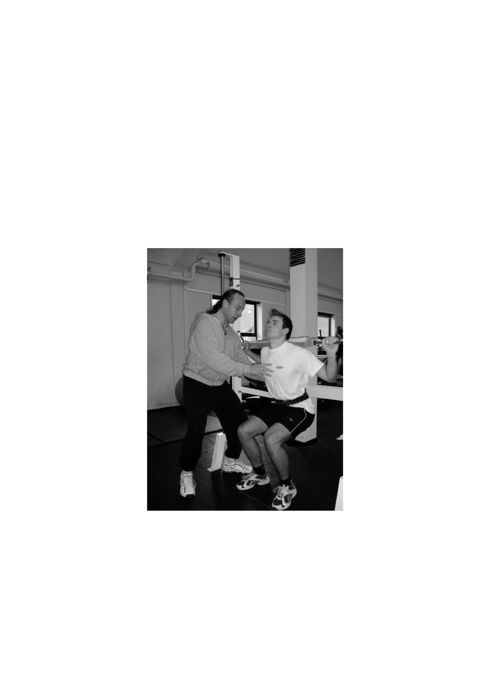
89
3.3 Exercise protocols that damage eccentric muscles
To trigger muscle damage, participants in each of the four studies were required to
perform 100 squats as 10 sets of 10 repetitions using the Smith machine (Figure 3.1).
Figure 3.1 A participant is guided in a correct and safe squatting technique beforehand

90
to complete 100 squats (Smith).
This procedure involves controlled isotonic resistance of the external load of the rod,
which was calculated according to about 70% of the body mass of each participant.
It is calculated in 5 kg of the predicted 70% body mass as the smallest available
It weighs 2.5 kg. Before commencing, participants are properly instructed and

91
safe lifting techniques (see appendix E for risk analysis). Before the bar is released, the bar
is positioned on the participant's shoulders and the legs are positioned under the bar.
During the movement, the participant's head remains advanced, the back is straight and the
legs are fully extended (knees = 180°). The foot remains flat on the floor, the toes pointing
forward, with the same weight distribution through the forefoot and heel. Before starting,
the corresponding leg position is marked on the floor with tape so that it can be reproduced
in each of ten sets of ten repetitions. The descent phase involves the eccentric action of the
knee extensor to lower the bar to the angle of the knee just past 90°. The lifting phase
involves concentric action to return the bar to its starting position. To facilitate consistent
90° squat production, plastic meter rules are affixed to the safety stop frame on the guide
bar using gaffer tape to provide visual and sound guidance for participants and
investigators. When participants reach the required angle, this can be seen in the mirror
facing and heard as the meter rules touch the base frame of the Smith machine.
During the pilot's work, the metronome is used in an attempt to control the speed of
contraction as it has been suggested that this can affect the magnitude of induced damage
(Chapman et al., 2006). However, participants struggled to maintain a set rhythm from
four to one upwards with a metronome set at 60 beats per minute without losing shape.
Therefore, this element of the protocol is abandoned and participants are encouraged to
perform a controlled and feasible phase and lift at their own pace while maintaining correct
and safe lifting techniques. After each set of ten repetitions, a minimum of 1 minute rest
time is allowed, with participants encouraged to take as much time as they need between
sets to ensure that correct and safe lifting techniques can be maintained.

92
3.4 Markers of muscle damage
In a review of measurement tools used to evaluate exercise-induced muscle damage,
Warren et al. (1999) proposed that maximum voluntary contraction torque (MVC)
provides the best measure of muscle damage due to eccentric contractions. These authors
underestimate the use of changes in perceived muscle pain or changes in blood levels of
myofibre proteins such as Creatine Kinase (CK) because they are reported to be poorly
correlated with the magnitude and time of changes in muscle function (Warren et al.,
1999). However, we chose to include measures of plasma CK pain and activity in addition
to MVC torsion, to provide a broader physiological perspective on induced damage. In
each study, these markers of muscle damage were assessed in the order listed below before
and after completing muscle-damaging eccentric exercises to measure the effectiveness of
the protocol.
3.4.1 Creatine Kinase Activity
Plasma CK activity was assessed from fingertip capillary samples. The samples were
centrifuged at 4000 RPM (2000 x g) for 5 min and two 20 μl plasma samples were then
added to 1 ml of the reagent composition supplied by Randox (CK-NAC 110, Randox
Laboratories Ltd., Crumlin, Co. Antrim, UK). After 1 min incubation at 37°C and during
follow-up incubation, absorbance at 340 nm was recorded by spectrophotometry (Jenway

93
6310 spectrophotometer, Jenway, Essex, UK) at 0, 1, 2 and 3 min. The CK value is
calculated using the formula CK(U/l) = 8095 x Δ absorbance 340 nm/min. The average
CK values of both samples were calculated and used for subsequent analysis. Normal
serum values of 24-195 U/l were reported for men using this method (Szasz et al., 1976).
Intra-

94
The test variation coefficient for duplicate samples using this procedure in our laboratory
is 6.6%.
3.4.2 Muscle pain felt
Participants assessed their knee extensor pain using an empty 0-10 visual analogue scale
(VAS) (Appendix G). The VAS consists of a 10 cm line labeled from left (no pain) to right
(worst pain ever). After squatting to approximately 90o knee flexion with hands on the
hips, participants were asked to mark the VAS to indicate their pain level. The pain felt
was then measured by measuring the distance to the mark on the line to the nearest 0.1 cm.
This procedure has been used in previous studies (Rowlands et al., 2001; Twist and Eston,
2005, 2007).
3.4.3 Isokinetic peak torque
After the habituation session, the peak isokinetic torque of the knee extensor was measured
using a Biodex B-2000 (Biodex Corp, Shirley, NY) isokinetic dynamometer, which was
calibrated prior to each data collection session in accordance with the manufacturer's
guidelines (Figure 3.2).
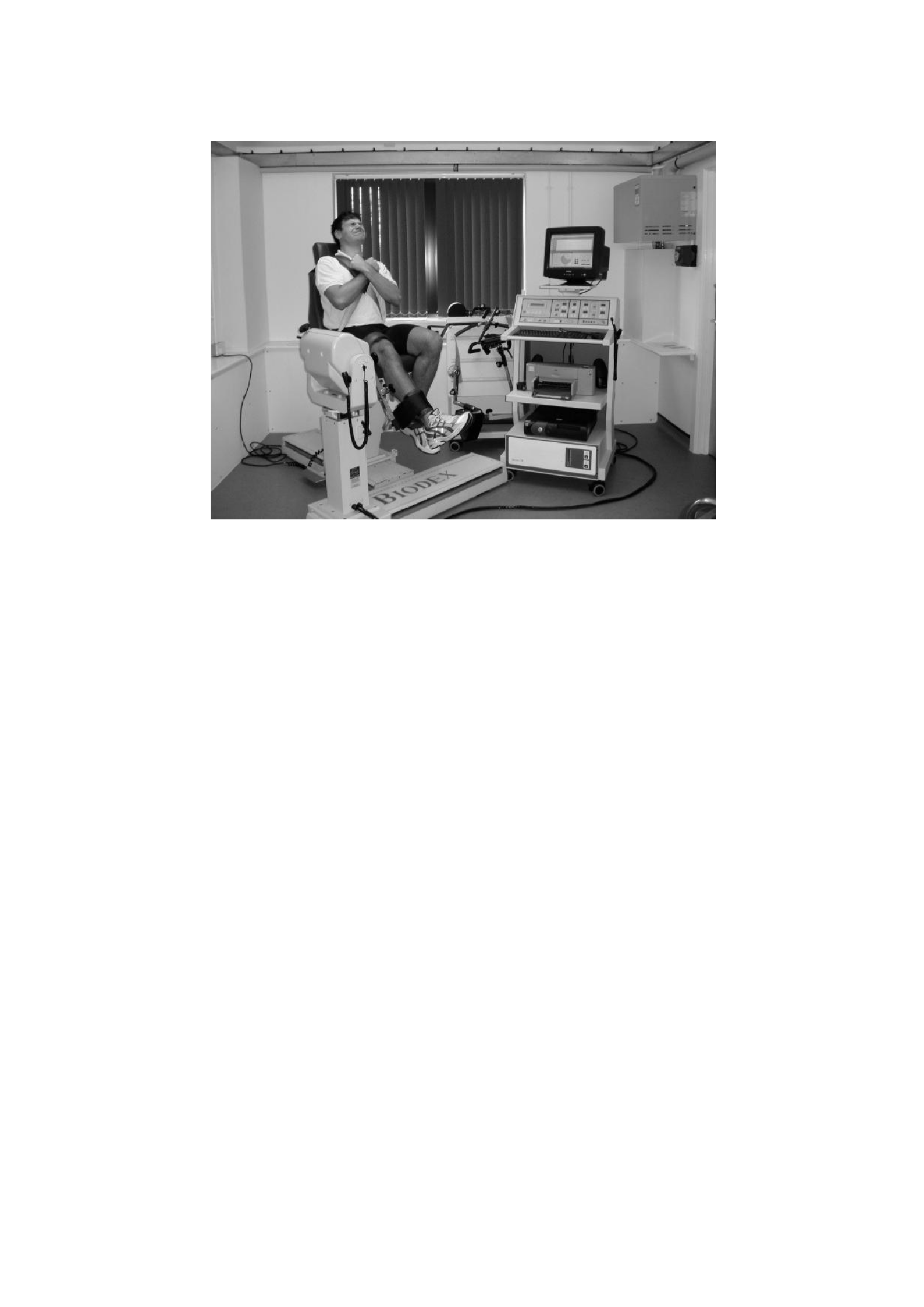
95
Figure 3.2 Assessment of isokinetic peak torque, using a Biodex B-2000 isokinetic
dynamometer (Biodex Corp, Shirley, NY)
The initialization, system diagnostics and Calibratio
n
The procedure is carried out
automatically by Biodex System by pressing the Start button
on
control panel.

96
Participants complete Standard heating 2 minutes of cycling at 50 W on
Electronic brake cycle ergometer (Lode Excalibur Sport, Groningen, Netherlands)
followed by static stretching exercises from the extensor muscles/flexors of the knee.
Participants completed a standard warm-up for 2 minutes of cycling at 50 W on the cycle
Ergometer followed by static stretching exercises for extensor muscles/knee flexors
Group. The participants then sit on an isokinetic dynamometer in an upright position
Position with seat angle set at 85°. Ankle secured to input arm
Dynamometer on tibia Allow Full
ankle
movement, with axis of rotation
Dinamomete
r
parallel to the lateral epicondyle femoralis. All Adjustments to

97
The dynamometer was recorded and replicated in subsequent tests. 80° motion range

98
For each subject (from 90º to 10º knee flexion (full extension = 0°)) it was manually
established by the researcher and confirmed goniometrically. The mass of the limbs is
recorded by the dynamometer to allow for gravitational correction of peak torque values.
The highest of the five maximum voluntary contractions (MVCs) at angular speeds of 30
degrees s-1 was recorded. A 30-second rest period is allowed between contractions. Visual
feedback, displaying real-time power and strong verbal nudges are used to promote
maximum effort. All data is collected using the Biodex Advantage Software package and
stored on a computer for further analysis. The coefficient of variation between tests for
duplicate actions using this procedure in our laboratory was 4.9%.

99
CHAPTER 4:
EFFECTS OF EXERCISE-INDUCED MUSCLE DAMAGE ON PERCEIVED
VENTILATION AND EXERTION RESPONSES TO MODERATE AND
VIGOROUS INTENSITY CYCLE EXERCISE

100
The content of this chapter forms the basis for the following publications:
Davies RC, Rowlands AV, Eston RG. Effect of exercise-induced muscle damage on
breathing and perceived exertion response to moderate and vigorous cycle exercise.
European Journal of Applied Physiology 2009;107(1):11-9.

101
4.1 Abstract
This study examined the effects of exercise-induced muscle damage (EIMD) on the
ventilation and exertion responses felt to cycle exercise. Ten healthy, physically active
men cycled for six minutes at moderate intensity and fatigue at a strenuous intensity before
and 48 hours after eccentric exercise (100 squats with a load corresponding to 70% body
mass). Changes in ventilation and perceived exertion ratings (RPE) are calculated for each
individual and expressed against time (moderate and vigorous exercise) and as a
percentage of fatigue time (vigorous exercise). Ventilation increases during moderate
exercise in
48 hours (V E; 34.5 ± 5.0 to 36.3 ± 3.8 L.min-1, P<0.05) but the increase in RPE is not
important. During strenuous training at 48 hours, the time of fatigue (TTE) is
reduced and
V
E
(87.1 ± 14.1 to 93.8 ± 11.7 L.min-1) and RPE (15.5 ± 1.3 to 16.1 ± 1.4) increased (P<0.05).
When expressed as a percentage of TTE, the difference between the vent value and the
RPE disappears. The findings suggest that the enhanced ventilation response to cycle
exercise after EIMD may be an important cue in informing the perception of effort during
high-intensity exercise but not during moderate-intensity exercise.

102
4.2 Introduction
Unusual sports, especially those with high eccentric components, have a significant impact
on muscle structure and function. However, the effect of EIMD on sub-maximal training
performance remains vague. During submaximal running, reported increased oxygen
consumption (V2), minute ventilation (V2), respiratory exchange ratio (RER), blood lactate
concentration ([La]) and heart rate (HR) (Braun & Dutto, 2003; Chen et al., 2007b, 2008)
was countered by observation of unchanged cardiorespiratory and metabolic responses
(Hamill et al., 1991; Scott et al., 2003; Paschalis et al., 2005; Marcora & Bosio, 2007).
During the submaximal cycle, the V
O2 response does not appear to be altered by the previous
eccentric attacks and muscle damage (Gleeson et al., 1995; Moysi et al., 2005; Schneider et al.,
2007; Twist & Eston, 2009). However, the effect of EIMD on others
Cardiorespiratory and metabolic responses are still debated. Elevated V E and HR
Response has been reported during high-intensity cycling 48 hours after inducing
damage, bench-stepping and has been associated with an increase in [La] (Gleeson et al.,
1995;
Schneider et al., 2007). During low-intensity exercise (62% V O2max) after squatting
exercises, V
E , HR and [La] do not seem to change (Moysi et al., 2005). Thus will

103
It appears that damage induction mode and intensity and training mode are important
factors in determining the response to submaximal exercise with EIMD. However,
regardless of the mode of damage, intensity of the exercise, or the exercise protocol used,
the weight of the evidence supports the premise that exercises that are eccentric and
muscle-damaging produce

104
peningkatan rasa usaha (Gleeson et al., 1995; Scott dkk., 2003; Marcora & Bosio, 2007;
Chen dkk., 2007b, 2008; Twist & Eston, 2009).
The impaired force generation resulting from EIMD results from reduced nerve input to
the muscles that serve as a protective mechanism to prevent further injury (Proske et al.,
2004). Similarly, higher perceived force deployment ratings (RPEs) reported during sub-
maximal exercises after EIMD may also contribute to centrally mediated protection
systems. Perception effort involves assimilating many afferent signals from various
perceptual cues originating from various body systems including the cardiorespiratory and
neuromuscular systems, which can be interpreted both in the way of feedback and forward
feedback (Hampson et al., 2001).
Cues that inform a perceptual response to exercise can arise from central or peripheral
sensations. Central cues, which reflect the aerobic demands of exercise (Åstrand &
Ryhming, 1954), originate in the cardiorespiratory system, while peripheral cues include
local muscle sensations (such as sore muscles) and sensations generated by stimulation of
mechanoreceptors and chemoreceptors (Watt & Grove, 1993; Robertson & Noble, 1997).
The influence of central signals (cardiorespiratory) is less important than local sensations,
especially at lower exercise intensities (Mihevic, 1981; Hampson et al., 2001). Thus, the
perceived activity reported during exercise with EIMD can be affected differently by
central and peripheral cues depending on the intensity of the exercise.

105
The exercise domains used during investigations are important in determining
cardiorespiratory responses and perceived activity. The three domains of sub-maximal
exercise intensity (moderate, heavy and strenuous) elicit very different metabolic,
physiological and perceived exertion responses. The physiological events that separate the
different training domains are the lactate threshold, which marks the transition from
moderate-intensity to vigorous exercise and the maximum latat of stable state or critical
power, which marks the transition from heavy-intensity to vigorous intensity training. As
such, it is important that the intensity of the exercise is accurately defined by depicting the
boundaries between these domains when investigating such responses (Jones et al., 2009).
Investigations that utilize exercise intensity determined by the percentage of maximum
working capacity can be misleading due to differences between individuals. For example,
exercising in
80% V−O2
max , the intensity typically used to investigate the effect of EIMD on
submaximal sports performance (e.g. Gleeson et al., 1995; Twist & Eston, 2009), can result
in one individual exercising in the weight domain while another may be in the weight
domain.
Therefore, the aim of this study was to investigate the changes in ventilation and exertion
responses perceived to cycling with and without exercise-induced muscle damage.
Specifically, to investigate these changes during 6 minutes of moderate-intensity cycle
training and thorough heavy-intensity cycle training. We hypothesize that exercises that

106
damage the eccentric muscles will increase the ventilation response to vigorous intensity
exercise but will not alter the response during moderate-intensity exercise. In addition, we
predict that after previous eccentric exercise, the perceived exertion response to

107
Cycle training at both training intensities will increase and that scalar time property of
exertion felt during exercise to fatigue will not be affected.
4.3 Method
Participants
Ten healthy, physically active men voluntarily participated in the study. All showed no
symptoms of pre-existing illness and injury and did not participate in lower limb endurance
training for at least six months prior to the assessment. Their characteristics are shown in
Table 4.1. Participants give written consent to participate in the research, which is
approved by the School of Sport and Health Sciences Ethics Committee at the University
of Exeter (See appendices B, C and D for sample participant information sheets, participant
consent forms and ethical consent certificates).
Table 4.1 Participant characteristics (N = 10).
Characteristic Installment (± SD)
Age (yrs)
21.5 (± 1.2)

108
Height ( m)
1.82 (± 0.06)
Massa (kg)
79.8 (± 8.2)
Maks V O2 (ml kg min)
. -1 -1
48 (± 3)
Max HR (tap.min-1)
194 (± 6)
WR Max (W)
353 (± 30)
WR at GET (W)
120 (± 14)
Medium WR (W)
96 (± 11)
WR Parah (W)
283 (± 24)
The maximum value (max), gas exchange threshold (GET) and other working rate
(WR) are those measured during the ramp test.

109
Participants were asked not to take anti-inflammatory medications during the study and
were instructed to report to the laboratory in a resting state, having not done strenuous
exercise in the previous 24 hours. Changes in the kinetics of V O2 and deoxyhemoglobin
Kinetics because EIMD is assessed in sub-samples and this data has been reported
elsewhere (Chapter 7).
Procedure
Exercise testing
Participants were asked to visit the laboratory at the same time (± 1 hour) on four occasions
over a 3-week period. All tests are carried out on an electronically braked cycle ergometer
(Lode Excalibur Sport, Groningen, Netherlands). During the first visit, after the
measurement of height and body mass (SECA, Hamburg, Germany) participants
completed an incremental cycle exercise test (ramp) until fatigue to establish maximum
oxygen absorption (V
O2max) and gas exchange threshold (GET) and to establish future
work intensity (Table 4.1). Participants cycled with a self-selected pedal speed (between
70 and 90 rpm) and the speed of these pedals along with the height and configuration of
the saddle and handlebar were recorded and reproduced in subsequent tests. The ramp test
consists of a 4-minute base cycle at 0 W followed by a continuous increase in the working
rate of 1 W every 2 seconds
(i.e. 30 W.min−1) until the subject is unable to continue. The V˙ O identified as

110
2 max
The highest 30-s average score recorded before the termination of the test by the
participant's will. The GET was determined independently by two experienced reviewers
using the V tilt method (Beaver et al., 1986). It involves a visual inspection of individual
plots
CO2 to CO2 to establish a disproportionate first increase in CO2. Work
rates that require 80% of GET (moderate exercise) and 70% of the difference ( )

111
between GET and V O2
max (strenuous training) is calculated, taking into account
the average response time of the V O
2 adaptation for ramp training (about 2/3 of the ramp rate
i.e. minus 20 W) (Whipp et al., 1981). At the second and fourth visits, respectively before
and 48 hours after eccentric training, participants cycled at a self-selected pedal speed
(between 70 and 90 rpm) for six minutes at a constant working speed (WR) of 80% GET
(moderate). After a six-minute break, participants cycled at a constant work rate of 70%
(severe intensity) and continued until the cessation of the will of the test when exhausted.
Participants received a strong verbal urge to continue exercising for as long as possible in
all complete tests.
Eccentric exercises
At the third visit, participants completed a muscle-damaging eccentric exercise protocol
consisting of 100 squats (Smith) performed as 10 sets of 10 repetitions. The load on the
bars was calculated according to 70% of the body mass of each participant. For more
details on this procedure, please refer to Chapter 3.
Measurement
Markers of muscle damage

112
Markers of muscle damage (muscle pain and isokinetic peak torsion) were measured
immediately before and then 30 minutes and 48 hours after eccentric exercise. In addition,
plasma creatine kinase (CK) activity was assessed immediately before and then 30
minutes, 24 and 48 hours after eccentric exercise. Due to availability at 24 hours, only 8
participants were measured for CK activities.

113
Pain felt in the knee extensor was assessed using an empty visual analogue scale of 0-10
(VAS). The VAS consists of a 10 cm line labeled from left (no pain) to right (worst pain
ever). Participants squatted down to 90o knee flexion with their hands on their hips and
then marked the VAS to indicate their pain level. The pain felt was then measured by
measuring the distance from 0 to the mark on the line to the nearest 0.1 cm.
Plasma CK activity was assessed from fingertip capillary samples. The sample was
centrifuged at 4000 RPM (2000 x g) for 5 min and two 20 μl plasma samples were then
added to 1 ml of reagent (Randox CK-NAC 110, Randox Laboratories Ltd., Crumlin, Co.
Antrim, UK). The solution was then incubated at 37°C and absorbance at 340 nm was
recorded by spectrophotometry (Jenway 6310 spectrophotometer, Jenway, Essex, UK) at
1,
2, 3 and 4 minutes. The value of CK was calculated using the formula CK(U/l) = 8095 x
Δ, absorbance of 340 nm/min. The average CK values of both samples were calculated
and used for subsequent analysis. Normal serum values of 24-195 U/l were reported for
men using this method (Szasz, 1976). The intra-test coefficient of variation for duplicate
samples using this procedure in our laboratory is 6.6%.
Isokinetic peak torque is measured using a Biodex B-2000 (Biodex Corp, Shirley, NY)
isokinetic dynamometer, which is calibrated prior to each data collection session in
accordance with the manufacturer's guidelines. After the habituation session, participants
performed five maximum voluntary contractions (MVCs) at 30 degrees 1 with a 30-second

114
rest period between contractions. The coefficient of variation between tests for duplicate
actions using this procedure in our laboratory was 4.9%. Visual feedback, displaying

115
real-time power, used to drive maximum effort. For more details on this
procedure, please refer to Chapter 3.
Steps of the sports test
Pulmonary gas exchange is measured breath-by-breath through an online gas analysis
system (Cortex MetaMax 3B, Biophysik, Leipzig, Germany) during all exercise tests.
Change
in respiratory frequency (fR), minute ventilation ( V
E ), oxygen uptake ( V
O2 ) and V
E / V
O2
recorded continuously during testing via Metasoft Cortex 3.1 software. The system is
calibrated prior to each test in accordance with the manufacturer's guidelines for known
cylinder gas concentrations (15% oxygen, 5% carbon dioxide) and a 3 l calibration syringe
(for flow volume). The heart rate (HR) was monitored using a wireless chest strap
telemetry system (Polar Electro T31, Kempele, Finland) and recorded continuously via a
link to the Cortex gas analysis system in all training tests.
Participants were familiarized with the 6-20 Borg Perceived Mobilization Rating Scale
(RPE) and were given standard instructions on how to use the scale (Borg, 1998).
Participants were encouraged to focus on their overall perception of activity when
reporting their RPE, which was recorded during the last 15 seconds of each minute of all
exercise tests (Figure 4.1).

116
Blood lactate responses were assessed from fingertip blood samples collected immediately
before and immediately after moderate and vigorous intensity exercise. Samples were
collected into Lithium heparin microvettes (CB300, Sarstedt AG & Co.,
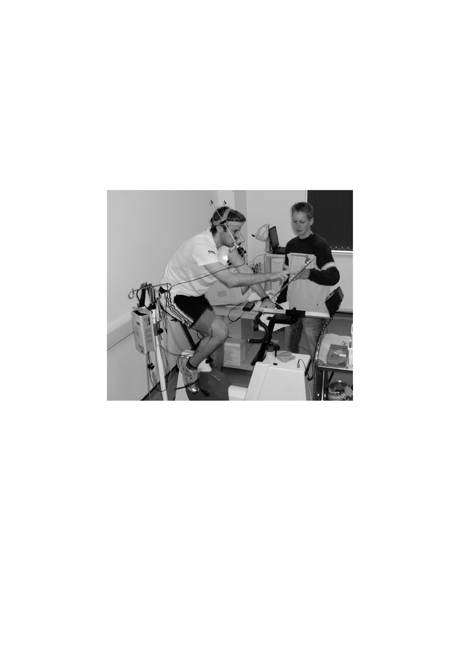
117
Nümbrecht, Germany) and then analyzed for blood lactate concentrations
using the YSI 2300 STAT plus analyzer (Yellow Springs, Ohio, USA).
Figure 4.1
RPE scale.
A participant reported his or her RPE during cycle training using Borg 6-20

118
Statistical analysis
Markers of muscle damage
Changes in Marker
s
muscle Damage (isokinetic peak torque, felt muscle
pain and creatine kinase activity) were analyzed using a series of repeated, one-way methods
measure the analysis of variance (ANOVA). The data is initially checked for assumptions
normality and because the CK activity data is not distributed normally; These values are log-

119
changed before statistical analysis. After the transformation, the CK activity data is distributed
normally (see appendix H).
Steps of the sports test
Changes in
fR,
V E , V
O2, HR, V
E/V
O2 and RPE are calculated for each individual and
expressed towards time (minute value) for moderate and vigorous intensity exercise, and
in the case of heavy exercise, as % time for fatigue (% time). These values are analyzed
through a series of two-factor (test x time) full repeated measurements of ANOVA. Where
the assumption of roundness is violated (P <.05), the Greenhouse-Geisser correction factor
(GG) is applied to adjust the degree of freedom. Where a significant x-time test interaction
was observed, a modified post-hoc Tukey test for repeated measurements (Stevens, 2002)
was run to determine where significant differences occurred. The paired t-test was used to
determine significant differences in blood lactate [La] concentrations and fatigue time
during strenuous intensity exercise before and after eccentric exercise. All data were
analyzed using the SPSS statistical software package for Windows (version 13). The
statistical significance was set at 0.05. A P value between 0.05 and 0.10 is considered a
trend.

120
4.4 Result
Markers of muscle damage
Eccentric exercise is effective in triggering significant changes in all markers of muscle
damage. Table 4.2 shows changes in isokinetic peak torque, muscle pain and CK activity
before and after eccentric exercise. Isokinetic peak torque (30 degrees-1) decreased to 86%
of the pre-damage value at 30 minutes recovered to 89% at 48 hours (F (2, 18) = 13.37 P <
0.001). Significant pain is reported 30 minutes after eccentric exercise with

121
the highest value was reported at 48 hours (F (2, 18) = 71.49 P < 0.001). CK activity increased
after eccentric exercise, with the highest value observed at 24 hours (FGG (1.4, 9.8) = 13.13 P <
0.05).
Table 4.2 Changes in markers of muscle damage. Average score (± SD) before
(before) and 30 minutes and 48 hours after eccentric exercise
Measurable variables
pre
30 minutes
24 hours
48 hours
CK Activity (U/l)
178 ±129
244 ± 181
794 ± 690*
88 ± 217
Pain
0.5 (0.5)
5.4 (1.6)*
N/a
6.8 (1.6)*
Peak Torque (Nm) 30
degrees-1
279(45)
239 (28)*
N/a
248 (35)*
n = 10, peak pain and torque; N = 8, CK* activity differs significantly from the pre value (P <
0.05).
Steps of the sports test

122
Moderate intensity training
Table 4.3 shows the mean change (± SD) of fR, V
E , V
O2 , HR, V
E / V
O2 and RPE respectively
during moderate-intensity cycling (80% GET) before and 48 hours after eccentric
training. The main effects for the test were fR ( F(1, 9) = 7.29, P = 0.024), V E (F (1, 9) = 6.36, P
= 0.033) and HR (F (1,
9) = 12.52, P = 0.006) indicates an increase in grades after eccentric exercise. There
there was no significant difference in V
O2, V
E/V
O2 or RPE as a result of eccentric exercise
(P > 0.05). Similarly, there was no significant difference in blood lactate concentrations
before and after exercise before and after eccentric exercise (before: 1.13 ± 0.43, 1.08 ±
0.29 and after: 0.98 ± 0.21, 1.10 ± 0.28 mmol.l-1 before and after eccentric exercise,
respectively) (P
> 0,05).

123
Table 4.3 Changes in Fri, S E , V O2 HR S E / V O2 and RPE during moderate-intensity exercise.
Average score (± SD) before (before) and 48 hours after (after) eccentric exercise.
Fri V
And
V O2 HR S E / V O2 RPE
(breath.Min-1) (T.Min-1) (ml.Medical history-1.Min-1) (Tap.Min-1)
To
20.6 (±
3.5)
34.5 (±
5.0)
19,9 (±
0,9)
109 (± 7)
21,8 (±
1,6)
9,9 (± 1,7)
Post
21.8 (±
3.2)*
36.3 (±
3.8)*
20.1 (±
1.8)
116 (±
8)*
22.7 (±
1.2)
10.6 (±
1.6)
* Significantly different from the pre value (P < 0.05) N = 10
Heavy intensity training to fatigue
As expected, the time to fatigue was significantly reduced after eccentric exercise (7.33 ±

124
2.26, 6.37 ± 2.16 minutes before and after eccentric exercise, respectively) (t (9) 2.64, P =
0.027). Before eccentric exercises, all 10 participants completed a strenuous intensity of at
least 5 minutes. However, 48 hours after eccentric training, the minimum time achieved
by all participants is reduced to 4 minutes. Pre-workout [La] did not change, but the final
workout [La] was significantly lower after eccentric training (t (9) = 3.31, P = 0.009) (pre-
training:
0.90 ± 0.34, 0.79 ± 0.34 and final training: 9.33 ± 1.73, 8.45 ± 1.51 mmol.l-1 before and
after eccentric training, respectively).
Minute-by-minute size
To investigate minute-by-minute changes in fR, V
E , V
O2 , HR, V
E / V
O2 and RPE,
The first 4 minutes of data are analyzed. Figure 4.2 a–f shows minute-by-minute changes
for 1-4 minutes in fR, V
E , V
O2 , HR, V
E / V
O2 and RPE respectively during cycling in
70%∆ eccentric before and after training. The main effect for time is observed for all

125
variable (P < 0.05). Main effect for fR test (F (1, 9) = 10.53, P = 0.010), V E (F (1, 9) =
9,50, P = 0,013), V E / V O2 (F (1, 9) = 8,47, P = 0,017), dan RPE (F (1, 9) = 8,15, P = 0,019)
and the trend for HR (F (1, 9) = 4.43, P = 0.065) indicates that the value has increased after
eccentric exercise. There is an interaction (test*time) for fR (FGG (1,3, 11,3) = 5.25, P =
0.036) and V E (F (3, 27) = 3.19, P = 0.039). Post hoc tests show that the following are eccentric
exercise, fR and V E were significantly higher for all but the first minutes of training. There
there was no significant difference in V O2 (P > 0.05).
Percentage time value
Figure 4.3 a–f shows the % time change in fR, V
E, V
O2, HR, V
E/V
O2 and RPE respectively
During cycling at 70%∆ eccentric before and after training. When expressed as % time to
willpower fatigue, the main effect for time remains but the main effect for the fR, V
E, HR
and RPE tests disappears. However, there was a major effect for the V O2 test (F(1, 9) = 5.25,
P = 0.048) which showed that the value had decreased after eccentric exercise. This was
accompanied by an increase in V
E/V
O2 (primary effect, F(1, 9) = 5.17, P = 0.049).
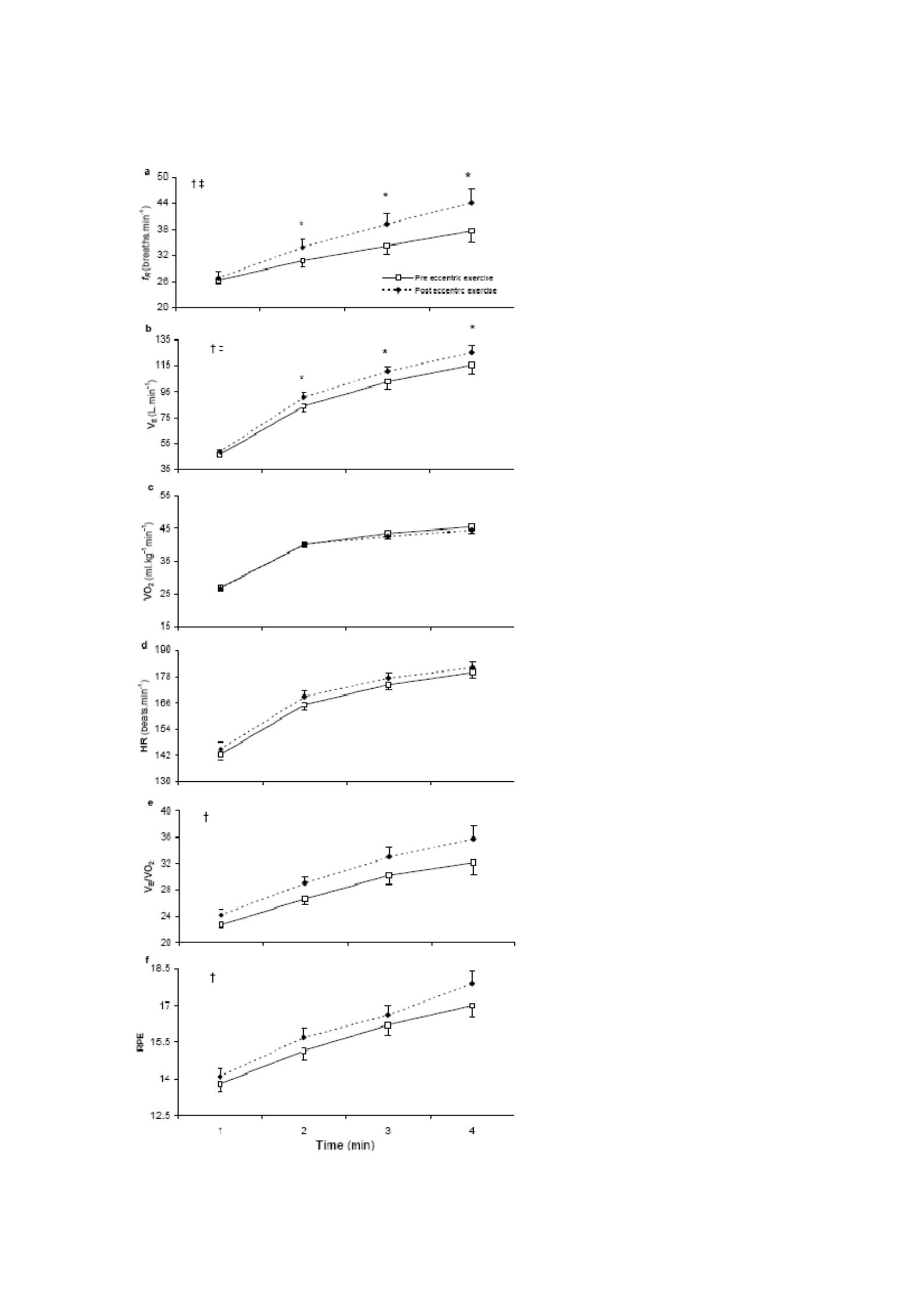
126
Figure 4.2
Minute value (heavy intensity
training). Minute-by-
minute changes in a: respiratory
frequency (Fr), b: minute
ventilation ( V E ), c: Oxygen
absorption ( V−O2 ), d: HR e: V E / V−O2 and f:
RPE during cycling at 70%∆ pre-
workout and 48 hours post-
eccentric.
The value is the average (± SEM).
The main effect was significant for
time for all measures (P < 0.05).
† Significant key effects for testing (P
< 0.05).
‡ Significant interaction effect
(test*time) (P < 0.05).
* Significantly different from the

127
pre score (P < 0.05).
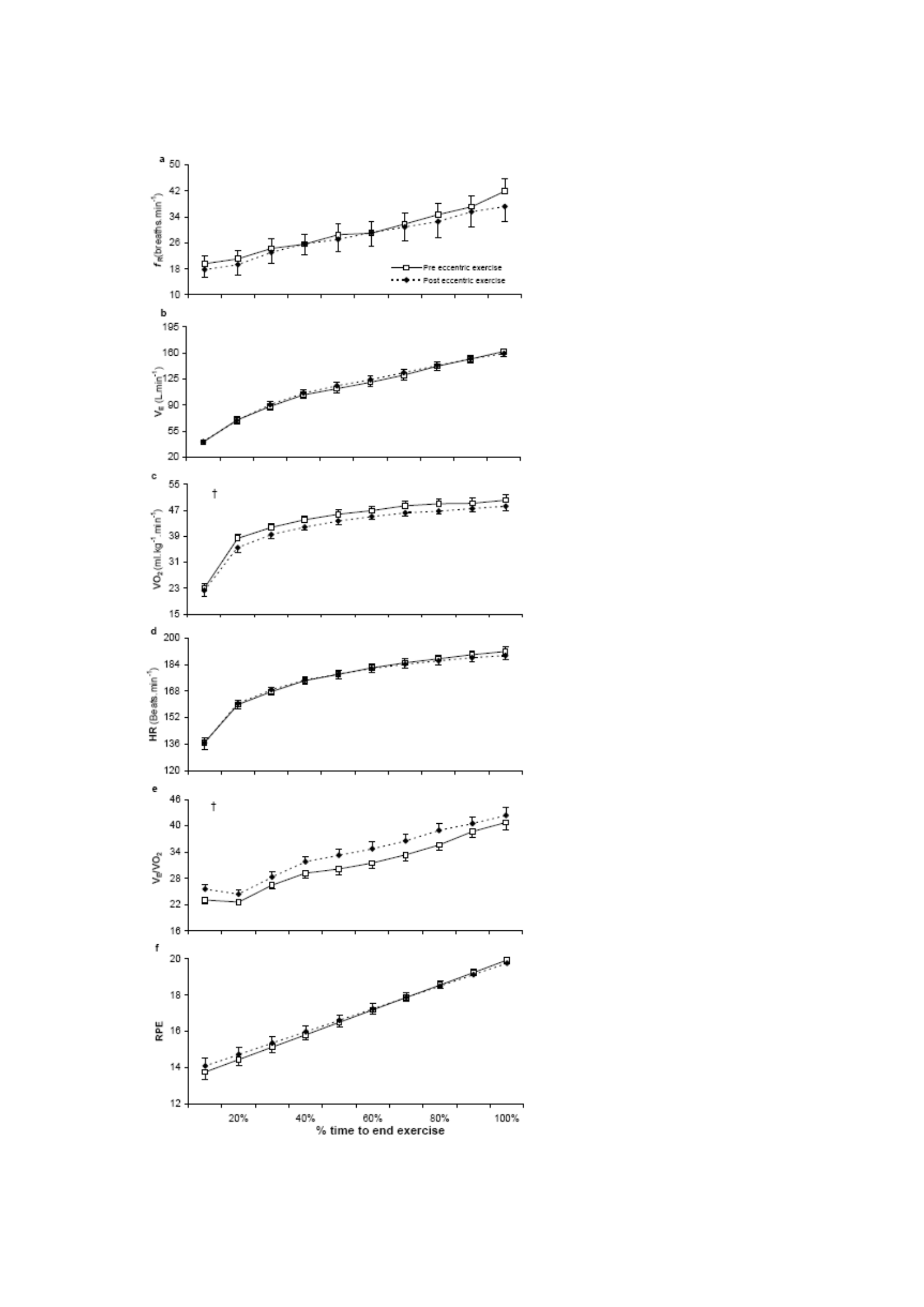
128
Figure 4.3
Percentage time value (heavy
intensity training). Change a :
respiratory rate (fR), b: minute
ventilation ( V E ), c: oxygen
absorption
( V O
2 ), d: HR, e : V
E / V
O2 and f:
RPE during cycling at 70%∆ pre-
workout and 48 hours post-
eccentric set against % time for
willpower fatigue.
The value is the average (± SEM).
Significant effect for time for all
measures (P < 0.05).
† Significant primary effect for
testing (P < 0.05).
‡ Significant interaction effect
(test*time) (P < 0.05).
* Significantly different from the

129
pre score (P < 0.05)

130
4.5 Discussion
This investigation showed that previous attacks of muscle-damaging eccentric exercises
improved ventilation response not only during vigorous-intensity cycle training (70% ∆)
but also during moderate-intensity cycle training (80% GET). In addition, the study
showed that the perception of exertion increased during vigorous intensity training but did
not appear to be affected by previous eccentric exercise attacks during moderate exercise.
Markers of muscle damage
As anticipated, the squatting protocol was effective in eliciting changes in markers of
muscle damage in all participants. The significant decrease in peak torque observed after
eccentric exercise is in line with previously reported findings (Byrne et al., 2001).
Perceived muscle pain increased above baseline size at 30 minutes after eccentric exercise
with the highest score reported at 48 hours. These findings are consistent with the
previously reported characteristic temporal profile of increased muscle pain after
eccentricly biased exercise (e.g. Twist & Eston, 2005). As expected, the course of CK
response time does not reflect changes in muscle function or perceived pain (Warren et al.,
1999). However, CK efflux, which was largest at 24 hours, did provide indirect evidence
of increased myocyte membrane permeability (Allen et al., 1995). Rapid increases and
peaks in CK activity similar to those observed here have previously been observed
following a similar eccentric exercise protocol (100 squats @ 70% body mass, Byrne &
Eston, 2002a).

131
Steps of the sports test
The increased ventilation response to the heavy-intensity cycle exercise observed in this
study is in line with previously reported observations during high-intensity fixed-load
cycles

132
muscle-damaging post-workout exercise (Gleeson et al., 1995; Schneider et al., 2007).
However, the observation that V E and fR also increase during moderate intensity
practice (<GET) is unexpected. Using a similar squatting protocol, which results in
comparable strength loss rate (-14% at 48 hours) and 62% V O2max training intensity
Moysi et al. (2005) reported no changes in ventilation or HR. In contrast, improvements in
ventilation and HR were observed in this study, despite the fact that the subjects
performs a much lower intensity, 80% GET, equivalent to ~27% V O2max for this
Individual.
The improved ventilation response observed when exercising with EIMD or DOMS has
been associated with changes in metabolic factors. Specifically, it has been assumed that
an increase in [La] contributes to improved ventilation response. An increase in [La] can
be attributed to increased dependence on type II fibers and shifts corresponding to
increased production of glycolytic energy after muscle-damaging exercise (Braun & Dutto,
2003; Chen et al., 2007b; Gleeson et al., 1995). However, no such changes in [La] were
observed in the current study at both training intensities. Importantly, the observation that
ventilation response after EIMD is higher during exercise under GET suggests that the

133
altered exercise response may not be due to changes in metabolic factors.
To support this proposition, Schneider et al. (2007) observed that phase II kinetics V O2
not altered by DOMS and concluded that the increased [La] was not caused by a change
in oxidative function. Instead, these authors suggest that higher [La] levels arise from an
increase in the rate of lactate efflux from damaged myocytes due to increased membrane
permeability. However, higher levels of lactate efflux can be counteracted with increased
clearance is facilitated by increased muscle blood flow (Laaksonen et al., 2006) and

134
thus giving rise to an unchanged [La] response. While the effect of conjecture [La] on
ventilation response should not be ignored; Given the findings of this study, other potential
stimuli demand consideration.
Improvement in V E
and fR was observed in this study during moderate and severe
intensity and in V E / V O2 during severe intensity can be associated with changes in nervous
factors. The complex mechanisms involved in ventilation control during exercise involve
a combination of central command and afferent feedback but this is poorly understood.
However, ventilation is known to increase in response to painful stimuli (Haouzi et al.,
2004). Thus local muscle pain that occurs as a result of eccentric exercise, as observed in
this study, is expected to have a stimulating effect on ventilation. It has been proposed that
group III and IV afferent fibers located in and around the blood vessels of exercising
muscles are involved in modulating the ventilation response (Haouzi et al., 2004). This
vascular distension, such as the change in the shape of the capillary lumen observed by
Kano et al. (2005) after eccentric exercise, will trigger the release of afferent fibers leading
to increased ventilation. Thus, nerve monitoring of peripheral blood vessels and local
muscle events could, in part, explain the observed increased ventilation response. Hotta et
al. (2006) suggested that changes in neural factors contribute not only to improved
ventilation response but also to changes in force formation. After eccentric exercise,
increased activation of motor units may be necessary to achieve a certain sub-maximal
force (Semmler et al., 2007). Similarly, a greater sense of effort is reported when
generating a certain strength after eccentric exercises (Proske et al., 2004).

135
Consistent with these observations, subjects in this study reported higher ratings of
perceived exertion (RPE) during fixed weight intensity cycle exercise after eccentric
exercise, although RPE did not appear to change during moderate exercise. Jameson and
Ring (2000) have suggested that during a workout cycle, the perceived exertion rating is
based on a combination of leg muscle pain and a feeling of shortness of breath. Thus, the
increased leg muscle pain experienced by participants in this study after eccentric exercise
may have provided peripheral cues that are important for informing the RPE response.
Similarly, the improved ventilation response may have provided an important central
signal. Although the relative contributions of various central and peripheral cues are poorly
understood, they may be less important than peripheral cues, especially during low-
intensity exercise (Mihevic, 1981; Hampson et al., 2001). It is interesting to note that the
increase in muscle soreness and increased ventilation response experienced by participants
in this study at 48 hours did not significantly affect the perception of activity during
moderate-intensity exercise. However, during heavy-intensity training, where central
ventilation cues may play a more influential role in informing the perception of exertion,
RPE increases.
The perceived higher activity reported in strenuous exercise at 48 hours may explain the
reduction in time to end training. Previous increases in RPE have been associated with
decreased time-trial performance in running (Marcora & Bosio, 2007) and in cycling
(Twist & Eston, 2009). In addition, the reinforced ventilation response can contribute to
the termination of premature exercise. Acute respiratory muscle fatigue may have
increased the severity of locomotor muscle fatigue through respiratory muscle

136
metaboreflex

137
improve the perception of effort and further centrally mediated reduction in motor output
(Romer & Polkey, 2008).
The complex interaction of central and peripheral fatigue factors is crucial in determining
the duration of each participant's athletic performance. However, the participant's decision
to end the exercise was ultimately a conscious behavior based on the perception of changes
in the subconscious homeostatic control system (St Clair Gibson et al., 2003). Thus, the
perception of exertion can be considered fundamental to an individual's exercise response
when a participant is required to exercise for 'willpower fatigue'. Related to this is the
observation that minute-by-minute differences in perceived activity values reported during
strenuous exercise before and after eccentric exercise are omitted when expressed as a
percentage of the total duration of exercise. Thus, further evidence is provided to support
the proposition that there is a scalar-linear relationship between perceived exertion ratings
and exercise duration (Eston et al., 2007, Crewe et al., 2008; Faulkner et al., 2008; Joseph
et al., 2008). Next
Observation that the difference in V E
and fR is also omitted when expressed as
The proportion of time to fatigue can provide some insight into the cues that inform the
exertion response felt during high-intensity cycling after a previous eccentric training
attack.

138
4.6 Conclusion
This is the first study to investigate the effects of muscle-damaging exercise on ventilation
responses and perceived activity against cycling in specific exercise domains above and
below the gas exchange threshold. The findings suggest that there is a strong association
between enhanced ventilation responses to cycling after eccentric exercise and higher
ratings of perceived activity reported during exercise above GET. Furthermore, additional
evidence was provided to support the observation that the perceived activity scale with the
duration of exercise during exercise to fatigue.

139
CHAPTER 5:
EFFECT OF ECCENTRIC EXERCISE-INDUCED MUSCLE DAMAGE ON
GAS EXCHANGE THRESHOLD

140
The contents of this chapter are currently under review for publication:
Davies RC, Rowlands AV, Poole DC, Jones AM and Eston RG. Muscle damage caused
by exercise separates the exchange threshold of Lactate and Gas. It is currently under
review.

141
5.1 Abstract
We tested the hypothesis that exercise-induced muscle damage (EIMD) would increase
the ventilation response ( V E ) to incremental/ramp cycle exercise (lowering the gas
exchange threshold), without altering the blood lactate profile thereby separating gas
exchange and lactate thresholds. It is thought that this intervention may provide a broader
understanding of the underlying mechanisms of the relationship between GET and the
lactate threshold (Tlac). Ten physically active men completed a maximum incremental
cycle test before (before) and 48 hours after (after) performing eccentric exercises
consisting of 100 squats with a load corresponding to 70% body mass. Pulmonary gas
exchange is measured breath-by-breath and fingertip blood samples at 1-minute intervals
for blood determination [La]. GET occurs at lower working rates (before, 136 ± 27 W;
post, 105 ± 19 W, P< 0.05) and V O2
(pre, 1.58 + 0.26; post, 1.41 + 0.14 l.min-1, P< 0.05) after eccentric exercise. However,
Tlac occurs at the same working rate (before, 161 ± 19 W; post, 158 ± 22 W, P> 0.05) and
V O2 (pre, 1.90 ± 0.20 l.min-1; post, 1.88 ± 0.15 l.min-1, P > 0.05) after eccentric exercise.
These findings suggest that EIMD separates V
E Response to Incremental/Ramp
exercise from the [La] response that indicates that V
E can be controlled by additional or
changes in neurogenic stimulation after eccentric exercise. Thus, proper consideration of
previous eccentric exercises must be made when using gas exchange thresholds to provide
a non-invasive estimate of lactate thresholds.

142
5.2 Introduction
During an incremental training protocol or dynamic ramp, carbon dioxide production expires
(V
CO2) increases disproportionately as a function of V
O2 above what has been called
gas exchange threshold (GET) (Beaver et al., 1986). Traditionally, GET has been thought
to be the result of a mandatory increase in non-metabolic CO2 production (and the
associated increase in ventilation, ( V
E ) mainly due to the plasma bicarbonate buffer of H+
derived from lactic acid (Beaver et al., 1986; Wasserman et al., 1990). Thus, the
identification of the GET provides the basis for non-invasive estimation of the lactate
threshold (Tlac) (Beaver et al., 1986; Caiozzo et al., 1982; Wasserman et al., 1973, 1990).
But
The responses of V
E and lactate to gradual exercise have been separated by various
experimental and clinical conditions including sports training (Poole & Gaesser, 1985),
glycogen depletion (Hughes et al., 1982; Sabapathy et al., 2006) and McArdle's disease
(Hagberg et al., 1982; Paterson et al., 1990; Riley et al., 1993). On the contrary, recently
dichloroacetate (DCA) studies reported reducing blood lactate [La] and V
E during
Additional exercises thus supporting the existence of a causal relationship between bicarbonates
Lactic acidosis buffer and increase V
E (Wilkerson et al., 2009). Today

143
appreciate that a wide variety of humoral and nervous control mechanisms (central commands,
afferent feedback from carotid body chemoreceptors and from contractions
muscles) contribute to the V
E response to incremental/ramp exercise (Dempsey et al.,
2006; Haouzi, 2006; Haouzi et al., 2004; Waldrop & Iwamoto, 2006). While the complex
integration of this mechanism is understood to mediate the V
E response, most likely
Under different conditions, the proportional contribution of each of these may change.

144
During rhythmic exercises, group III and IV afferents are known to give some V
E drives
through mechanical stimuli such as local muscle distortion, vascular distension and
increased intramuscular pressure as well as through local humoral mediators including H+,
lactate and bradykinin (Ward, 2000; Whipp et al., 1981). Eccentric exercise causes
profound structural and functional disorders of muscles collectively referred to as exercise-
induced muscle damage (EIMD) (Clarkson et al., 1992). EIMD adds release from group
III and IV afferents (Avela et al., 1999; Komi, 2000; Taguchi et al., 2005) and this, rather
than an increase in blood lactate concentrations which is not a mandatory consequence of
EIMD
(Twist & Eston, 2009), most likely contributing to the greater V E response to the constant.
load cycle exercise induced by EIMD (Gleeson et al., 1995; Schneider et al., 2007;
Twist & Eston, 2009) (Chapter 4).
Effects of previous eccentric training and resulting EIMD on V
E and
The gas exchange response to gradual exercise has received very limited attention. Only
one previous study examined the physiological response to the maximum incremental
cycle; unfortunately the bench-stepping protocol used in the investigation only produced
mild pain (Gleeson et al., 1998). Thus, the bench-stepping protocol (Gleeson et al., 1998)
may not increase the release from group III and IV afferents as much as procedures
specifically designed to induce more severe EIMD. To our knowledge, the effect of

145
exercise damaging eccentric muscles on GET has never been investigated before. It is our
opinion that such eccentric exercise protocols are used
previous in our lab (Chapter 4) is likely to exacerbate the V
E response to

146
additional drills/ramps and, thus, potentially separating the V
E , gas exchange
and lactate response.
The purpose of this investigation is to determine the effect of EIMD on the GET and Tlac
responses specified during ramp training. Specifically, we tested the hypothesis that the
completion of 100 squats (performed as 10 sets of 10 reps at ~70% body mass) would
adds the V
E response (lower GET) to the next incremental/ramp workout in
the absence of altered blood lactate profiles thus separating the GET from the lactate
threshold. It is speculated that this intervention may provide mechanistic insights into the
relationship between lactate accumulation and ventilation control in the presence of
apparent muscle damage.
5.3 Method
Subject
Ten healthy, physically active male subjects (age, 25 ± 7 years; mass, 80.1 ±9.9 kg; height,
1.80 ± 0.08 m) voluntarily participated in the study. All participants showed no symptoms

147
of pre-existing diseases and injuries and had not done resistance training on the lower limbs
in the previous 6 months. Participants give written consent to participate in research
approved by the School of Sport and Health Sciences Ethics Committee at The University
of Exeter (See appendices B, C and D for sample participant information sheets, participant
consent forms and ethical consent certificates).

148
Experimental Design
Incremental ramp training test
Participants conducted two incremental ramp training tests until willpower fatigue on an
electronically braked bicycle ergometer (Lode Excalibur Sport, Groningen, Netherlands)
(Figure 5.1). The height of the chair and handlebar position preferred by each participant
were recorded during their first test and replicated in subsequent tests. The height and mass
(SECA, UK) of each participant were also recorded. The exercise test was performed at
the same time ± 1 hour for each participant, before and 48 hours after performing muscle-
damaging eccentric exercises. After 3 minutes of disassembled baseline cycling, the
working rate is ramped up by 1 W every 2 seconds (30 W.min−1) until the subject is unable
to continue despite receiving a strong verbal impulse. The participants were asked to
maintain a pedal speed of 80 rpm during both additional tests.
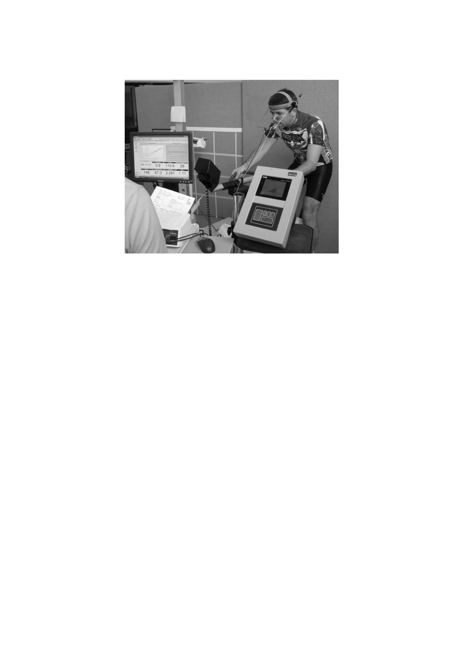
149
Figure 5.1 A participant who performs an incremental ramp cycle exercise test.

150
Eccentric exercise protocols that damage muscles
Participants completed 100 squats (Smith), performed as 10 sets of 10 repetitions with
weights on the bars corresponding to ~70% of each participant's body mass. For more
details on this procedure, please refer to Chapter 3.
Measurement
Assessment of muscle damage
All indicators of muscle damage (perceived muscle pain (using a visual analogue scale of
0-10 (VAS)), creatine kinase (CK) activity and isokinetic peak torque (30 degrees s-1)) were
measured in the listed order, just before, 30 minutes after and 24 and 48 hours after
performing the muscle-damaging eccentric exercise protocol. For more details on this
procedure, please refer to Chapter 3.
Additional training
Throughout the cycle exercise test, before and 48 hours after the muscle-damaging
protocol, lung gas exchange was measured breath-by-breath through an online gas analysis
system (Cortex MetaLyzer 3B, Biophysik, Leipzig, Germany). Participants wore nose pins
and breathed through a low-dead-chamber, low-resistance mouthpiece. Gas exchange was

151
measured during testing using Metasoft Cortex 3.1 software. The system is calibrated prior
to each test in accordance with the manufacturer's guidelines for known cylinder gas
concentrations (15% oxygen, 5% carbon dioxide) and a three-liter calibration syringe
(Hans Rudolph, Kansas City, USA) for gas flow. Inter-test coefficient
variations for V
E , V−O2
and V CO2
Responses using this equipment in our laboratory
respectively 4.1%, 3.1% and 4.2% (respectively for V
E , V
O2 and V
CO2 ).

152
Gas exchange datasets are blindly surveyed to determine peaks
and V
O2 gases
exchange threshold (GET). The peak of V O
2 is determined as the highest 30-second average value
recorded prior to the termination of the test at the participant's will. The GET is determined
from a group of measures that include 1) a disproportionate first increment
in the production of CO2 (CO2
) from visual inspection of individual plots of CO2 versus
V O
2 (V tilt method (Beaver et al., 1986)) and 2) increased ventilation
equivalent to O2 (V E / V O2
) with no concurrent increase in ventilation equivalent
untuk CO2 ( V
E / V CO2 ) (Caiozzo et al., 1982).
The heart rate was continuously monitored using a wireless chest strap telemetry system
(Polar Electro T31, Kempele, Finland) and recorded via a link to the Cortex gas analysis
system. Fingertip blood samples are collected into capillary tubes before, after, and at 1-
minute intervals during each gradual exercise test. The sample was then analyzed for whole
blood lactate (YSI 2300 Sport, Yellow Springs, Ohio, USA). Blood lactate concentration
dataset [La] was blindly reviewed for
determine the lactate threshold (Tlac). Tlac is defined as V O2 which is associated with

153
the level of work before the first clear and continuous improvement in [La] above the
resting rate of visual inspection of the individual plot [La] vs. V O
2 .
Subjects were also asked to report their perceived exertion rating (RPE) at 1-minute intervals
during additional tests. All subjects are familiar with Borg 6-

154
20 RPE scales and comes with standard instructions on how to use the scale prior to testing
(see Appendix F).
Statistical analysis
Indicators of muscle damage
Changes in muscle damage indicators (perceived muscle pain, creatine kinase activity and
isokinetic peak torque) were analyzed using a series of one-way iterative measurement
variance analyses (ANOVAs). A modified post-hoc Tukey test for repetitive actions
(Stevens, 2002) was run to determine where significant differences occurred. Since CK
activity data is not normally distributed, these values are changed log prior to statistical
analysis (Twist & Eston, 2005) (see appendix H).
Ramp Supplement Training
The paired t-test was used to determine significant differences in fatigue time, peak values
and gas exchange threshold values (GET) and lactate threshold values (Tlac) before and
after eccentric exercise. The paired t-test is further used to determine significant
differences in
physiological response in V−O2
in accordance with the pre-eccentric exercise of GET.

155
Pearson product moment correlation is used to check the relationship between GET and
Tlac values. All data were analyzed using the SPSS statistics software package for
Windows (version 13) with statistical significance set at 0.05.

156
5.4 Result
Indicators of muscle damage
There were significant changes in all indicators of muscle damage after eccentric exercises.
Table 5.1 shows changes in muscle pain, CK activity, and isokinetic peak torque before
and after eccentric exercise.
Table 5.1 Changes in muscle damage indicators. The average ± elementary school grades
before (before) and at 30 minutes, 24 hours and 48 hours after eccentric exercise.
Measurable variables
pre
30 minutes
24 hours
48 hours
Nyeri (1-10 VASES)
0.4 ± 0.4
4.4 ± 2.2*
5.8 ± 2.0*
6.4 ± 3.0*
CK Activity (U/L)
180 ± 61
224 ± 103
637 ± 413*
350 ± 151*
Peak Torque (Nm) 30 degrees-
1
300 ± 53
240 ± 68*
248 ± 62*
254 ± 72*
* significantly different from the pre-eccentric exercise value (P < 0.05).

157
Muscle pain increased 30 minutes after eccentric exercise with the highest value reported
at 48 hours (F (3, 27) = 21.01 P < 0.001). CK activity increased after eccentric exercise, with
the highest value observed at 24 hours (FGG (1,38,12,39) = 9.61 P < 0.05). The isokinetic peak
torque (30 degrees-1) decreased by 20% at 30 min and was still 15% lower than the
baseline value at 48 h (F (3, 27) = 17.96 P < 0.001).
Ramp Supplement Training
The peak values achieved during the ramp test before and after eccentric training are shown
in Table 5.2. Fatigue time and associated WRpeak values decreased after
eccentric exercise (t(9) = 2.62, P < 0.05). No significant change in Puncak V
E,

158
A
n
d
V O 2
puncak,
V CO2
peak, HRpeak, RERpeak, [La] peak or RPEpeak (all P > 0.05). Table 5.3 shows
variable changes associated with GET before and 48 hours after eccentric exercise. GET
occurs earlier and thus at lower WRs (t(9) = 3.74, P = 0.005) and V O2 (t(9) = 2.57, P =
0.030)
.
The value of V CO2 in GET was also significantly lower (t(9) = 2.54, P = 0.032).
Table 5.2 Peak values achieved during the ramp practice test. Previous average ±
elementary school grades
(before) and 48 hours after (after) eccentric
training
To
post
Time for fatigue
682 ± 71
635 ± 86*
WRPeak (W)
341 ± 36
318 ± 43*
Puncak V (l.min-1)
145 ± 22
140 ± 35
3,34 ± 0,38
3,27 ± 0,35
4,47 ± 0,45
4,33 ± 0,56

159
2
2
Puncak V O
(l.min-1)
V Puncak CO
(l.min-1)
HR peak (beat.minute-1)
182 ± 12
179 ± 13
RERpuncak
1,35 ± 0,10
1,33 ± 0,12
[La]puncak (mmol.l-1)
6.05 ± 1.36
5,64 ± 1,72
RPEpeak (Borg, scale 6-20)
19,4 ± 0,5
19.1 ± 1.0
Peak minute ventilation (peak
V E), peak
V O2, peak V CO2
HRpeak and RERpeak are the highest
30-second average values achieved during the ramp training test.
* significantly different from the pre-eccentric exercise value (P < 0.05).

160
Table 5.3 Values achieved at the gas exchange threshold (GET) during the ramp
practice test. Elementary school grades ± average before (before) and 48 hours after
(after) eccentric exercise
To
post
Time
262 ± 55
200 ± 37*
WR (W)
136 ± 27
105 ± 19*
V
E
(l.min-1)
34.2 ± 6.1
31.7 ± 4.6
-1
V O2 (l.min)
1,58 ± 0,26
1,41 ± 0,14*
V CO2
(l.min-1)
1,35 ± 0,24
1,20 ± 0,18*
[La] (mmol.l-1)
1,71 ± 0,38
1,55 ± 0,21
RPE (scale Borg 6-20)
10.5 ± 0.9
10.6 ± 1.4
* significantly different from the pre-eccentric exercise value (P < 0.05).
Figure 5.2 shows the response of V CO2 versus V O2 (panel A) and blood [lactate] (panel B)
Representative participants during the incremental ramp exercise before and after eccentric

161
exercise; note the GET is shifted to a much lower
V
O2 after eccentric exercise while
Tlac is essentially unchanged. There was no significant difference in V
E , [La] or RPE in
GET before and 48 hours after eccentric exercise (P > 0.05).
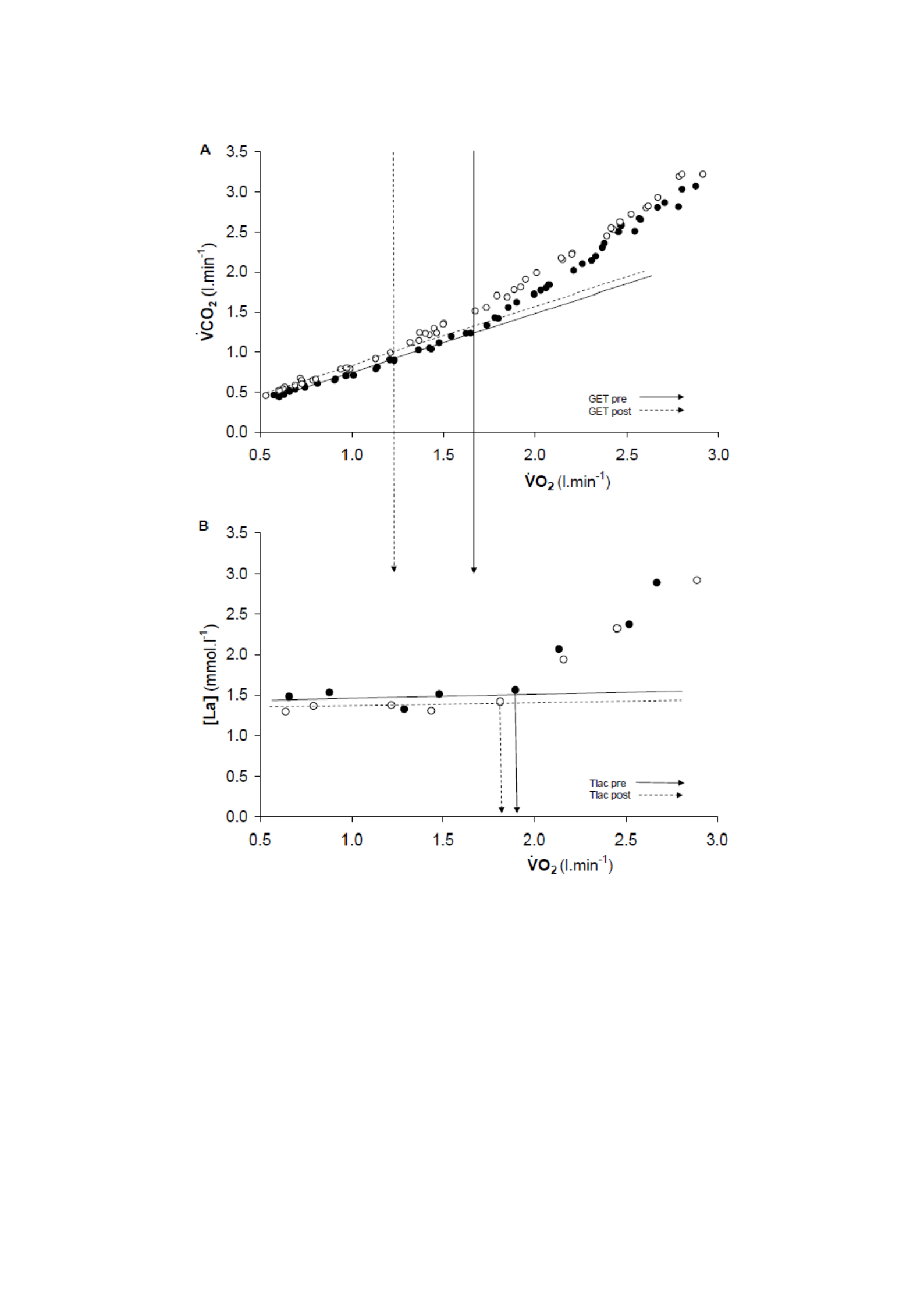
162
Figure 5.2 Representative responses
from
V CO2 vs V−O2
(panel A) and blood [lactate]
(panel B) indicates the region of interest, pre (●) and post-48 hours (○) eccentric exercises.
The most suitable slope of S1 and the vertical arrow showing the gas exchange threshold

163
(GET, panel A) and the lactate threshold (Tlac, panel B) illustrate the change in GET but
not the Tlac response, the eccentric exercises before (solid line) and post (dotted line).
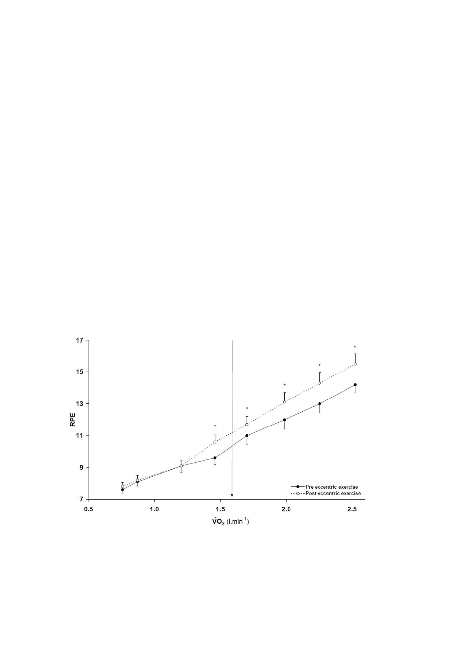
164
2
Figure 5.3 shows the RPE response before and after eccentric exercise. Tlac occurs in
the same working rate (before, 161 ± 19 W; post, 158 ± 22 W, P> 0.05) and V O2 (before, 1.90 ±
0.20
l.min-1; tiang, 1,88 ± 0,15 l.min-1, P > 0.05) after eccentric training. The V O in GET is
significantly correlated with V O2 on Tlac before (r = 0.78, P < 0.05) but not after eccentricity
exercise (r = 0.50, P > 0.05). There was no significant correlation between the change in
GET and Tlac values before and after eccentric exercise (r = 0.53, P > 0.05).

165
Figure 5.3 Changes in the perceived exertion rating (RPE) as a function of V O
2 ,
eccentric exercises before (●) and 48 hours after (○) respectively. The vertical arrows
indicate a pre-eccentric exercise of the gas exchange threshold (GET). There is an
increase of 7% (P < 0.05) in RPE
48 hours post-eccentric exercise at V O2 value from GET pre-eccentric exercise.
* Significantly different from the pre-eccentric exercise value (P < 0.05)

166
However, the RPE increased significantly at V O2 according to pre-eccentricity
GET exercises. V
O2 does not change by the breakdown protocol and, as anticipated, increases
as a linear function of the working rate before and 48 hours after eccentric exercise. Comparison
response before and 48 hours after eccentric exercise at V O2 values where pre-eccentric
GET exercises occur revealing some significant differences (Figure 5.4). Special
ada peningkatan V
E (t(9) = -2,88, P < 0,05), V CO2 (t(9) = -3,31, P < 0,05), RER (t(9)
= -2,92, P < 0 ,05), V
E / V O2 (t (9) = -2,63, P < 0 ,05), fR (t (9) = -2,49, P < 0 ,05), dan RPE
(t(9) = -2.40, P < 0.05). There is no difference in V E / V CO2, VT or [La] (P > 0.05).
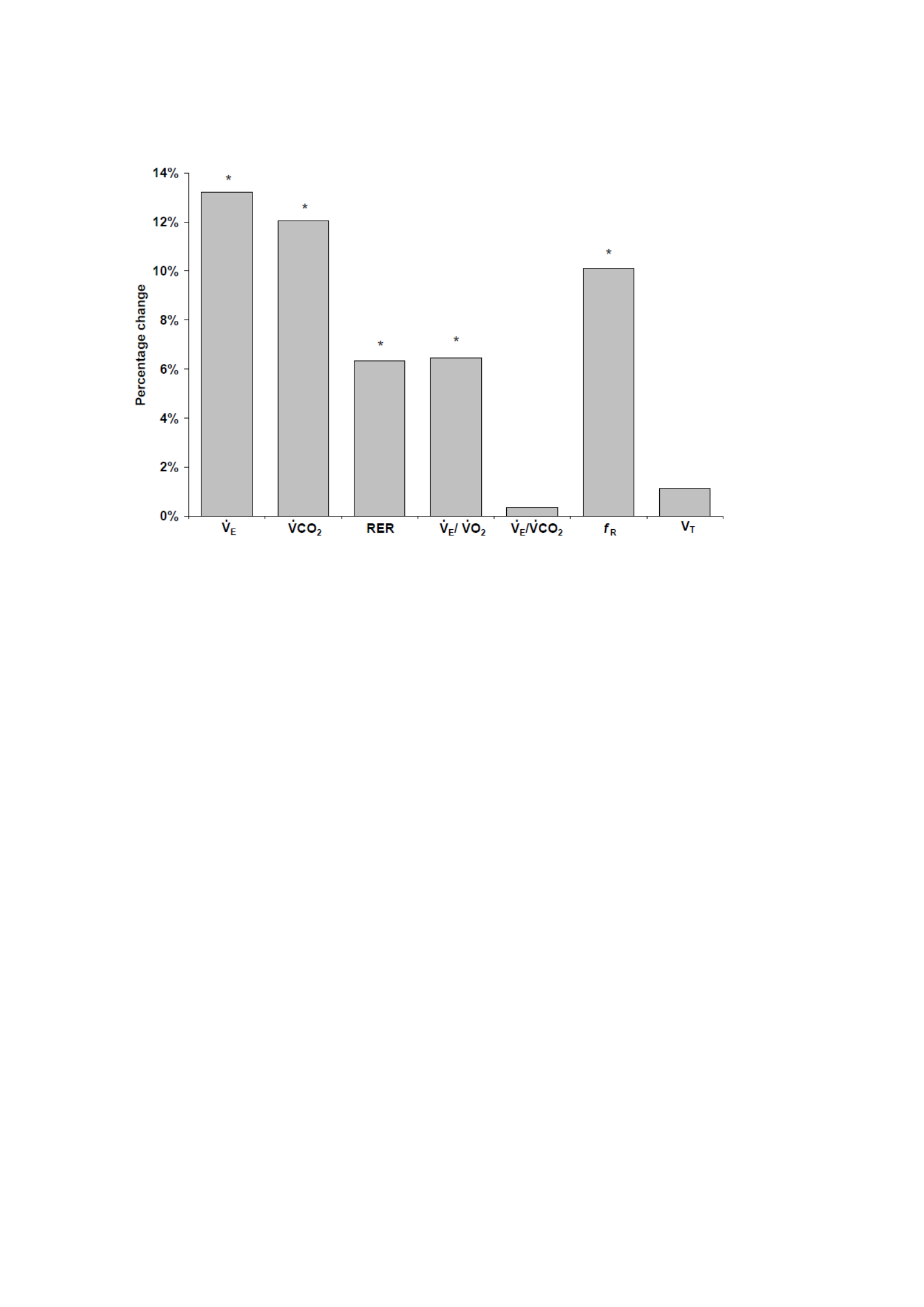
167
Figure 5.4 Percentage change in minute ventilation ( V
E ), CO2 expiration ( V
CO2 ), respiratory
exchange ratio (RER), ventilation equivalent to O2 ( V
E / V
O2 ), ventilation equivalent to CO2 (
V
E / V
CO2 ), respiratory frequency (fR) and tidal volume (VT) at V
O2 GET pre-eccentric exercise
values. The grade is average (± SD).
* Significantly different from the pre-eccentric exercise value (P < 0.05)

168
5.5 Discussion
The main original finding of this investigation is that it is eccentric and damaging to the
muscles
The drill separates V E and the gas exchange response for incremental ramp training from the
blood lactate response. While we believe these findings are new with respect to exercise-
induced muscle damage (EIMD), previous studies have shown that GET and lactate
threshold (Tlac) can be separated by certain experimental protocols including exercise
(Poole & Gaesser, 1985) and glycogen depletion (Hughes et al., 1982). Thus our findings
provide evidence to suggest that after eccentric exercise, pulmonary ventilation control is
affected by altered or additional stimuli, possibly of neurogenic origin.
As anticipated, the [La] response to incremental ramp cycle training was not altered by the
EIMD intervention. These findings support the argument that increased blood [La] is not
an inevitable consequence of EIMD and, therefore, is unlikely to be causal to
The added V E response
is observed here and elsewhere (Twist & Eston, 2009) (Chapter 4).
An example of increased blood [La] after eccentric exercise has been associated with a
suspected switch to more glycolytic energy production possibly as a result of increased
reliance on type II motor unit recruitment (Braun & Dutto, 2003; Chen et al., 2007b;
Gleeson et al., 1995, 1998). However, it has been shown that EIMD does not interfere with

169
the oxidative function of skeletal muscles (Walsh et al., 2001) or alter the lungs
Kinetika V O2 (Schneider et al., 2007).
Our findings contrast with those reported in the only previous study to investigate the response
to gradual exercise after eccentric exercise (Gleeson et al., 1998). Gleeson

170
and co-authors (1998) reported no changes in endurance time, V
O2, V
E , or RER response
but found [La] higher 48 hours after completing the bench-stepping intervention that
triggered only mild pain. Differences in damage interventions, incremental protocols used
and levels of induced muscle pain may cause differences
in V
E and [La] responses between investigations.
There was a small and statistically insignificant difference between the working rate of
the pre-eccentric exercise for the lactate threshold and gas exchange, with the gas
exchange threshold occurring slightly earlier. It must be acknowledged that the
dynamics of blood lactate production and clearance (and therefore the appearance of
lactate in the blood) may differ from more direct stimuli for changes in gas exchange
and ventilation during incremental ramp training. In addition, gas exchange was
measured breath-by-breath continuously whereas blood samples for determination
samples [La] were collected at discrete 1-minute intervals. This can increase the
potential for disagreement between the lactate and gas exchange thresholds.
The causal relationship between Tlac (causation) and GET (causation) is fundamental to
the traditional "anaerobic threshold" hypothesis (Beaver et al., 1986; Wasserman et al.,
1973, 1990). However, certain experimental conditions have been shown to give rise to
dissociation of responses [La] and V
E . In conditions of acute hypoxia (Ozcelik &

171
Kelestimur, 2004) and after previous volitional hyperventilation (Ozcelik et al., 1999)
GET was reduced compared to normal, control conditions, without concomitant decrease
in Tlac. It has been proposed that the 'pseudo-threshold' (Whipp, 1987) be resurrected
by 'washing' CO2 to depleted body stores (hyperventilation) or enhanced carotis

172
the body's sensitivity to CO2 (hypoxia). However, the reduction in GET observed in this
study does not appear to result from humoral stimuli arising from changes
blood [La]. Previous investigations reported dissociating the responses of V
E and [La] to
practice has shown that V
E may have been affected by differences or changes in
levels of neurogenic control. Patients suffering from McArdle's disease, a condition in
which a lack of muscle phosphorylase blocks the increase [La] during exercise,
showed the dissociation of V
E and [La] responses during the incremental cycle (Hagberg
et al., 1982; Paterson et al., 1990). Similarly, in conditions of reduced muscle glycogen
content (Hughes et al., 1982; Sabapathy et al., 2006) and after a period of training
training (Poole & Gaesser, 1985) V
E and [La] responses to sport are separated.
However, there is a causal relationship between the lactic acidosis bicarbonate buffer and
An increase in V
E was recently supported in a study that used DCA to reduce [La]
during gradual training (Wilkerson et al., 2009). While lactic acidosis can provide
The stimulus is important for V
E it is just one of the many factors that can affect the V
E response
and therefore modulates GET (Dempsey et al., 2006; Bangsal, 2000; Whipp et al., 1981).
It is worth mentioning here that while under normal control conditions it is considered that
the increase in non-metabolic CO2 production of the lactic acid bicarbonate buffer

173
stimulating an increase in V
E (Wasserman et al., 1973, 1990), in this study
Relative hyperventilation after eccentric exercise may have increased V CO2 by
'blowing' CO2 from the body's stores (Ozcelik et al., 1999) thereby reducing the GET as
determined using the V tilt method (Beaver et al., 1986). So we speculate that
The amplified V
E response and reduced GET observed here are likely the result of non-humoral
stimuli that come from damaged muscles and exercise.

174
The eccentric exercise protocol used in this study is known to be effective in inducing
damage (Byrne & Eston, 2002a, 2002b) (Chapter 4). The main events that cause muscle
damage from unusual eccentric exercises involve disorders of the sarcomere and myocyte
membranes that cause dysfunction of the excitation-contraction (EC) mechanism (Proske
& Morgan, 2001). As a consequence, the immediate and prolonged peak torque reduction
observed in this investigation may reflect a malfunction in the EC mechanism. In addition,
a significant increase in plasma CK activity is an indication of increased membrane
permeability. While EIMD does not appear to interfere with intrafusal fibers as indicated
by the unaltered sensitivity of muscle spindles or Golgi tendon afferents (Gregory et al.,
2002, 2004), disturbances in extrafusal fibers may provide mechanical stimulation of fine
myelinated (group III) and nonmyelinated (group IV) afferents (Avela et al., 1999; Komi,
2000; Taguchi et al., 2005). The afferent of this thin fiber
can exert a significant influence on V
E during dynamic training (Matieka & Duffin, 1995)
and may have provided an important additional impetus to V E leading to a reduction in
GET was observed in this study. Similarly, structural and functional impairment of the local
microvascular after eccentric exercise (Kano et al., 2005) has been implicated in the
augmenting the V
E response through stimulation of groups III and IV afferents (Haouzi et al.,
We have previously used an eccentric exercise protocol identical to the one used here to
induce impairment and have observed disturbances in matching O2 delivery (Q2) with O2
utilization , consistent with such microvascular dysfunction, in

175
conjunction with the amplified V
E response (Chapter 4).

176
Muscle pain is understood to provide an important stimulus to the ventilation response
through mechanical stimulation of nociceptive muscle afferent (Duranti et al., 1991). This
effervescent excitation through painful electrical muscle stimulation or ischemic muscle
pain gives rise to an increase in reflexes in respiratory frequency (fR) and minute
ventilation (Duranti et al., 1991) while simultaneously suppressing the central nervous
impulse to ventilation (Waldrop et al., 1982). Due to the afferent mechanical sensitivity of
group III and IV thin fiber muscles coupled with experimental muscle pain as described
above, it can be enhanced by mechanical hyperalgesia, muscle pain sensation and
movement-induced pain, resulting from unusual eccentric exercises. Such is the pain
experienced by
participants during muscle activation may have contributed to enhanced fR and
V E
Observed after eccentric exercise through afferent nerve reflexes.
The increased perceived exertion rating (RPE) reported by our participants corroborates
previously reported findings during constant load cycle exercise after eccentric exercise
(Twist & Eston, 2009) (Chapter 4) and may explain the reduction in time to fatigue. This
decrease and the associated decrease in WRpeak values are consistent with the observation
of impaired endurance performance caused by the effects of eccentric exercise (Marcora
& Bosio, 2007; Twist & Eston, 2009) (Chapter 4). Submaximum
differences in V
E , V CO2 , RER and RPE during ramp training completed to the maximum

177
exercising even at a lower level of work and training time. These observations support the
proposition that the rating of the perceived exertion scale with the duration of the exercise
(Crewe et al., 2008; Eston et al., 2007; Faulkner et al., 2008; Joseph et al., 2008) and also
that
there is a relationship between V
E and RPE responses to cycling after eccentric exercise (Chapter
4).

178
5.6 Conclusion
In conclusion, eccentric exercise attacks that damage the previous muscles add to
the
V
E
response to the ramp incremental cycle leading to a reduction in GET in the absence of
altered blood [La]. The resulting dissociation between GET and Tlac shows that the two
phenomena are not causally related to the existence of EIMD. We propose that the
in V
E and a reduction in GET 48 hours after muscle-damaging eccentric exercise is aroused
most likely with increased activation of group III and IV afferents stimulated through
mechanical disruption of local muscle fibers and microvessels and not as a result of blood
alterations [La].

179
ARTICLE 6
31P-MRS METABOLIC RESPONSE TO GRADUAL EXERCISE AFTER ECCENTRIC,
EXERCISES THAT DAMAGE MUSCLES

180
The contents of this chapter are currently under review for publication:
Davies RC, Eston RG, Fulford J, Rowlands AV and Jones AM. Muscle damage alters
metabolic responses to dynamic exercise in humans: a 31P-MRS study. It is currently under
review.

181
6.1 Abstract
The study used 31P magnetic resonance spectroscopy (31P-MRS) in an attempt to reveal the
relationship between changes in muscle metabolism and exercise tolerance limitations in
humans with and without exercise-induced muscle damage (EIMD). Ten healthy,
physically active men performed gradual knee extensor exercises inside the full-body MRS
hole before (before) and 48 hours after (after) performing 100 squats with a load
corresponding to 70% body mass. The time to fatigue was significantly reduced after
EIMD (519 ± 56 and 459 ± 63 seconds, before and after EIMD, respectively). End pH
training (pre: 6.75 ± 0.04 post: 6.83 ± 0.04) and [PCr] (pre: 7.2 ±1.7 post: 14.5 ± 2.1 mM)
higher value after EIMD (both P <.05). However, the final practice [Pi] was not
significantly different after the EIMD (pre: 19.7 ± 1.9 post: 21.1 ± 2.6 mM, P > 0.05). The
resting value [Pi] (pre: 4.7 ± 0.8 post: 6.7 ± 1.7 mM) and consequently the value of
[Pi]:[PCr] (pre: 0.12 ± 0.02 post: 0.18 ± 0.05) increased significantly after EIMD and this
mean difference was maintained during incremental exercise (all P <.05). In contrast, the
[PCr] and pH values did not differ before and after EIMD at rest or during gradual exercise
(all P >.05). The findings suggest that changes in phosphate metabolism, specifically the
increased resting [Pi] maintained during exercise, may contribute to the decrease in
exercise tolerance experienced with EIMD.

182
6.2 Introduction
Unusual eccentric refraction exercises result in substantial changes in skeletal muscle
structure and function. Morphological changes include cytoskeleton disorders,
sarcolemma and T-tubules (Fridén and Lieber, 2001), and changes in capillary geometry
(Kano et al., 2004). Loss of sarcolemmal integrity results in the release of intramyocyte
proteins into the bloodstream (Hortobágyi & Plan, 1989) and the entry of extracellular
Ca2+ into the sarcoplasm (Armstrong, 1984). These changes are associated with acute
inflammatory response and delayed-onset muscle pain (DOMS) (MacIntyre et al., 1996)
and are collectively referred to as exercise-induced muscle damage (EIMD).
Performance degradation associated with EIMD includes a reduction in maximum force
generation capacity (Clarkson et al., 1992) and a shorter time to fatigue (Asp et al., 1998;
Carmichael et al., 2005, 2006). Carmichael et al. (2005, 2006) have reported a decrease in
treadmill running time to fatigue in rats after downhill running attacks. Decreased
performance is associated with an increase in the inflammatory cytokine, interleukin 1 β
(IL-1β) within the brain regions responsible for movement, motivation, perception of effort
and pain (Carmichael et al., 2005). Central fatigue factors such as increased production of
inflammatory cytokines have also been implicated in the reduction of time trials (Marcora
and Bosio, 2007) and cycling performance in humans (Twist and Eston, 2009,) where the
perception of increased exertion seems to mediate performance after eccentric exercise.
Peripheral fatigue factors derived from damaged muscle tissue can also be involved in
reduced fatigue time after muscle-damaging eccentric exercises. Using a muscle biopsy

183
procedure, Asp et al. (1998) reported

184
A decrease in muscle glycogen content in human subjects after muscle-damaging exercises
was associated with a 23% decrease in maximum working capacity during additional knee
extensor exercises. Other changes observed in metabolic function after eccentric exercise
include impaired muscle glycogen resinsynthesis (Asp et al., 1995, 1998) and increased
blood lactate response during exercise (Gleeson et al., 1995, 1998; Asp et al., 1998). It is
possible that this change reflects a shift in the metabolic profile of muscles to an increased
dependence on non-oxidative metabolism, which contributes to a decrease in endurance
capacity after EIMD (Asp et al., 1998). However, the exact nature of the accelerated
fatigue progression experienced with EIMD is poorly understood.
Non-invasive evaluation of muscle metabolism can be achieved by using 31P magnetic resonance
spectroscopy (31P-MRS). Several studies have used this technology to show an increase in the ratio
of inorganic phosphate (Pi) to phosphocreatin (PCr) at rest after EIMD, showing an
increase in resting muscle metabolism (McCully 1992, Rodenburg et al., 1995; Lund et
al., 1998a, 1998b). Several candidate mechanisms for accelerated fatigue progression and
reduction in peak power observed after eccentric exercise, including increased Pi and ADP
and decreased pH, can be measured using the 31P-MRS. Rodenburg et al. (1995) reported no
difference in Pi:PCr, pH or peak power ratio during multi-level knee extensor exercises
performed 24 hours after an attack of stepping exercises designed to induce EIMD.
However, the authors conclude that the lack of changes in some markers of muscle damage
suggests that the muscle damage protocols used are not severe enough to alter muscle
metabolism (Rodenburg et al., 1995).

185
It has been shown that muscle-damaging protocols effectively reduce fatigue time during
high-intensity cycling (Chapter 4). However, the mechanism underlying the decrease in
endurance capacity after muscle-damaging exercise remains to be determined. Central
mechanisms that have the potential to influence increased fatigue development, such as
increased production of inflammatory cytokines, are difficult to evaluate during dynamic
exercise in human models. Suspected peripheral fatigue mechanisms including increased
depletion rate [PCr], increased accumulation [Pi] and [ADP] and decreased pH decline rate
can all be assessed using 31P-MRS. After the resistance training period, the tolerance of additional knee extensor training
is enhanced by the relationship of [Pi]: [PCr], [PCr] and pH to the time indicating a shift to the right (Jones et
al., 2007). It is possible that after a muscle-damaging exercise attack, the observed reduced
exercise tolerance may be attributed to a shift to the left in the time profile of this 31P metabolite.
Alternatively, an unchanged muscle metabolite response would suggest that another
potentially central mechanism may be responsible for the decrease in endurance capacity.
Therefore, the purpose of this study was to investigate the effects of well-defined muscle-
damaging eccentric exercises (Chapter 3) on changes in muscle metabolism during
dynamic incremental knee extensor exercises. We used the 31P-MRS to test the hypothesis that exercise-
induced muscle damage (EIMD) alters the metabolic response of muscles to dynamic exercise and thus limits exercise tolerance in
humans.

186
6.3 Method
Subject
Ten healthy, physically active male subjects (age, 22 ± 4 years; mass, 78.2 ± 8.8 kg; height,
1.79 ± 0.08 m) voluntarily participated in the study. All participants showed no symptoms
of pre-existing diseases and injuries and did not do lower limb resistance training in the
previous six months. Participants give written consent to participate in research approved
by the Institutional Ethics Committee (See appendices B, C and D for sample participant
information sheets, participant consent forms and ethical consent certificates).
Assessment of muscle damage
All indicators of muscle damage, perceived muscle pain (using a visual analogue scale of
0-10 (VAS)), creatine kinase activity (CK) and isokinetic peak torque (30 degrees s-1),
were measured in the listed order, immediately before, and 24 and 48 hours after
performing the muscle-damaging eccentric exercise protocol. For more details on this
procedure, please refer to Chapter 3.
Experimental procedure
After the assessment of muscle damage, the single-legged knee extension exercise test was

187
completed at the same time of day ± 1 hour for each participant, before and 48 hours after
performing muscle-damaging eccentric exercises. The dynamic knee extensor exercise test
is performed in a prone position with the subject positioned inside the full-body MRI
system. A 6 cm 31P transmission-receive surface coil is placed inside the base of the subject, and the subject is asked
to lie on it in such a way that the coil is in the center of the quadriceps

188
leg muscles to be trained. The subject was then secured to an ergometer bed with Velcro
straps on the thighs, buttocks, and lower back to minimize foreign movements during the
protocol. The leg of the leg to be trained is connected to a pulley system that allows
nonmagnetic loads to be lifted and lowered and the working rate calculated (Figure 6.1).
The exercise was performed at a rate of 40 repetitions/minute with the subject lifting and
lowering the mass at a distance of ~0.22 m according to the visual cues projected onto the
front wall of the scanner room. The contraction phase of the knee extensor and the
interrogation of the quadriceps 31P-MRS occurred simultaneously. After a 2-minute rest period, the
subject begins knee extension exercises with an initial basket weight of 1 kg. After that,
the basket load is increased by 0.5 kg every 30 seconds until the subject can no longer
maintain the kick frequency at 40 repetitions/minute. The subject receives a strong verbal
urge to continue for as long as possible while maintaining the appropriate shape.
Eccentric exercise protocols that damage muscles
Participants completed 100 squats (Smith), performed as 10 sets of 10 repetitions with
weights on the bars corresponding to ~70% of each participant's body mass. Before
starting, all participants are instructed in correct and safe lifting techniques. The bar is
positioned on the participant's shoulders and the legs are positioned under the bar, with the
back straight and the legs fully extended (knees = 180°). The descent phase involves the
eccentric action of the knee extensor to lower the bar to the angle of the knee just past 90°.

189
The
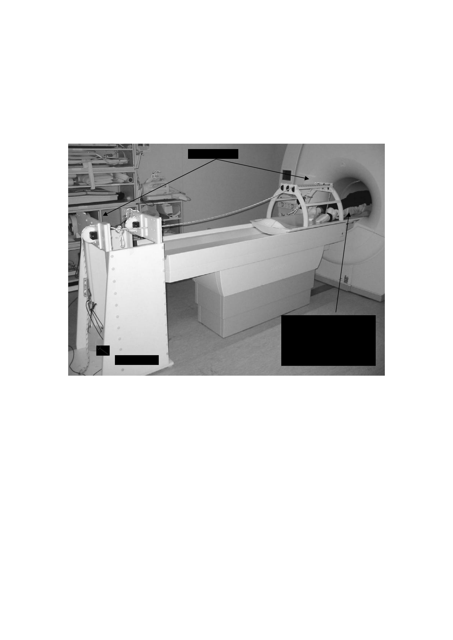
190
The lifting phase involves concentric action to return the bar to its starting position. For
more details on this procedure, please refer to Chapter 3.
Figure 6.1 The MRS knee extensor ergometer shows the pulley and load basket system.
Pulley system
Load basket
The subject lies in a prone
position inside the core of
the full-body scanner. The
right leg is attached to the
pulley system.

191
Measurement
MRS Measurement
The MRS was carried out at the Peninsula Magnetic Resonance Research Center using a
1.5-T superconducting magnetic resonance scanner (Philips Gyroscan Clinical Intera,
Philips Medical Systems, Best, Netherlands). Initially, a quick field echo image was
obtained to

192
Determine if the muscles are positioned correctly relative to the coil. This is aided by
placing a capsule of cod oil, which generates a high-intensity signal point inside the image,
adjacent to the coil, allowing its orientation relative to the volume of muscle being
examined to be assessed. A number of pre-acquisition steps are performed to optimize the
signals from the muscle under investigation. Coil tuning and matching is then performed,
followed by an automated shimming protocol performed in volumes that define the
quadriceps muscles. The volume of the muscle from which the signal comes is determined
by the sensitive volume of the coil itself. It is close to the physical size of the coil and thus
corresponds to a cylinder of 6cm diameter and 6cm depth adjacent to the coil. Beyond this
volume, the muscles will still contribute to the signal but to a limit that drops rapidly as
you move away from the sensitive area. To ensure that the muscles examined are
consistently at the same point relative to the coil during the exercise, the subject visually
queues through a screen made up of two vertical rods, one that moves at a constant speed
with a frequency of 0,67 Hz and another that monitors the movement of the legs through
sensors present inside the connected pulley. Thus the subject seeks to match the
movements of these two bars. The work performed by the subject is recorded through a
nonmagnetic strain gauge built into the pulley mechanism. Before training, during training,
and during recovery, data was obtained every 1.5 seconds, with a spectral width of 1,500
Hz and 1,000 data points. A phase cycle with eight phase cycles is used, which causes the
spectrum to be acquired every 12 seconds. The next spectrum was measured through peak
pairing, assuming prior knowledge, using the jMRUI software package (version 2) and the
AMARES pairing algorithm (Vanhamme et al., 1997; Naressi et al., 2001).

193
The spectrum was fitted assuming the presence of the following peaks: Pi, phosphodiester,
phosphocreatin (PCr), α-ATP (2 peaks, amplitude ratio 1:1), γ-ATP (2 peaks, amplitude
ratio 1:1), and β-ATP (3 peaks, amplitude ratio 1:2:1). In all cases, the relative amplitude
was corrected for partial saturation due to the short repetition time relative to the
longitudinal relaxation time constant of T1. The absolute concentration was determined by
calculating the size of the peak area relative to β-ATP set at 8.2 mM. The ratio of Pi to PCr
is determined from the spectral area of Pi and PCr respectively obtained during the
quantitative procedure. Intracellular pH is calculated from the chemical shift of the peak
spectral Pi relative to PCr (Moon and Richards, 1973). The concentration of ADP was
calculated as described by Kemp et al., (2001) by considering the pH dependence of H+,
K+ and Mg2+ binding. To determine the intracellular (TI) threshold during incremental
exercise, piecemeal linear regression is used. Briefly, the combination of two different
rows is adjusted to the relationship of the working rate [Pi]/[PCr] and the working rate of
pH until the lowest amount of the quadratic residue is found (Hogan et al., 1983; Marsh et
al., 1991). The point at which the combination of these two special lines intersects is
accepted as TI.
Statistical analysis
Changes in muscle damage indicators (perceived muscle pain, creatine kinase activity and
isokinetic peak torque) were analyzed using a series of one-way repeating measurements
of ANOVA. Since CK activity data is not normally distributed, these values are changed

194
log prior to statistical analysis (Twist & Eston, 2005) (see appendix H).
The paired t-test was used to determine the significant difference in final exercise values
between the two conditions. Where the data is not distributed normally, the Wilcoxon test
is also run. Changes in MRS measurements (Pi, PCr, Pi:PCr and pH ratio) at rest and

195
after 2, 4 and 6 minutes of additional exercise are examined using ANOVA separate two-
way measurements (condition x time). The assumption of roundness is evaluated using the
Mauchly test. Where roundness is violated (P < 0.05), the Greenhouse-Geisser correction
factor (GG) is applied. All data were analyzed using the SPSS statistics software package
for Windows (version 13) with statistical significance set at 0.05. Grades are meant ±
elementary.
6.4 Result
Muscle damage
There were significant changes in all indicators of muscle damage after eccentric exercises.
Table 6.1 shows changes in muscle pain, CK activity and isokinetic peak torque before
and after eccentric exercise. Muscle pain increased 24 hours after eccentric exercise with
the highest score reported at 48 hours (F (2, 18) = 26.22, P < 0.05). Plasma CK activity
increased after eccentric exercise, with the highest value observed at 24 hours (FGG (1,1,
9,9) = 15.02, P < 0.05). Isokinetic peak torque (30 deg.s-1) decreased by 15% at 24 hours
and
remained 11% lower than the baseline value at 48 hours (F (2, 18) = 14.33, P < 0.05).

196
Table 6.1 Changes in indicators of muscle damage. Elementary school grades ±
average and (range) before and 24 hours and 48 hours after eccentric exercise.
Measurable Variables before 24 hours 48 hours
Nyeri (0-10 VAS)
0.5 ±
0.3
(0.1 –
1.1)
5.0 ±
2.0*
(1.8 –
8.2)
5.5 ± 3.2*
(1.6 – 9.7)
CK Activity (U/L) 178 ±
61
(97 – 270)
798 ±
692*
(196 –
2481)
409 ± 264*
(213 – 1088)
Peak Torque
(Nm)
30
degrees-
1
310 ±
47
(227 – 366)

197
265 ± 66*
(113 – 340)
275 ± 77*
(115 - 361)
Pain, visual analogue scale (VAS) 0–10. CK, creatine kinase.
* Significantly different from the pre-eccentric exercise value (P < 0.05).
MRS Measurement
As anticipated, the fatigue time (519 ± 56 and 459 ± 63 seconds, before and after muscle
damage, respectively) and the associated peak work rate values (29 ± 4 and 25 ± 4 W,
before and after muscle damage, respectively) achieved during the gradual knee extensor
exercise were significantly reduced after muscle damage (t(9) = 4.85, P < 0.05 and t(9) =
5.21, P < 0.05, respectively). The final practice values are presented in Table 6.2. The
data for the final exercise [Pi]: [PCr] and ADP were not normally distributed therefore the
Wilcoxon test was used to analyze this data. However the results are no different from the
t-test results, therefore for consistency, paired t-test results are presented.

198
Table 6.2 Muscle metabolic response at rest and during gradual exercise before (Pre)
and 48 hours after (After) muscle-damaging eccentric exercise. Grades are meant ± SD.
Measurable
Variables
Rest
2 minutes
4 minutes
6 minutes
Ujung
[PCR] (mjuta) † To
38.3 ± 1.2
35.0 ± 2.1
30.7 ± 3.1
24.3 ± 4.0
7.2 ± 1.7
Post
37,9 ± 1,4
34.1 ± 2.6
31.3 ± 3.0
25.5 ± 4.8
14.5 ± 2.1*
[Pi] (mjuta) † To
4.7 ± 0.8
5.6 ± 1.1
8.0 ±1.9
10.6 ± 3.0
19.7 ± 1.9
Posting ‡
6.7 ± 1.7
7.5 ± 2.1
10.4 ± 4.0
14.8 ± 6.5
21.1 ± 2.6
Rasio [Pi]:[PCr]† To
0,12 ±
0,02
0,16 ±
0,03
0,26 ±
0,05
0,45 ±
0,13
2.09 (1.74-14.4) ††
Posting ‡
0,18 ±
0,05
0,22 ±
0,07
0,34 ±
0,15
0,62 ±
0,34
1,46 (0,85-2,29)
††*
Ph
†
To
7.02 ±
0.03
7,06 ±
0,04
7,03 ±
0,04
6,96 ±
0,06
6,75 ± 0,04
Post
7.02 ±
0.03
7,07 ±
0,03
7,04 ±
0,05
6,99 ±
0,09
6,83 ± 0,04*
† Significant primary effects for time (rest, 2, 4 and 6 minutes) (P < 0.05)
‡ Significant primary effect for conditions (Pre and Post) (P < 0.05)

199
* Significantly different from Pre-eccentric exercise (P < 0.05)
†† The data of the final training ratio [Pi]:[PCr] is not distributed normally so that the
value is presented as a median (interquartile range).
Table 6.3 Intracellular Threshold (IT) values before (Pre) and 48 hours after (Post)
muscle-damaging eccentric exercise. Grades are meant ± elementary.
Measurable Variables
To
Post
Time @ IT (s)
326 ± 77
316 ± 65
Rasio [Pi]:[PCr] @ IT
0,30 ± 0,14
0,31 ± 0,10
pH @ IT
7,07 ± 0,02
7,08 ± 0,02
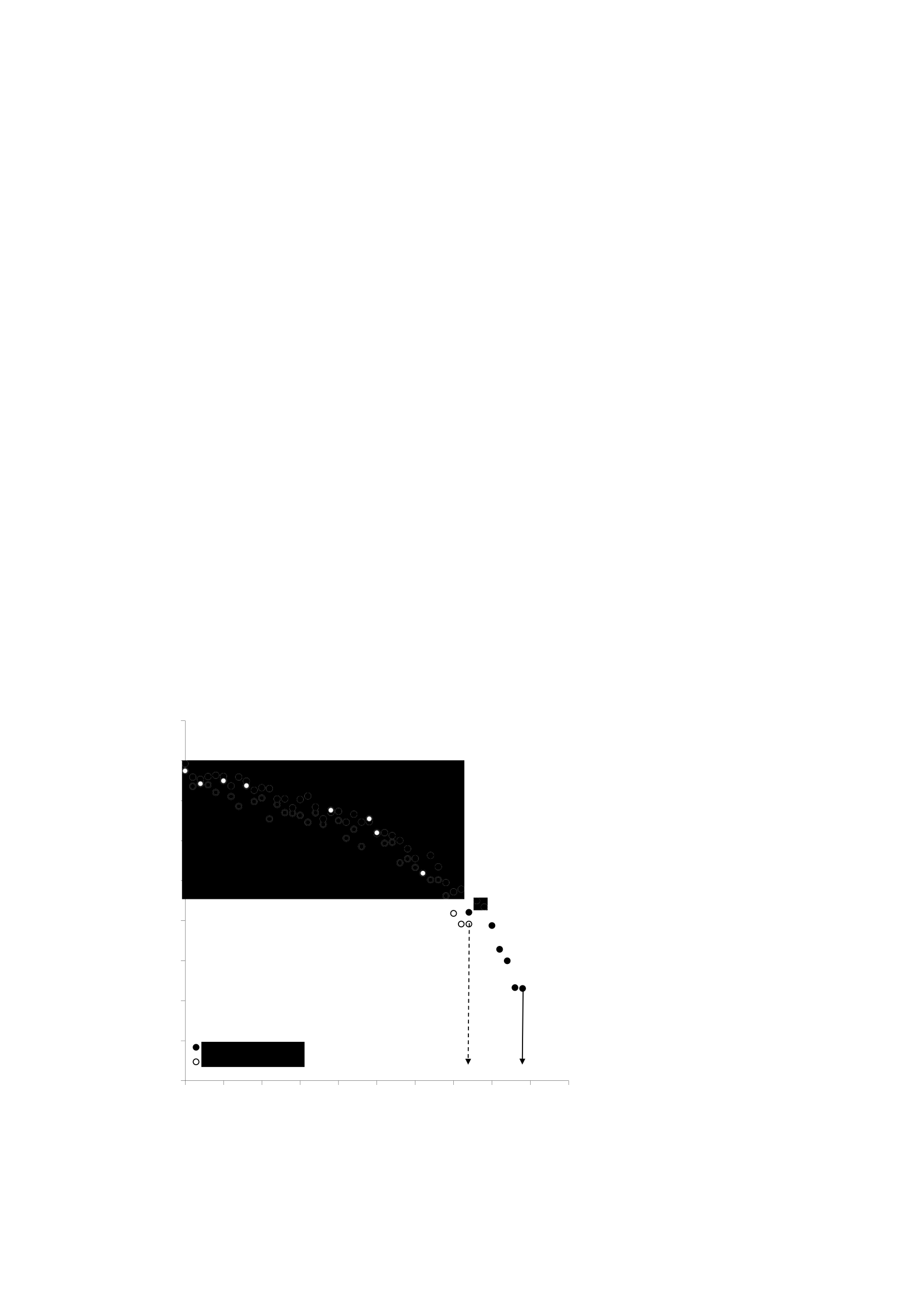
200
The pH and [PCr] values of the final exercise were higher (t(9) = -2.34, P < 0.05 and t(9) =
-4.49, P < 0.05, respectively) and the final exercise values [Pi]:[PCr] were lower (t(9) =
2.346, P < 0.05,) after eccentric exercise. However, the final exercise [Pi] and ADP did
not differ significantly after eccentric exercise (t(9) = -0.496, P > 0.05 and t(9) = 1.82, P >
0.05, respectively). TI was not significantly altered by eccentric exercises (t(9) = 0.51, P >
0.05). The time, [Pi]:[PCr] and pH values when IT occurs are presented in Table 6.3.
Before eccentric training, all ten participants completed a minimum of 7 minutes of
complete incremental exercise. However, 48 hours after eccentric training, the minimum
time achieved by all participants is reduced to 6 minutes. Thus, the first 6 minutes of
gradual exercise were analyzed to examine changes in muscle metabolic response during
exercise.
45
40
35
30
25
[PCR]
(mjuta)

201
20
15
10
5
0
0 60 120 180 240 300 360 420 480 540 600
Time
Figure 6.2 [PCr] responses of representative participants, muscle damage exercises before
and after eccentrics. The vertical arrows indicate the time of fatigue in two training
conditions. The solid line represents the time for pre-eccentric exercise fatigue; The dotted
line represents the time for eccentric post-workout fatigue.
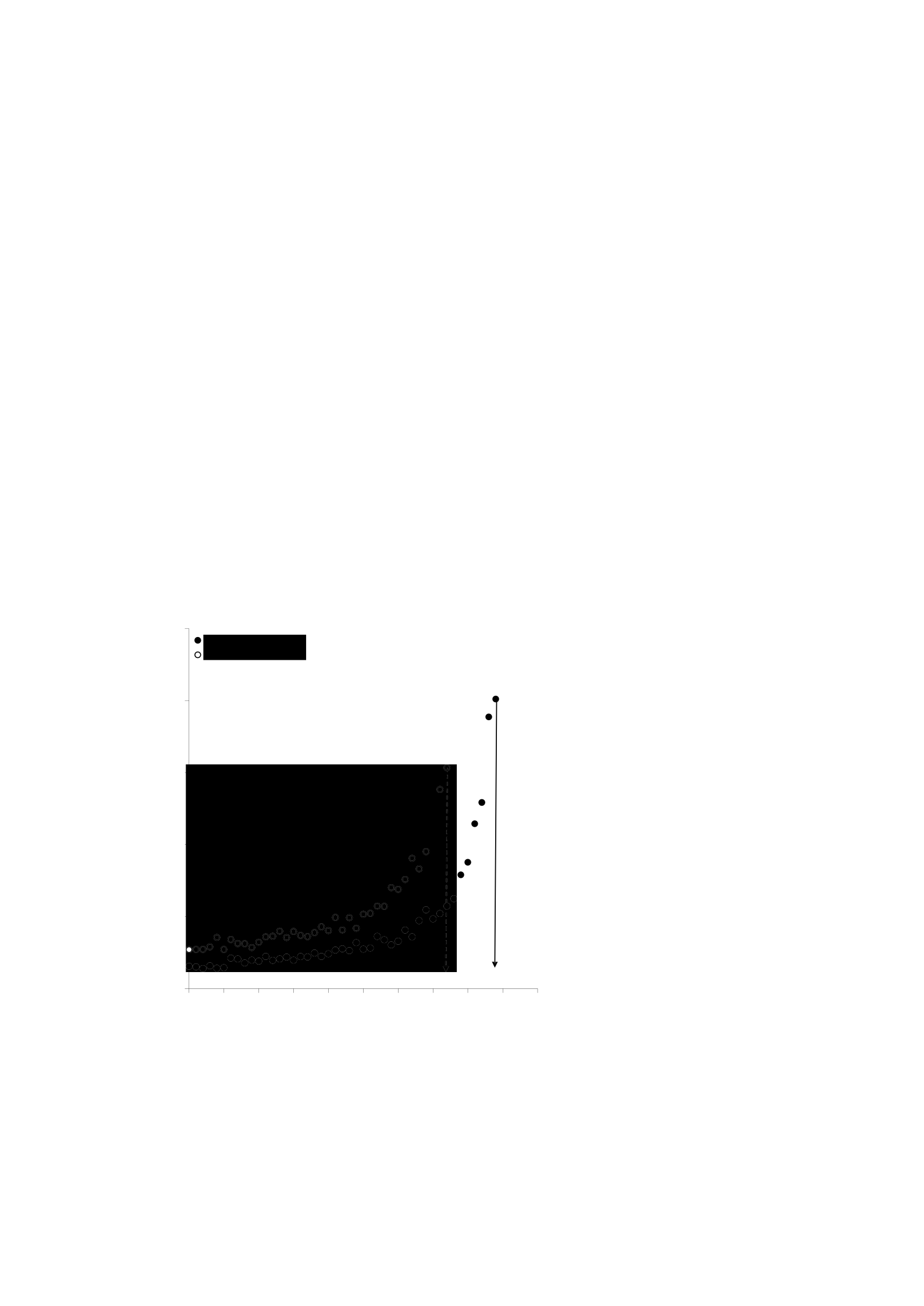
202
The metabolic response of muscles at rest and during gradual exercise is presented in the Table
6.2 (see appendix I for a graphical representation of this data). There was a significant
primary effect for time for all muscle metabolic responses, with [PCr] and pH values
decreasing, and [Pi] and [Pi]:[PCr] values increasing with the course of additional exercise
(all P < 0.05). There was a significant primary effect for the condition for [Pi] (F(1.9) =
7.080, P < 0.05). The resting value [Pi] increased significantly and this average difference
was maintained during the first 6 minutes of incremental exercise.
2.5
2
1.5
[Pi]:[PCr]

203
1
0.5
0
0 60 120 180 240 300 360 420 480 540 600
Time
Figure 6.3 [Pi]:[PCr] representative participant responses, exercises before and after
eccentric, muscle damage. The vertical arrows indicate the time of fatigue in two training
conditions. The solid line represents the time for pre-eccentric exercise fatigue; The dotted
line represents the time for eccentric post-workout fatigue.
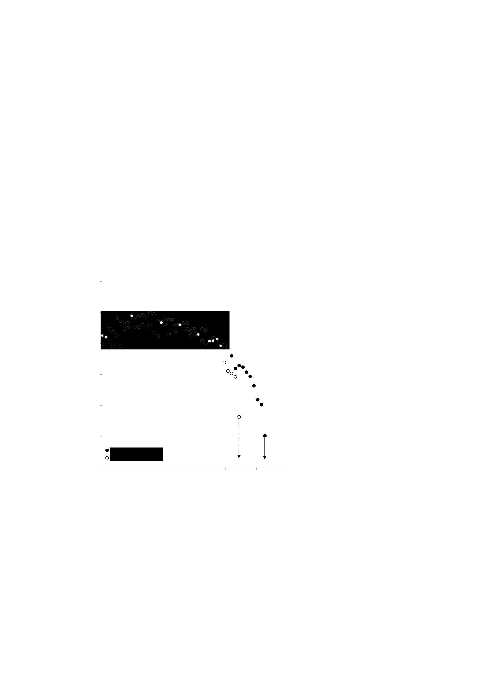
204
As a direct consequence, the value of [Pi]:[PCr] also increases at rest and during the first
6 minutes of training (F(1.9) = 5.908, P < 0.05). In contrast, the [PCr] and pH values did not
differ between the conditions at rest and during gradual exercise (both P > 0.05). There
was no significant interaction between condition and time for muscle metabolic responses.
Changes in [PCr], [Pi]:[PCr] and pH are illustrated for typical participants in Figures 6.2
– 6.4.
7.15
7.1
7.05
7
6.95
Ph

205
6.9
6.85
0 100 200 300 400 500 600
Time
Figure 6.4 pH response of representative participants, exercise before and after eccentric,
muscle damage. The vertical arrows indicate the time of fatigue in two training conditions.
The solid line represents the time for pre-eccentric exercise fatigue; The dotted line
represents the time for eccentric post-workout fatigue.

206
6.5 Discussion
To our knowledge, this is the first study to investigate changes in muscle metabolism with
31P-MRS during additional knee extensor exercises following an effective muscle-damaging protocol . The main original
finding of this investigation is that the reduction in fatigue time as a consequence of
eccentric attacks and muscle-damaging exercises is not associated with accelerated [PCr]
depletion or a faster decrease in pH. Specifically, fatigue time was reduced by 12% after
muscle-damaging exercise but temporal changes in [PCr] and pH were similar in the two
experimental conditions so that the final workout [PCr] and pH were significantly higher
48 hours after muscle-damaging exercise (Figures 6.1 and 6.3). In contrast, the final
exercise [Pi] did not differ significantly between the conditions before and after EIMD.
These results suggest that reduced exercise tolerance after EIMD may be related to an
increase in [Pi] observed at rest and during gradual exercise, or with other non-measurable
peripheral or central factors. Importantly, however, the results allow us to ignore the
greater levels of non-oxidative energy metabolism (as inferred from the changes in [PCr]
and pH) as important mediators of reduced exercise tolerance after EIMD.
Our findings provide new insights into the changes in the response of the 31P metabolite to
dynamic exercise after muscle-damaging exercise. Previous studies have reported Pi:PCr, pH
and peak power ratios that do not change during additional exercise after previous
eccentric exercise attacks (Rodenburg et al. 1995). However, these authors speculate that
the bench-stepping protocol used to induce damage is not severe enough to induce changes
in exercise metabolism. Indeed, the lack of changes in some markers of muscle damage

207
including maximum power output indicates that their exercise is eccentric

208
ineffective protocols (Rodenburg et al. 1995). Participants in the study completed muscle-
damaging eccentric exercises, which consisted of 100 squats with weights on bars set to
70% body mass (see chapter 3). This eccentric exercise protocol is known to be effective
in inducing damage (Byrne and Eston 2002a, 2002b). Indeed, in this study, there were
changes in all the symptoms of muscle damage measured. Disturbances of the sarcomere
and myocyte membrane during unusual eccentric exercises lead to a direct and prolonged
reduction in peak torque due to dysfunction of the excitation-contraction (EC) mechanism
(Proske and Morgan 2001). The 11% reduction in isokinetic peak torque observed at 48
hours is consistent with previous studies that have used this eccentric exercise protocol in
reflecting damage to the EC mechanism (Byrne and Eston 2002a, 2002b). In addition, a
significant increase in plasma CK activity is an indication of increased membrane
permeability. Impaired maximum force production can, in part, explain the reduction in
time to fatigue and the associated 14% reduction in peak work rate values. The accelerated
level of fatigue experienced by our participants was consistent with the observation of
impaired endurance performance caused by the effects of eccentric exercise (Asp et al.
1998; Marcora and Bosio 2007; Twist and Eston 2009).
Peripheral fatigue factors originating within damaged muscle tissue have been implicated
in the reduction of endurance capacity associated with muscle damage caused by exercise.
In particular, reports of increased blood lactate (Asp et al., 1998; Braun & Dutto, 2003;
Chen et al., 2007) and increased utilization of glycogen stores (Asp et al., 1998) after
eccentric exercise have been associated with an alleged shift towards increased
dependence on anaerobic energy production leading to impaired endurance performance

209
(Asp et al., 2007).

210
et al., 1998; Braun & Dutto, 2003; Chen et al., 2007). However, in this study, the PCr
response for a specific level of work did not change significantly, although the
performance of the knee extensor exercise was gradually impaired. It is recognized that
the PCr muscle and
V O2 exhibits a similar kinetic profile during the transition to higher training intensity
(Barstow et al., 1994; Mahler, 1985; Marsh et al., 1993), showed that the rate of oxidative
phosphorylation is closely related to the hydrolysis of PCr (Mahler, 1985, Meyer, 1988).
Thus our observations of the unchanged PCr profile during incremental exercise were
consistent
with a report of unchanged V
O2 kinetics after eccentric exercise (Schneider et al.,
2007). Collectively these data show that exercise-induced muscle damage does not
compromise oxidative function during dynamic exercise. The reduction in fatigue time
resulted in a significant change in the final exercise value [PCr]. Prior to muscle-damaging
exercises, the average thinning of the PCr pool was more than 80% at the end of the
supplemental exercise, with some subjects almost completely depleting their muscle PCr.
But 48 hours after the induced muscle damage, participants achieved fatigue faster with an
average thinning of the PCr pool of about 62%. Thus the final exercise [PCr] is higher
when the gradual exercise is performed in the condition of the damaged muscles.
Similarly, intracellular pH shows a drop rate that does not change during gradual exercise
after muscle damage is induced. There was no significant difference in the pH value for a

211
given work rate at 48 hours even though the pH of the end of the workout was higher due
to the shorter fatigue time. In addition, the unchanged [PCr] and pH responses to phased
exercise resulted in no significant difference in IT scores after eccentric exercise. [PCr]
and low pH have been involved in the fatigue process

212
(Westerblad and Allen 2003; Wilson et al. 1988). However, based on our observational
evidence, this effect does not appear to be related to the reduction of exercise tolerance
that accompanies exercise-induced muscle damage.
At rest, an increase in [Pi], resulting in a 50% increase in the ratio of [Pi]:[PCr] resting 48
hours after eccentric exercise. These findings are consistent with several previous studies
using 31P-MRS in reporting an increase in resting [Pi]:[PCr] after eccentric exercise (McCully 1992, Rodenburg et al., 1995; Lund
et al., 1998a, 1998b). Exalted rest [Pi] as observed here and elsewhere (McCully 1992, Rodenburg
et al., 1995; Lund et al., 1998a, 1998b) can be interpreted as an increase in muscle
metabolism. Repair and renovation of damaged tissue through eccentric exercises as
reported by Yu et al. (2002) and Yu and Thornell (2002) can lead to an increase in resting
muscle metabolism. In addition, the glycogen content of resting muscles depleted after
eccentric exercise, especially the content of specially-damaged type II fibers, has been
associated with increased glycogen utilization of resting muscles (Asp et al., 1998). An
alternative explanation for the increase in [Pi]:[PCr] which, importantly, results from an
increase in [Pi] but not a decrease in [PCr], can be attributed to muscle tissue damage as a
consequence of eccentric exercise. Disruption of intracellular Ca2+ after eccentric exercise
has been reported with concurrent activation of calcium-activated proteolytic pathways
(Belcastro, 1993; Belcastro et al. 1998). Thus, the observed increase in resting [Pi] may
not be due to increased muscle metabolism but due to the degradation of muscle proteins.
Increased resting [Pi] and [Pi]:[PCr] after a period of cast immobilization has been

213
implicated in the loss of muscle strength resulting from periods of unused (Pathare et al.,

214
2005, 2008). Similarly, an increase in the resting ratio [Pi]:[PCr] of patients with postpolio
residual paralysis, a condition characterized by decreased endurance capacity and muscle
weakness, was associated with the severity of paralysis (Sharma et al., 2007). Intracellular
increase [Pi] can inhibit force production through direct action on the formation of cross
bridges or at sites in the excitation-contraction pathway and may play a key role in the
development of muscle fatigue (Westerblad et al., 2002). Thus the reduction in peak torque
and exercise tolerance observed in this study after muscle damage, may be related to the
increased [Pi] and [Pi]:[PCr] observed not only at rest but also during gradual exercise.
The new observation that a significant increase in rest [Pi] and [Pi]:[PCr] at 48 hours was
maintained during the dynamic exercise test, is very interesting. The rate of increase [Pi]
during additional exercise does not change as a result of muscle damage. However, the
premature termination of testing at 48 hours resulted in no significant difference in the
final exercise [Pi] value. It may be tempting to speculate that the limits to exercise
tolerance are moderated by [Pi]. However, wide inter-subject variability is observed with
the value of [Pi], so we cannot reliably conclude that [Pi] is indeed a limiting factor. In
contrast, the [Pi] final workout is likely to make an important contribution to the reduction
of fatigue time experienced after muscle-damaging exercise.
Central fatigue factors including inflammatory cytokine production may be involved in the
reduction of fatigue time after eccentric exercise observed here. Carmichael et al. (2005)
have reported an increase in IL-1β of the brain in the areas responsible for movement,
motivation, perception of effort and pain, which has been associated with decreased
treadmill running to fatigue in rats. In human subjects, it has been proposed that the

215
increase in

216
The sense of effort reported during dynamic training mediates the performance of the time
trial after eccentric exercise (Marcora & Bosio 2007; Twist & Eston 2009) The duration
of individual additional training for 'willpower fatigue' is regulated by the complex
interaction of central and peripheral fatigue factors. However, the decision to end exercise
is ultimately conscious behavior based on the perception of changes in the unconscious
homeostatic control system (St Clair Gibson et al., 2003). It is beyond the scope of this
study to determine whether central or peripheral factors contribute more to the acceleration
of the progression of fatigue experienced with EIMD.
6.6 Conclusion
In conclusion, the results of this study suggest that reduced exercise tolerance after EIMD
cannot be associated with greater levels of non-oxidative energy metabolism (as inferred
from changes in [PCr] and pH). While we cannot rule out an important role for centrally
mediated fatigue, our results suggest that increased rest [Pi], which is maintained during
exercise, may be a contributing factor to the decrease in exercise tolerance observed after
EIMD.

217
ARTICLE 7
EFFECT OF ECCENTRIC EXERCISE-INDUCED MUSCLE DAMAGE ON
THE DYNAMICS OF MUSCLE OXYGENATION AND PULMONARY
OXYGEN UPTAKE

218
The content of this chapter forms the basis of the following publications/presentations:
Publication
Davies RC, Eston RG, Poole DC, Rowlands AV, Dimenna F, Wilkerson DP, Twist C,
and Jones AM. Effect of eccentric exercise-induced muscle damage on the dynamics of
muscle oxygenation and pulmonary oxygen uptake. J Appl Physiol (14 August 2008).
doi:10.1152/japplphysiol.90743.2008
Presentation
Davies R, Eston R, Poole D, Rowlands A, Dimenna F, Wilkerson D, Twist C, and
Jones A. Effect of exercise-induced muscle damage on pulmonary oxygen uptake and
muscle deoxygenation kinetics during high-intensity exercise. 13th Annual Congress of
the European Congress of Sport Science, Abstract Book, edited by Cabri J, Alves F,
Araujo D, Barreiros J and Veloso A, Estoril, 2008, p. 630.

219
7.1 Abstract
Unusual eccentric exercises have a major impact on muscle structure and function.
However, it is not known whether related microvascular dysfunction interferes
matching the delivery ofO2 ( Q2O2 ) with the utilization of O2 ( V2 ). Near infrared spectroscopy
(NIRS) was used to test the hypothesis that muscle damage caused by eccentric exercise
will increase the ratio of Q O2 : V O2 muscle during temporary heavy intensity training
maintains the speed of the V O2 kinetics at the beginning of the exercise. Nine men are physically active
Complete the 'step' test for heavy intensity training from the baseline unloaded on the cycle
ergometer before and 48 hours after eccentric exercise (100 squats with a weight corresponding
to the
70% of body mass). NIRS and lung breath by breath V O
2 Measured
continuously during the exercise test and then modeled using standard non-linear
regression techniques. No change in the kinetics
of phase II pulmonary V O 2
after the start of the exercise (time constant, pre: 25 ± 4; post: 24 ± 2 seconds; amplitude,
pre: 2.36 ± 0.23; post: 2.37 ± 0.23 L/min; all P>0.05). However, the primary (pre: 14 ± 3;
post: 19 ± 3 sec) and overall (pre: 16 ± 4; post: 21 ± 4 sec) mean response times of [HHb]
responses were significantly slower after eccentric exercise (P<0.05). Slower kinetics

220
[HHb]
observed after eccentric exercise consistent with an increase in
the ratio of Q O2 : V O2
during the transition to heavy-intensity training. We propose that the primary phase of the
unchanged
Kinetics V O2 is associated with an increase in the ratio of Q O2 : V O2 which conserves blood-
fluks miosit O2.

221
7.2 Introduction
Unusual eccentric exercises have a major impact on muscle structure and function. After
the exercise, myocytes exhibited ultra-structural changes including sarcomical disorders
described as 'popping' (Morgan, 1990), 'Z-band streaming' (Fridén & Lieber, 2001; Stupka
et al., 2001) or smearing (Kano et al., 2005), and damage to the t-tubules, reticulum
sarcoplasm and sarcolemma (Fridén & Lieber, 2001). This disorder leads to increased
entry of extracellular Ca 2+ into the sarcoplasm, leading to increased proteolytic enzyme
activity (Proske et al., 2004) and the accompanying inflammatory response (Fielding et al.,
1993). In addition, intramyocyte content such as creatine kinase and myoglobin are
released into the bloodstream (Warren et al., 1999). These degenerative changes are
associated with delayed-onset muscle pain (DOMS) and a reduction in maximum force-
generating capacity (Byrne et al., 2004; Clarkson, 1992; Cleak & Eston, 1992).
Because myocyte degeneration is known to cause a decrease in maximal force production,
the associated damage to microcirculation has the potential to have adverse effects on sub-
maximal locomotor activities such as running or cycling. Activities that require repetitive
low-force contractions depend on effective vascular function that ensures adequate blood
supply and O2 to the muscles. Therefore, Kano et al. (2005) have reported substantial
microvascular dysfunction in rat spinotrapezius muscles after unusual eccentric exercise
(downhill running). Specifically, these authors reported an increase in the proportion of
capillaries that do not support red blood cell flux (RBC) and an increase in mean capillary
diameter in resting muscle. Furthermore, an accelerated decrease in microvascular oxygen

222
pressure is observed at the beginning of electrical stimulation

223
Contraction. Microcirculation dysfunction like this can cause disruption in the delivery
and distribution of O2 within the capillary bed. Similarly, matching O2 delivery (Q
O2) and
O2 utilization (V
O2) at the beginning of exercise may be disrupted, thereby sacrificing muscle-
blood O2 flux and, if severe enough, slowing down the kinetic adaptation of V
O2 at the beginning
of exercise (Kano et al., 2005). Although the interesting weight of
evidence supports the premise that the kinetics of V O2 in healthy individuals are not restricted by
O2 delivery, per se, in disease conditions such as chronic heart failure (Richardson et al.,
2003) and type II diabetes (Padilla et al., 2006) where blood vessels and capillaries
function
hemodynamics are disturbed, the Kinetics of V O2 is slowed down (Poole et al., 2005). It is possible
that microcirculation dysfunction caused by previous eccentric exercise may result in
in the Kinetics V O2 healthy individuals become slower due to the delivery of muscle O2
Limitation. That is, muscle damage can cause the individual to pass the so-called 'tipping
point' at which the reduction in the availability of muscle O2 begins to prolong the time
constant describing the phase II response of V O2 (Poole et al., 2008).
Near infrared spectroscopy (NIRS) facilitates the assessment of muscle oxygenation
(hemoglobin + myoglobin) and thus can be used to determine the dynamic balance
between Q O2 and V O2 after the onset of exercise. Specifically, the NIRS signal of

224
deoxyhemoglobin (HHb) concentration ([HHb]) can be used to estimate the non-invasive
extraction of O2 in skeletal muscle microcirculation. Thus, the [HHb] signal derived from
NIRS is expected to exhibit a slower kinetic response at the beginning of exercise if, as
anticipated, eccentric exercise does harm muscle-blood O2 flux.

225
A recent study by Schneider et al. (2007) reported no change in the kinetics
of V
O2in
Onset of 48 and 72 hours of strenuous intensity training after bench-stepping training
designed to cause damage. The explanation for this obvious paradox is that V O2 is normal
Kinetics in the face of severe muscle damage and microvascular hemodynamic disorders,
can be found in the work of Laaksonen et al. (2006) who reported that muscle damage
increases muscle blood flow during exercise. If the microvascular function and therefore
ability to match Q
O2:V
O2 is effectively compromised after being eccentricly performed
muscles, it is possible that an increase in the ratio of Q2
: V2O2 will serve to increase the pressure of Capillary
O2 and restore the O2 flux of blood tissue in the face of capillary hemodynamic disorders.
Therefore, this investigation used NIRS to test a novel hypothesis that eccentric exercise-
induced muscle damage would lift the Q O2 muscle:
Ratio of V O2 (as indicated by changes in kinetics [HHb]) during severe intensity
exercises and thus maintain the kinetic velocity of V O
2 at the beginning of the exercise.
7.3 Method
Participants

226
Nine healthy men (average ± S.D. age 22.7 ± 2.8 years; height 1.83 ± 0.06 m; mass 76.7 ±
7.0 kg), with no symptoms of pre-existing disease and injury, voluntarily participated in
the study. All were physically active but not trained and did not do resistance training on
the lower limbs for at least six months prior to the assessment. Participants give written
consent to participate in research approved by the School of Sport and Health Sciences
Ethics Committee at the University of Exeter and in accordance with the Helsinki
Declaration (See annexes B, C
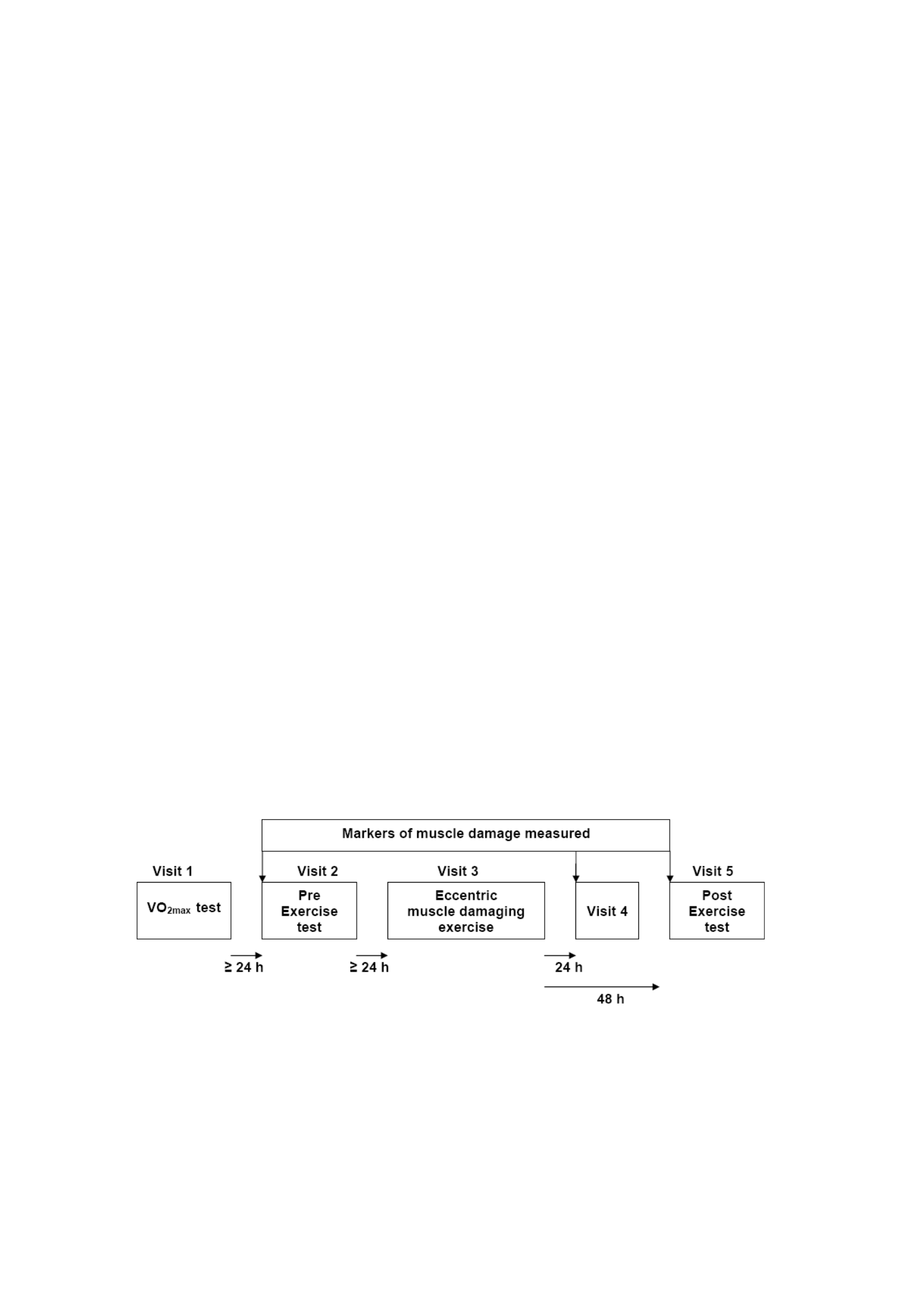
227
and D for exemplary participant information sheets, participant consent forms and ethical
approval certificates). The participants were asked not to take anti-inflammatory
medications during the study and to refrain from strenuous exercise for 24 hours before
each visit.
Procedure
All tests are carried out on an electronically braked bicycle ergometer (Lode Excalibur
Sport, Groningen, Netherlands). The participants were instructed to report to the laboratory
at the same time (± 1 hour) on five separate occasions over a period of 2-3 weeks (Figure
7.1). On the first visit to the laboratory, the seat height and handlebar position are
individually adjusted for comfort and adjustments are recorded and replicated in
subsequent tests. The height and mass (SECA, UK) of each participant were also recorded.
Figure 7.1 Schematic overview of the experimental procedure. V−O2 max,

228
maximum oxygen absorption.

229
Participants then completed additional exercise tests until fatigue of the will to determine
the maximum oxygen uptake (V O2
max) and gas exchange threshold (GET) and to establish
the intensity of future work. This entails cycling at a self-selected pedal speed (between
70 and 90 rpm) for 4 minutes of basic cycling at 0 W. The working rate is then ramped up
by 1 W every 2 seconds (i.e. 30 W.min−1) until the subject is unable to
continue. V O2
max is defined as the highest 30-s average value recorded before
termination of the test by the will of the subject. The GET was determined independently by
two experienced reviewers using the V-slope method (Beaver et al., 1986). The working rate
required 70% of the difference ( ) between the peak of GET and V O2 (strenuous exercise) is
calculated, taking into account the average response time of V O2 adaptation to ramp
training (about 2/3 of the ramp rate, which is minus 20 W) (Whipp, 1984). At the second
and fifth visits, respectively before and 48 hours after exercise damaging the eccentric
muscles, participants cycled at a self-selected pedal speed (between 70 and 90 rpm) for 4
minutes at 0 W, after which a warm-up phase at moderate intensity (~80% GET) was
applied for another 6 minutes. After 2 minutes of passive rest, the subject pedals for 4
minutes of basic cycling at 0 W after which a severe working rate is suddenly applied for
6 minutes. The test is repeated after a 2-hour break. In the second test, the strenuous
training battle continued until the subject voluntarily ended the test when exhausted.
Eccentric exercises

230
To trigger muscle damage, participants performed 100 squats (Smith) as 10 sets
10 repetitions. The load on the bars was calculated according to 70% of the body mass of each
participant. For more details on this procedure, please refer to Chapter 3.

231
Measurement
Markers of muscle damage
Markers of muscle damage (muscle pain and isokinetic peak torsion) were measured
immediately before and then 30 minutes and 48 hours after eccentric exercise. In addition,
plasma creatine kinase (CK) activity was assessed immediately before and then 30
minutes, 24 and 48 hours after eccentric exercise. Due to availability at 24 hours, only 8
participants were measured for CK activities.
Assessment of muscle damage
All indicators of muscle damage, perceived muscle pain (using a visual analogue scale of
0-10 (VAS)), creatine kinase activity (CK) and isokinetic peak torque (30 degrees s-1),
were measured in the listed order, immediately before, and 24 and 48 hours after
performing the muscle-damaging eccentric exercise protocol. For more details on this
procedure, please refer to Chapter 3.
Steps of the sports test

232
Pulmonary gas exchange and ventilation were measured breath-by-breath in all tests with
participants wearing nasal tongs and breathing through a low-dead-chamber, low-
resistance mouthpiece through an online gas analysis system (Cortex MetaMax 3B,
Biophysik, Leipzig, Germany). This system is calibrated prior to each test in accordance
with the manufacturer's guidelines against known cylinder gas concentrations (5% oxygen,
15% carbon dioxide) and a 3 l calibration syringe (Hans Rudolph, Kansas City, MO). The

233
V−O2
Data collected from pre- and post-workout tests are then modeled into
provides an estimate of the kinetic parameters of V
O2 (see Data modeling of V
O2 and [HHb]).
Heart rate (HR), blood lactate concentration ([La]) and Perceived Exertion Rating (RPE)
(Borg, 1998) were recorded at 2 minutes, 4 minutes and at the end of the exercise. HR and
[La] measures are also recorded during the base cycle. HR is monitored using a wireless
chest strap telemetry system (Polar Electro T31, Kempele, Finland) and measured
continuously via a link to the Cortex gas analysis system. Fingertip blood samples were
collected and analyzed for [La] using the YSI 2300 STAT plus analyzer (Yellow Springs,
Ohio, USA). Participants were familiarized with the 6-20 Borg RPE Scale and given
standard instructions on how to use the scale (Borg, 1998). Participants were encouraged
to focus on their overall perception of exertion when reporting their RPE.
Near infrared spectroscopy (NIRS)
The oxygenation profile of the right lateral vastus muscle was recorded in all exercise
tests using a continuous near-wave infrared spectrometer (NIRS) (Hamamatsu NIRO 300,
Hamamatsu Photonics K.K., Japan) (Figure 7.2). The system monitors changes in
oxyhemoglobin (HbO2) and deoxyhemoglobin (HHb) concentrations calculated from

234
changes in light attenuation by utilizing a modified Beer-Lambert law. The HHb
concentration signal ([HHb]) obtained from NIRS is considered to be relatively insensitive
to changes in blood volume during exercise and thus reflects the balance between oxygen
delivery and utilization (Ferrari et al., 1997).

235
Figure 7.2 Measurement principle and structure of the NIRO 300 probe (Hamamatsu,
Hamamatsu Photonics KK, Japan)
Pulsating light emitted at 1 second intervals from the emission probes at
four
different
wavelengths (775, 810, 850 and 910nm) and detected, as a function of distance, using
a three-segment photodiode detection probe that receives NIRS signals at 2 Hz. Probe
be Located
in
The
black
one
Silicon Handles are
provided. The
Distance between
optodes
between

236
the transmitter and receiver are 4 cm, and the penetration depth is approximately half of the
the distance between the transmitter and the receiver, which is 2 cm. Before placement on the
right
Vastus lateralis, the site is shaved and cleaned using an alcohol swab. NIRO300
system is then calibrated and Probe mount secured by from two sides
Adhesive
Sheet
~12 cm above the lateral epicondyle of the right leg, with place
marked with letters that
cannot be erased
markers to allow reproduction The position of the probe
in the
next test (48 hours). The thigh with the probe mount attached is then wrapped in a dark color-
colored elastic bandage to better secure the probe and to eliminate the ambient light that

237
may contaminate NIRS signals (Figure 7.3)
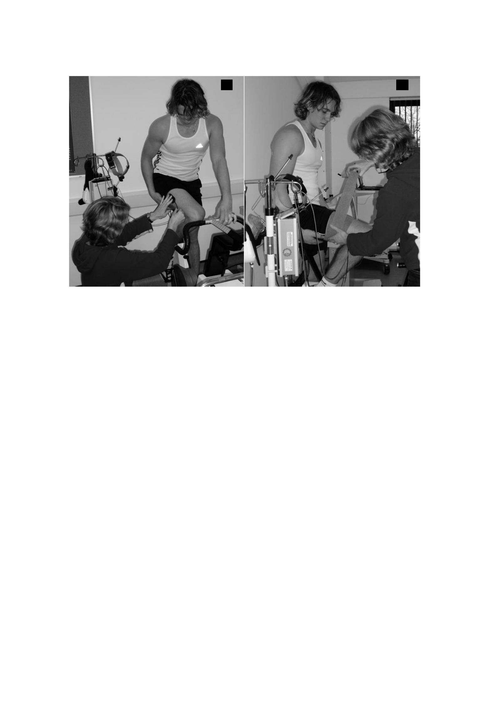
238
Figure 7.3. Installation of NIR probes. The NIRS probe mount is secured with double-
sided adhesive (a). The thigh with the NIRS probe mount attached is wrapped in an elastic
bandage (b).
The NIRS data collected represent changes in relative concentrations on hemoglobin
chromophores and, therefore, do not represent absolute tissue O2 values. Since [HHb] is
measured as a change from the baseline value, the probe gain is a set zero prior to the test
with the subject stationary in a seated position. Differences in the thickness of the adipose
tissue above it can affect the amplitude of the NIRS signal. However, the same subjects
(a)
(b)

239
were used before and after eccentric exercises and the probe position was strictly
maintained for each subject in each test, so no correction was required for adiposity
between sites. After the exercise test, the data is downloaded and the generated text file is
saved for further analysis.

240
V O2 and [HHb] data modeling
Breath-by-breath data from each exercise test was manually filtered to remove outside
breath, which is defined as a breath ± 3 SD of five adjacent breaths. The data for each
individual were then interpolated to provide a value of 1 s and two datasets of each test
before and after the exercise were aligned with the time and average. The first 20 seconds
of data after exercise onset (Phase I response) are deleted and the biphenial model is
used to analyze the V O2 response
to strenuous exercise, as described by the following
equation:
V O2 (t) = V O2
garis dasar
+ Ap[1 – e
– (t – Tdp)/ τp
] (phase 2 / main component)
+ As[1 – e
– (t – Tds)/ τs
] (phase 3 / slow component)
where t is time; The V O2
baseline is the V O2 baseline; Ap and As are the main and slow ones

241
the amplitude of the components, respectively; Tdp and Tds are the main and slow
component time delays, respectively; and τp and τs are the time constants of the primary
and slow components, respectively. Model parameters are determined using
nonlinear least squares algorithm. In the above equation, V O2 (t) represents the absolute
V O2 at a given time t, and V O2
baseline represents the average V O2 through the baseline
cycling period. Since the fatigue time is not identical on the first and second attacks of
heavy training, we adjusted the data 1) to the end of the exercise in both attacks and 2) to
the same time point (given by the fatigue time in the shortest fight). Primary

242
The "gain" component (i.e., Ap/∆WR) is calculated from the asymptotic projection of V O2. In
addition, the "actual" profit achieved at the end of the exercise is calculated.
To provide information on the effects of exercise damaging eccentric muscles on the
dynamics of muscle oxygenation, we also modeled the response ∆[HHb] to strenuous
exercise. The NIRS-derived [HHb] data were time-aligned and averaged to provide a
single response for each exercise subject before and after eccentric. The delay in time
before the increase [HHb] after the onset of exercise is defined as the first point greater
than one standard deviation above the baseline mean (DeLorey et al., 2003). The [HHb]
data is then mounted with a bi-exponential model similar to that described by the equation
above, with the exception that the pairing window starts at the beginning of the exercise
(i.e., at time 0). Furthermore, the [HHb] data are supplemented with a mono-exponential
model from the beginning of the exercise to a time point representing the interface of the
primary and slow components to determine the rate of adaptation of muscle deoxygenation
during the primary phase (MRT1). In addition, the dynamics [HHb] for the entire response
was modeled with a similar mono-exponential function (MRTt).
Statistical analysis
Changes in markers of muscle damage (peak torque, pain and CK activity) were analyzed
using a series of ANOVA unidirectional repeated measurements (RM). All data is checked
for normality assumptions. Because the CK activity data was found to be not normally

243
distributed, the values were changed in the log prior to statistical analysis (Twist & Eston,
2005) (see appendix H).

244
After the transformation, the CK activity data is distributed normally. Changes in HR,
RPE, [La] and ventilation were analyzed using a separate 2-way ANOVA RM (test x time).
The assumption of roundness is evaluated using the Mauchly test. Where roundness is
violated (P< 0.05) the Greenhouse-Geisser correction factor (GG) is applied. A modified
post-hoc Tukey test for repetitive actions (Stevens, 2002) was run to determine where
significant differences occurred. The paired t-test is used to determine
the difference in fatigue time and V−O2
and [HHb] kinetic response to
intensity training before and after eccentric exercises. All data were analyzed using the
SPSS statistical software package for Windows (version 13). The statistical significance
was set at 0.05.
7.4 Result
Markers of muscle damage
Eccentric exercise is effective in triggering significant changes in all markers of muscle
damage. Table 7.1 shows changes in isokinetic peak torque, perceived muscle pain and
plasma CK activity before and at 24 hours and 48 hours after eccentric exercise.

245
Table 7.1 Changes in markers of muscle damage. Average ± elementary school grades
before (pre)
and at 24 hours and 48 hours after eccentric training.
Measurable variables
pre
24-hour post
48-hour post
Peak Torque (N.m) 30 degrees-
1
287 ± 39
227 ± 44*
228 ± 60*
Pain
0.6 ± 0.5
6.4 ± 1.8*
7.1 ± 1.5*
CK Activity (U/L)
172 ± 123
740 ± 666*
373 ± 208
The grades were the average ± elementary school before (secular) and 24 hours and 48
hours after eccentric exercise (post). Pain, visual analogue scale 0-10. CK, creatine
kinase. * significantly different (P < 0.05) from the pre score
Isokinetic peak torque (30 degrees-1) decreased by 21% at 24 hours post-eccentric exercise
and remained depressed at 48 hours (F (2.16) = 21.85 p < 0.001). Significant pain was
reported 24 hours after eccentric exercise with the highest score at 48 hours (F (2.14) = 80.50
p < 0.001). Plasma CK activity increased after eccentric exercise, with the highest activity
observed at 24 hours (F (2.16) = 17.15 p < 0.001). Changes in markers of muscle damage
were detected in all subjects, although considerable inter-subject variability was observed.
The decrease in isokinetic peak torsion peak (30 degrees s-1) ranged from 12 - 44%, and

246
the increase in plasma pain peak and CK activity ranged from 53 - 95% and 146 - 1176%,
respectively.
Response to strenuous intensity training
Table 7.2 shows the response of V O2 to strenuous intensity exercise. No change in
kinetic phase II V O2 after eccentric exercise, also no slow change
component time delay (TD) (P > 0.05). However, the amplitude of the slow component
was significantly reduced (t (8) = 3.84, P < 0.05) and the overall mean response time (MRT)
was significantly faster (t (8) = 4.01, P < 0.05) after eccentric exercise. A much shorter time
to fatigue was observed in the 2nd attack of strenuous intensity training (before:

247
7:24 min:s (± 2:41) post 6:14 min:s (± 2:46)) (t (8) = 2.58, P < 0.05). V O2 response from
representative subject is illustrated in Figure 7.4.
Table 7.2 Response of pulmonary O2 absorption to strenuous intensity exercise before and after
exercises damage eccentric muscles.
To
48-hour post
Main components
TDp (s)
12 ± 4
12 ± 2
p (s)
25 ± 4
24 ± 3
Ap V O2 (L·min-1)
2,36 ± 0,23
2,37 ± 0,23
Gain (mL·min-1· W-1)
9,02 ± 0,45
9,04 ± 0,52
Slow components
TD2 (s)
102 ± 19
107 ± 29
A2ref V O2 (L·min-1)
0,63 ± 0,33
0,37 ± 0,17*
A2end V O2 (L·min-1)
0,67 ± 0,33
0,38 ± 0,17*
Overall response
MRTref
61 ± 15
51 ± 9*
MAR
64 ± 15
51 ± 9*

248
Puncak V O2 (L·min-1)
3,89 ± 0,44
3,70 ± 0,36
Grades are ± means elementary school. TDp, p. Ap and gain are the time delay, time
constant, amplitude and increase in V O2 per unit of increase in working rate for phase II
kinetics respectively. TD2, A2ref and A2end are time delays, amplitudes up to 6 minutes
and amplitudes until the end of training for the slow components, respectively. MRTend
and MRTref are the average response times that correspond to the end of the workout in
both fights and at the same time point
(given by the fatigue time in the shortest fight), respectively. The peak of V O2 tends to be lower after
eccentric exercises but the difference is not statistically significant. Significant difference
(P < 0.05) from the pre-score.
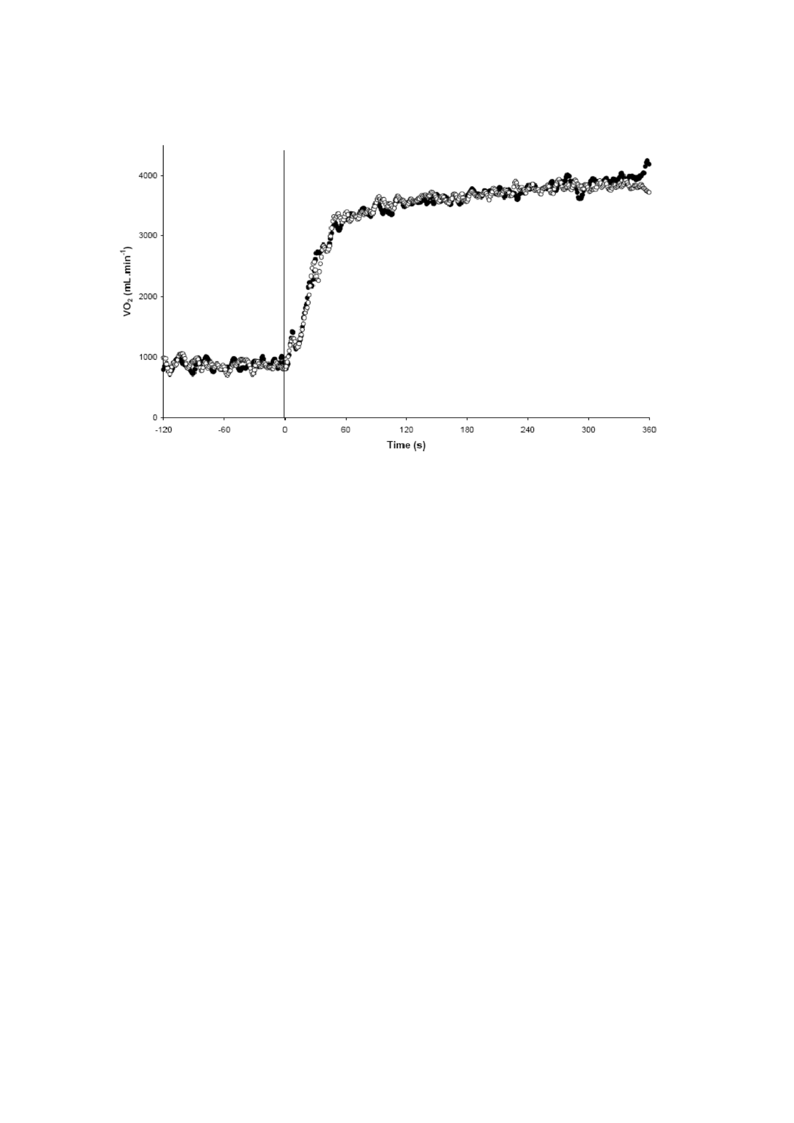
249
Figure 7.4 Representative subject V O2 Response to heavy cycle exercise before (●)
and 48 hours after (○) eccentric exercise. The vertical line represents the transition
from the 'unloaded' to the 'loaded' cycle.
There was a significant increase in RPE values reported during strenuous intensity exercise
after eccentric exercise (F(1.8) = 6.7 P < 0.05). However, there was no significant difference
in blood lactate response or heart rate before and after eccentric exercise (Table 7.3) (P >
0.05). In addition, there was a significant increase in ventilation equivalents for O2 (/V O2)
after eccentric exercise (pre: 29.3 (± 3.5) post: 32.6 (± 5.2))
(F (1,8) = 7,45, P< 0,05).

250
Table 7.3 Perceived Exertion (RPE), Heart Rate (HR) and blood lactate
concentration ([La]) ratings during strenuous intensity training before and 48 hours
post-eccentric exercise.
Basis 2 minutes 4 minutes End Workout
To
48 jam
To
48 jam
To
48 jam
To
48 jam
RPE*
ON
ON
15 ± 1
16 ± 1
17 ± 1
18 ± 2
19 ± 1
19 ± 1
SDM (b.min-
1)
83 ± 9
86 ± 6
159 ± 8
163 ±11
174 ±11
176 ±11
184 ± 9
181 ±11
[La] (mmol.l-
1)
0,9 ±
0,5
1.0 ±
0.4
3.5 ±
1.0
3.8 ±
1.0
6.9 ±
1.3
6.8 ±
1.8
8.4 ±
1.4
8.7 ±
1.8
Grades are a means of ± elementary school. RPE, a perceived level of mobilization
based on the scale of 6-20 Borg (Borg, 1998); HR, heart rate; [La], blood lactate
concentration; NA, not applicable.
* Significant primary effect for time (P < 0.05).

251
There was also a significant interaction of the time test x (F(9.72) = 3.15, P< 0.05) at V
E/V
O2.
Post-hoc Tukey tests showed that V
E / V
O2 was significantly greater post-eccentric
training during the last 70% of the training with the average score increasing from 25.3 to
36.0 (pre) and from 26.7 to 39.9 (post) (P< 0.05).
The results of the kinetic response [HHb] to strenuous intensity exercise before and 48
hours after eccentric exercise are shown in Table 7.4. Most importantly, in relation to our
experimental hypothesis, both [HHb] MRT1 and MRTt were significantly slower after
eccentric exercise (P < 0.05). There was no significant correlation between either marker
of muscle damage and the muscle oxygenation index in post-injury conditions (P > 0.05).
The [HHb] response of the representative subject is illustrated in Figure 7.5.

252
Table 7.4 [HHb] response to severe intensity constant weight training before and 48 hours
post-eccentric exercise.
To
48 jam
MRT1 (s)
14 ± 3
19 ± 3*
Main Amp (%)
91 ± 8
88 ± 8
Ampli Utama (AU)
309 ± 102
297 ± 72
MRTi
16 ± 4
21 ± 4*
SC Amp (%)
9 ± 8
Dec ± 8
SC Amp (AU)
33 ± 25
41 ± 25
The grade is the average ± elementary school. [HHb], deoxyhemoglobin concentration;
MRT1 and MRTt are the mean response time of the primary phase (the rate of
adaptation of muscle deoxygenation during the primary phase) and the overall response
(the rate of adaptation of muscle deoxygenation for the entire response), respectively.
Primary Amp is the change [HHb] during initial and rapid increases expressed as a
percentage of the overall response (%) and as arbitrary units (AU). SC Amp is the
change [HHb] during the slow component (an incremental increase [HHb] that
progresses slowly when the training intensity is above the gas exchange threshold)
expressed as a percentage of the overall response (%) and as an arbitrary unit (AU)
*48 hours of post value is significantly slower than pre score (P < 0.05).
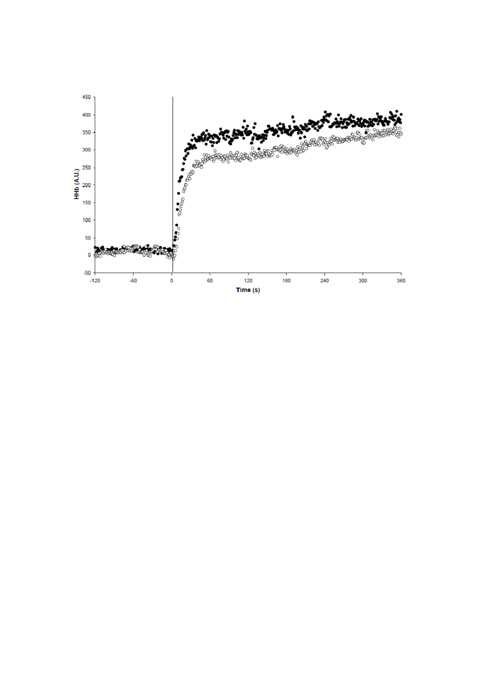
253
Figure 7.5 Response of deoxygenated Hb ([HHb]) to heavy cycle exercise before (●) and
48 hours after (○) eccentric exercise in representative subjects. The vertical lines represent
the transition from unloaded cycling to loaded cycling. Note the slower overall kinetics
[HHb] after eccentric exercise (slower rate of adaptation of muscle deoxygenation or
fractional O2 extraction, for the whole response),. AU, an arbitrary unit.
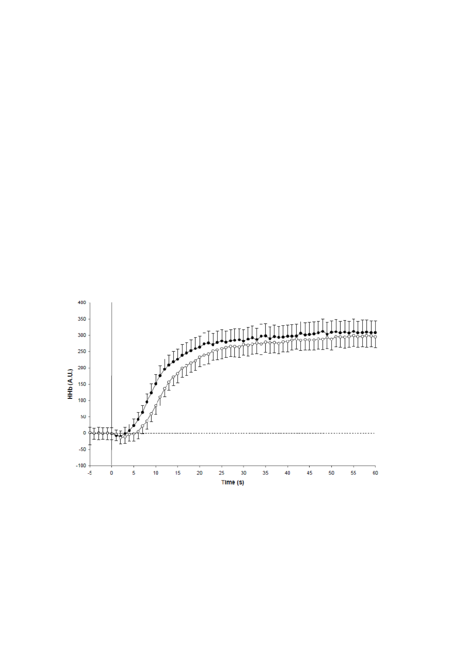
254
Change Q O2 : V O2 balance is most prominent during the initial response after
The onset of strenuous intensity training (Figure 7.6), with the largest mean difference
observed during the first 5-20 seconds (Figure 7.7). There was no significant difference in
pre and post-condition between response amplitude in either the primary phase or the slow
component (P > 0.05). Similarly, there was no difference between the total hemoglobin
response in the two conditions (Figure 7.8).
Figure 7.6 Mean initial response [HHb] to heavy cycle exercise before (●) and 48 hours
after (○) eccentric exercise. The vertical line represents the transition from the 'unloaded'

255
to the 'loaded' cycle. The data is normalized for the amplitude of the response at baseline.
Average score (± SE). AU, an arbitrary unit.
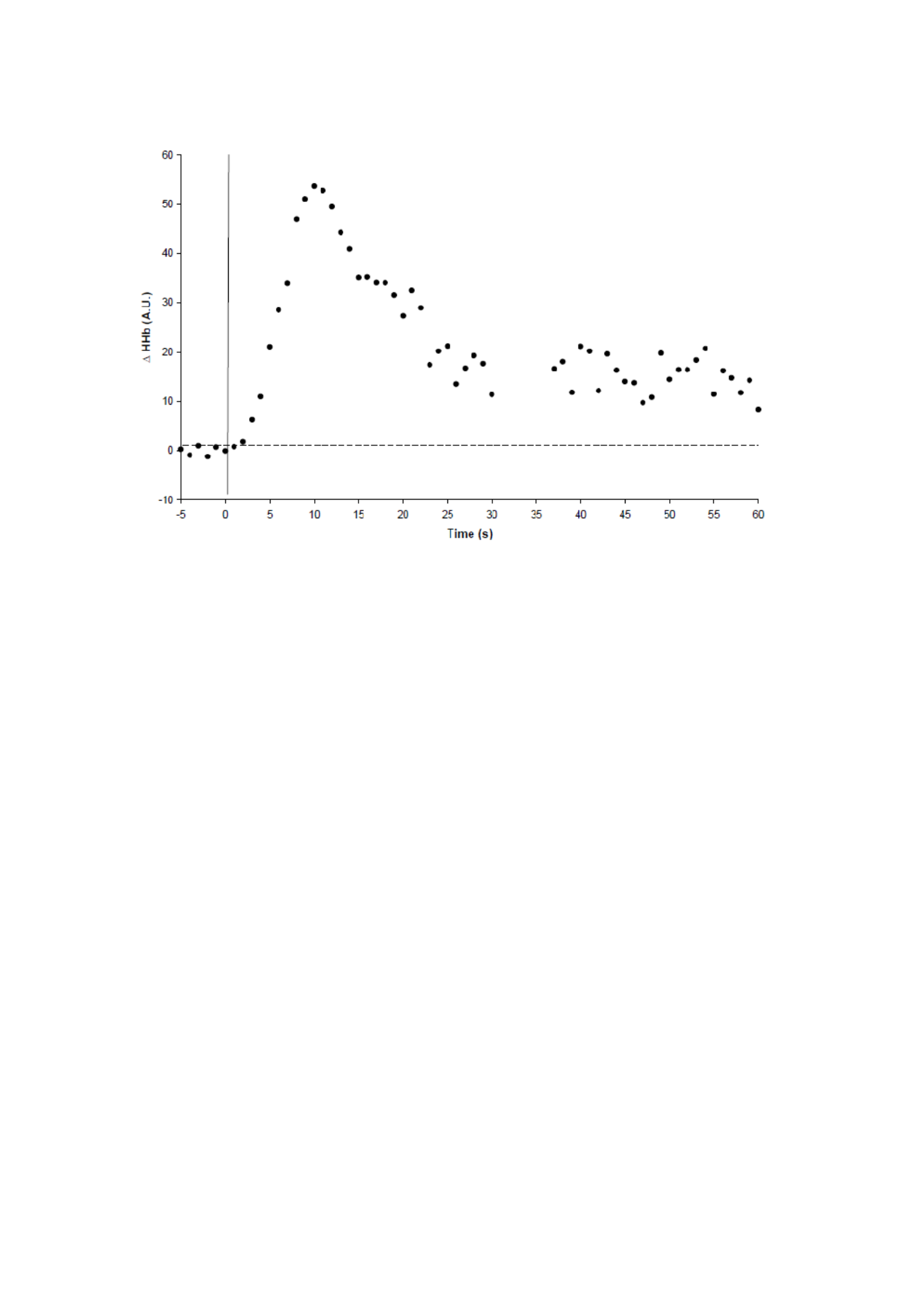
256
Figure 7.7 Mean difference in initial response [HHb] to strenuous intensity exercise
before and 48 hours after eccentric exercise (i.e. post-eccentric pre-minus exercise data).
AU, an arbitrary unit.
7.5 Discussion
The main original finding of this investigation is that it is eccentric and damaging to the muscles
exercise results in slowing down muscle kinetics [HHb] without altering the lungs V−O2

257
Kinetics during high-intensity cycle exercise in humans. We interpret slower kinetics
[HHb], in the face of unchanged pulmonary
V
O2, as a consequence of local improvement
the Q O2 : V O2 ratio. Observation that kinetics [HHb] is more than 30% slower 48 hours
after doing eccentric exercises, it shows that matching Q O2 and V−O2
was greatly altered as a consequence of the intervention.

258
Figure 7.8 Total hemoglobin response to heavy cycle exercise before (●) and 48 hours
after (○) eccentric exercise. AU, an arbitrary unit. The change in total hemoglobin does
not differ between conditions in such a way that the change in [HHb] provides a valid
measure of muscle deoxygenation (fractional O2 extraction). Increased blood flow, as has
been observed by Laaksonen et al. (2006) can compensate for the decrease in the
proportion of capillaries that favor red blood cell flow as observed by Kano et al. (2005)
and therefore maintain the total hemoglobin response in the interrogation area.
The dynamic balance between O2 delivery and O2 utilization has been sharply debated

259
With regard to possible muscle limitations V−O2
kinetics (Poole et al., 2008). Selama
transition to exercise intensity under GET, a convincing weight of evidence supports the
premise that metabolic inertia is a major constraint for muscle O2 uptake (Bangsbo et al.,
2000; Grassi et al., 1998, 1996). However, for the transition to exercise intensity above
GET, O2 delivery can provide additional simple constraints on the Kinetics of the V O2 muscle
(Grassi et al., 2000, 2003; Tschakovsky & Hughson, 1999). Current data shows that
after eccentric exercise-induced muscle damage, V−O2
Kinetics are maintained by

260
increased local muscle blood flow that may be necessary to compensate for
microcirculation dysfunction (Kano et al., 2005). Increased blood flow, as has been
observed by Laaksonen et al. (2006) after eccentric exercise, can compensate for a decrease
in the proportion of capillaries that favor red blood cell flow as observed by Kano et al.
(2005). To support this thesis, we observed no change in the total hemoglobin response,
indicating local muscle blood flow in the interrogation area. Thus, the change in response
[HHb] provides a valid measure of muscle deoxygenation (fractional O2 extraction).
Therefore, these data imply that the subject operates to the right of the 'tipping point' (Poole
et al., 2008) during the training fight, so that the compensatory changes in local muscle
bloodflow are able to prevent a measurable slowdown in the kinetic V O2 after muscle damage.
The squat protocol used here (Byrne & Eston, 2002a, 2002b), is effective in inducing
muscle damage as shown by a significant decrease in peak torque and increased plasma
CK activity. However, there were no significant changes in phase II
V O2 kinetic after eccentric exercise. This data is consistent with Schneider et al.
(2007) which reported that V O2 and HR responded to strenuous intensity cycling training
not affected by previous eccentric exercises and suspect that muscle levels are moderate
damage does not interfere with the delivery of O2 to active muscles or alter the ratio of Q2 O2 / V2O2.
However, the muscle oxygenation index was not measured by this author (60). Our
findings contrast with Ahmadi et al. (2008), who reported faster oxygen

261
desaturation (analogue to a lower ratio of Q O2 / V O2) during isometric contraction at 30%,
50% and 80% MVC after walking downhill thoroughly. However, the difference in

262
The experimental protocols and exercise modalities used make it very difficult to compare
the two studies. The high intramuscular pressure generated during isometric contractions
is known to inhibit muscle blood flow and this, or increased demand for contraction
energy, may explain the faster oxygen saturation reported by Ahmadi et al. (2008).
The experimental protocol used in this study is fundamentally different from the one used
by Kano et al. (2005). These authors electrically induced twitch muscle contractions (1 Hz,
3-5 V, pulse duration 2 ms) in rat spinotrapezius muscles, whereas in our study, human
subjects transitioned to a dynamic, high-intensity exercise cycle. While considering these
differences, it is important to note that the spinotrapezius muscle of rats does provide a
very acceptable comparative model for the analysis of human microvascular and myocyte
damage because it shows a fiber composition (Delp & Duan, 1996) and oxidative capacity
(Leek et al., 2001) that are very similar to those of the human quadriceps. Thus the
disorders observed in the microvascular rats after eccentric exercises, most likely, will also
be present in the damaged muscles of the subjects interrogated here. Kano et al. (2005)
reported impaired capillary geometry and substantial microvascular dysfunction after
eccentric exercise. Specifically, an increase in the luminal area of capillaries of damaged
muscles was reported resulting in a decrease in PO2mv at the beginning of electrically
stimulated contractions. In addition, a 27-34% increase in capillaries that did not flow red
blood cells was found 1-3 days after one attack of eccentric exercise.

263
Structural and functional changes in the microvascula can act to change the
matching Q O2 with V O2 both spatially and temporally with respect to energetic networks
Requirement. Specifically, lower [HHb] was observed at certain V
O2s
across
The eccentric post-exercise transition suggests that achieving the required blood myocyte
O2 flux may require higher microvascular O2 driving pressure. The muscle-blood O2 flux
is primarily determined by the number of erythrocytes located adjacent to the active
myocytes at any given time (Federspiel & Popel, 1986; Groebe & Thews, 1990). A
decrease in the proportion of capillaries that support the flow of red blood cells will lead
to a decrease in the O2 flux of blood muscles. Similarly, an increase in the diameter of the
free-flowing capillaries will increase the carrier-free diffusion distance and lead to a
reduction in the diffusion capacity of O2.
Fick's law of diffusion states that:
V O2 m = DO2
(PO2mv –
intramiositis PO2)

264
where DO2 is the dispersion capacity for O2 and PO2mv is the microvascular O2 pressure.
The reduction in DO2 is expected to have a negative impact on V O2 , especially in
the concurrent change in PO2mv was similar to that reported by Kano et al.
(2005). The O2 pressure gradient from blood to myocytes that promotes O2 diffusion is
determined primarily by changes in PO2mv (Poole et al., 2006; Poole & Ferreira, 2007).
Thus the acceleration of PO2mv decrease observed by Kano and his colleagues during the
first 20-40 seconds of electrically stimulated muscle contractions is expected to have
profound effect on the muscle spread capacity of O2 and resulting in a slowdown of V O2

265
Kinetics (Behnke et al., 2004, 2007, 2006). However, the PO2mv here (as assessed by the
[HHb] response) appears to increase during the dynamic rest-exercise transition after
eccentric exercise and the kinetic response of V O2 remains unchanged.
The given V
O2 is achieved through the interaction of O2 delivery (Q2) and diffusing O2
property (Behnke et al., 2003). In addition, changes in the lungs of V O2 in the rest-
The exercise transition is known to directly reflect the V O2 leg during cycling training (Poole et
al., 1991), and showed a good estimate of the Kinetics of the V O2 muscle (Grassi et al.,
1996). Therefore, the increased ratio of Q O2 :
V O2 is evidenced by slower kinetics [HHb]
response (i.e. lower [HHb] at a given
V O2 throughout the training transition) should, in this case
for example, due to a change in compensation on O2 deliveries (see Appendix J). With
regard to this issue, an increase in blood flow (and therefore an increase in O2 delivery)
has been observed by Laaksonen et al. (2006) who found that blood flow to exercised
Quadriceps femoris increased by 25% after a previous all-out eccentric exercise attack.
Importantly, they
also reports that V O2 remains unchanged and indicates that Q O2 : V O2 is
altered

266
The observed balance may be due to impaired oxygen extraction. Thus, after exercise that
damages eccentric muscles, an adaptive, compensatory mechanism can act to increase
PO2mv throughout the rest-exercise transition, maintaining the O2 flux of blood myocytes as indicated by the
unchanged phase II Kinetics V O2.
Higher ratings of perceived activity reported for a specific exercise intensity (70%∆) after
eccentric exercise may be due, in part, to the increased ventilation response that
accompanies increased muscle soreness. It has been proposed that changes to the

267
Sense of effort may be part of the central protective mechanism in which nerve inhibition
serves to reduce the generation of force to prevent further injury (Kyrolainen et al., 2000;
Michaut et al., 2002; Proske et al., 2004). In addition, Hotta et al. (2006) suggest that
changes in neural factors contribute not only to changes in force generation but also to
improved ventilation responses. Group III and IV afferent fibers located in and around the
blood vessels of exercising muscles are involved in modulating the ventilation response.
This vascular distension triggers the release of afferent fibers leading to increased
ventilation (Haouzi et al., 2004, 1999). Thus, peripheral nerve monitoring
vascular events may explain, in part, the increase in / V
O2observed in this study if
There are changes in the microvascular system as a result of eccentric exercises. In relation
to this problem, in a study using male Wistar rats, Kano et al. (2004) have reported a
change in the shape of the capillary lumen (luminal ellipsicity) that increases the luminal
cross-sectional area by up to 62% after eccentric exercise. A similar increase in the cross-
sectional area of microvessels in the muscles recruited in this investigation may serve to
augment ventilation responses through neural modulation.
It has been proposed that, after eccentric training, a change in the pattern of recruitment of
motor units arises to meet the energetic demands of a certain level of work (Clarkson,
1992). Reports of increased blood lactate concentrations after eccentric exercise have been
associated with the additional recruitment of type II fibers and concomitant increases in
glycogenolysis rates (Chen et al., 2007b; Gleeson et al., 1998). However, Chen (2003)
reported that neural adaptation to motor unit activation patterns occurs after a single attack

268
of eccentric exercise, so additional type I motor units are recruited. Extra fiber
Recruitment has been involved in the development of the slow component
V O2in

269
intensity training (Krustrup et al., 2004). In particular, it has been proposed that
the development of the slow component V O2 may be related, in part, to the hiring
serat tipe II tambahan (Barstow et al., 1996; Pringle dkk., 2003; Whipp, 1994). Si
reduction in the amplitude of the slow component V O2 observed here is possible therefore
Indications of altered motor unit recruitment patterns as a consequence of muscle damage
(Pringle et al., 2003). However, the unchanged primary V
O2 response and
The observed concentration of lactate in the blood is arguably inconsistent with this
suggestion. In addition, the full expression of the slow component may have been inhibited
as a result of muscle fatigue and the reduction in maximum working capacity caused by
the effects of eccentric exercise. Such performance inhibitions have previously been
reported to result in shorter fatigue times in mice (Carmichael et al., 2005, 2006) and
reduced time-trial running distances in human subjects (Marcora & Bosio, 2007). Thus,
The observed reduction of the slow component V O2 post-eccentric exercise can be attributed
to a shorter time for fatigue and a tendency for lower V
O2 peaks
to be achieved in this case
condition.

270
Experimental considerations
As mentioned earlier, our findings contrast with Kano et al. (2005) because of the
fundamental differences in the process of muscle activation used rather than species
variation. We studied the transition of dynamic training to severe intensity cycle training
(70%∆), while Kano and colleagues used a set rate of electrical stimulation (1 Hz, 3-5 V,
pulse duration 2 ms) to induce muscle contraction. Electrical stimulation induces the
recruitment of all fibers, while voluntary sports recruit fibers and certain types of fibers

271
depending on the intensity and duration of the exercise (Kindig et al., 2002). Thus, the
muscle activation and fiber recruitment patterns studied here present a more ecologically
valid model for investigation. Thus, this study may provide more realistic insights into the
effects of exercise damaging eccentric muscles on human functional performance.
NIRS is a well-established technique for the measurement of muscle oxygenation (e.g.
Ferrari et al., 2004; Jones et al., 2006). However, the reliability and reproducibility of the
[HHb] signal derived by NIRS depends on the proper placement of the optode. In this
study, the location of the optome was marked on each individual subject during the first
visit to the laboratory and the placement was carefully reproduced on subsequent visits.
Koga et al. (2007) have revealed the presence of significant heterogeneity with respect to
the dynamics of muscle oxygenation in the quadriceps muscles of healthy subjects after
the onset of exercise. These findings are not surprising given muscle blood flow,
distribution and recruitment of motor units and consequently vascular responses are known
to be heterogeneous within and across muscles. However, it is important to acknowledge
that the NIRS data reported here represent changes in the superficial muscle area that is
only interrogated and thus may not represent the entire muscle mass affected by eccentric
exercise.

272
7.6 Conclusion
In conclusion, this investigation shows that eccentric exercises damage muscles
changing the matching of Q O2 and V O2 during heavy intensity training. In particular, across the
rapid metabolic transition after the onset of exercise, for the given V
O2 , [HHb]
signal is reduced. We propose that structural and possibly functional changes in
microvascular acts to increase the ratio of Q O2 : V O2 both spatially and temporally with
Respect the energetic needs of the network. Therefore, after exercises that damage the
eccentric muscles, the compensation mechanism acts to improve microvascular motility
pressure for the O2 flux of blood myocytes, allowing preservation of V
O2 kinetics throughout the
transition to rest to exercise.

273
CHAPTER 8
CONCLUSION

274
8.1 Key findings
8.1.1 Ventilation response to dynamic exercise with EIMD
Previous investigations have reported that ventilation responses to running training did not
change after EIMD (Paschalis et al., 2005; Scott et al., 2003; Marcora and Bosio, 2007).
Another investigation that reported an increase in ventilation response and an increase in
blood response [La] during running linked the changes to changes in lower limb kinematics
(Braun & Dutto, 2003; Chen et al., 2007b, 2008). During cycle exercises, where potentially
confusing kinematic influences are avoided, improvements in ventilation and blood [La]
have been reported (Gleeson et al., 1995; Schneider et al., 2007). Before this thesis study
was conducted, Schneider et al. (2007), using 40% exercise intensity∆, provided the only
investigative example using a specific exercise domain to study the effect of EIMD on
physiological responses to dynamic exercise.
Study findings 1 (Chapter 4) and 4 (Chapter 7) showed that the ventilation response to
cycle exercise increased 48 hours after eccentric exercise (100 squats with a load
corresponding to 70% of body mass). Importantly, ventilation was improved not only
during vigorous-intensity cycle training (70% ∆) but also during moderate-intensity cycle
training (80% GET) without significant changes in blood response [La]. Previously, it had
been assumed that the increase in [La] contributed to the improved ventilation response.
However, our findings, specifically the observation that ventilation response after EIMD
is higher during exercise under GET, suggest that the altered exercise response may not be

275
due to changes in metabolic factors. Instead, we propose that neural monitoring of
peripheral blood vessels and local muscle events could, in part, explain

276
Improved ventilation response is observed. The mechanisms involved in ventilation
control during exercise are complex and involve a combination of central command and
afferent feedback. However, the structural and functional changes that characterize EIMD
are understood to increase the discharge from group III and IV afferents. This change
rather than an increase in blood [La] which is not a mandatory consequence of EIMD, is
likely to contribute to a greater ventilation response to dynamic cycle exercise induced by
EIMD.
8.1.2 Gas Exchange Threshold (GET) and EIMD
During dynamic incremental training protocols, GET identification provides the basis for
non-invasive estimation of lactate threshold (Tlac) (Beaver et al., 1986; Caiozzo et al.,
1982; Wasserman et al. 1973, 1990). However, ventilation and lactate responses to gradual
exercise can be separated by various experimental and clinical conditions (e.g. Poole &
Gaesser, 1985; Hughes et al., 1982; Hagberg et al., 1982). It is our opinion that EIMD is
likely to worsen the ventilation response to incremental/ramp exercise and, as such,
potentially separate the ventilation response, gas exchange, and lactate.
The findings of study 2 (Chapter 5) showed that 48 hours after GET eccentric exercise
occurs at lower working rates (before, 136 ± 27 W; post, 105 ± 19 W, P< 0.05) and V O2 (before,

277
1.58 + 0.26; post, 1.41 + 0.14 l.min-1, P< 0.05). However, the lactate threshold occurs at
similar working rates (pre, 161 ± 19 W; post, 158 ± 22 W, P> 0.05) and V O2 (previous, 1.90 ±
0.20 l.min-1; post, 1.88 ± 0.15 l.min-1, P > 0.05) after eccentric exercise. These findings
suggest that EIMD separates the ventilation response to gradual exercise from the blood
response [La]. Thus, these findings provide further evidence to support the thesis

278
That ventilation can be controlled by additional neurogenic stimuli or altered after
eccentric exercise.
8.1.3 Activity perception during dynamic training with EIMD
Regardless of the mode of damage, intensity of the exercise, or the exercise protocol used,
muscle-damaging eccentric exercises are known to evoke an increased sense of effort
(Gleeson et al., 1995; Scott et al., 2003; Marcora & Bosio, 2006; Twist & Eston, 2009).
Signals that inform perceptual responses to exercise can arise from central or peripheral
sensations, with the influence of central (cardiorespiratory) cues being less important than
local sensations (muscles), especially at lower exercise intensities (Mihevic, 1981;
Hampson et al., 2001). Thus, the perceived activity reported during exercise with EIMD
may be affected differently by central and peripheral cues depending on the intensity of
the exercise.
During ramp/incremental cycle training in study 3 (Chapter 5) participants reported a
perception of higher exertion at 48 hours, with a reported increase in RPE of 7% observed
in
the V−O2
GET pre-eccentric exercise values. In lessons 1 and 4 (Chapters 4 and 7)
Participants reported higher ratings of perceived exertion (RPE) during constant weight

279
cycle exercise, heavy intensity (70% ∆) 48 hours after eccentric exercise, although RPE
did not appear to change during moderate exercise (80% GET). It was interesting that the
increase in muscle pain and the increase in ventilation response experienced by participants
at 48 hours did not significantly affect the perception of exertion during moderate-intensity
exercise. However, during heavy-intensity training, where central ventilation cues may
have played a more influential role in informing the perception of exertion, RPE is

280
much higher. In study 1 (Chapter 4) the minute-by-minute difference in RPE values
reported during strenuous exercise to willpower fatigue before and 48 hours after eccentric
exercise was eliminated when expressed as a percentage of the total duration of exercise.
These observations provide further evidence to support the proposition that there is a
scalar-linear relationship between perceived exertion ratings and training duration. In
addition, ventilation differences are also eliminated when expressed as a proportion of time
to fatigue. These findings suggest that there is a strong association between ventilation
response and perceived activity towards cycling after eccentric exercise.
8.1.4 Time to burn out
The perception of exertion can be considered fundamental to an individual's exercise
response when a participant is required to exercise for 'willpower fatigue'. Previous
observations of increased RPE have been associated with decreased time trial performance
in running and cycling after EIMD (Marcora & Bosio, 2007; Twist & Eston, 2009).
During the incremental cycle exercise in study 2 (Chapter 5) and the incremental knee
extensor exercise in study 3 (Chapter 6), the observed decrease in time for willpower
fatigue and a reduction in peak work rate values was consistent with the observation of
impaired performance of the time test caused by the effects of eccentric exercise. Similarly,
in studies 1 and 4 (Chapters 4 and 7) the fatigue time was reduced after eccentric exercise,
during a constant load cycle at an intensity of 70% Δ. The exact nature of the accelerated

281
fatigue progression experienced with EIMD is poorly understood. Central and peripheral
fatigue factors have been involved in the observed decline in endurance capacity after
EIMD.

282
8.1.5 Metabolic response to dynamic exercise with EIMD
Central fatigue factors such as increased production of inflammatory cytokines within
brain regions responsible for movement, motivation, perception of effort and pain have
been associated with decreased operating time to fatigue (Carmichael et al., 2005).
Similarly, peripheral fatigue factors derived from damaged muscle tissue such as changes
in metabolic function have been implicated in the decreased endurance capacity observed
after EIMD (Asp et al., 1998)
In study 3 (Chapter 6), we used 31P magnetic resonance spectroscopy (31P-MRS) to evaluate
changes in muscle metabolism during dynamic incremental knee extensor exercises after
eccentric exercises. The reduction in fatigue time after EIMD was not associated with
accelerated [PCr] depletion. The fatigue time during knee extensor exercises is gradually
reduced by 12% after muscle-damaging exercises. However, the rate of depletion [PCr]
was similar in the two experimental conditions with the final exercise [PCr] remaining
13% higher 48 hours after muscle-damaging exercise. An increase in [Pi] and [Pi]:[PCr]
is observed not only at rest but also during gradual exercise. However, the rate of
improvement [Pi] did not change as a result of muscle damage however premature
termination of the test at 48 hours resulted in no significant difference in the final
exercise score [Pi]. It is tempting to speculate that the limit to exercise tolerance is
moderated by [Pi]. However, wide inter-subject variability is observed with the value of
[Pi], so we cannot reliably conclude that [Pi] is indeed a limiting factor. In contrast, [Pi]
final workouts may have made an important contribution to the reduction of fatigue time

283
experienced after muscle-damaging workouts.

284
8.1.6 Kinetics of oxygen absorption and muscle oxygenation
While the increase in [Pi] produced by EIMD may contribute to decreased dynamic
exercise performance, microvascular dysfunction observed after EIMD (Kano et al., 2005)
may also contribute to impaired performance due to impaired delivery and distribution of
O2 within the base of active muscle capillaries. We used Near Infrared Spectroscopy
(NIRS) to assess muscle oxygenation (hemoglobin + myoglobin) and to determine the
dynamic balance between oxygen delivery (Q
O2) and oxygen uptake (V
O2) after the onset of
exercise. Specifically, the NIRS signal of deoxyhemoglobin (HHb) concentration ([HHb])
was used to estimate the non-invasive extraction of O2 in skeletal muscle microcirculation.
Study findings 4 (Chapter 7) suggest that EIMD results in a slowdown in
Kinetics of muscle deoxyhemoglobin [HHb] concentrations without altering lung V O2
Kinetics. The observation that kinetics [HHb] was more than 30% slower 48 hours after
eccentric exercise performance, suggesting that there was an increase in the ratio of oxygen
delivery to oxygen uptake ( Q O2 : V O2 ) during the transition to heavy intensity
exercise. It is proposed that the increased ratio of Q O2 :V O2 is indicated by a slower
[HHb] kinetic response (i.e. lower [HHb] at a specific V
O2 throughout the exercise transition)
caused by a change in compensation in O2 delivery. In summary, this data shows that

285
after EIMD, V
O2 kinetics are maintained with increased compensation in local muscles
blood flow that is able to prevent a measurable
slowdown of V
O2 kinetics.

286
8.2 Limitations
During studies 1 and 4 (Chapters 4 and 7) we used a complete and full training protocol to
study human responses to dynamic exercise. The use of time or distance trials such as those
used by Marcora & Bosio (2007) or Twist and Eston (2009) can provide a more
ecologically valid model for studying the effects of EIMD on human responses to dynamic
exercise. However, the transition to constant load training provides the only proper
platform for modeling kinetic responses. Therefore, while we appreciate the low ecological
validity of using a complete constant load training protocol, we believe we justified the
need to use the training protocol we chose to precisely examine the human response to
dynamic training with and without EIMD.
The relatively small sample size used for the investigation comprising this thesis was
approved as adequate to detect clinically or biologically beneficial outcomes by the Ethics
Committee of the School of Sport and Health Sciences at the University of Exeter
following precise and rigorous power calculations (See Appendix A).
All participants were young men who were active and recreationally healthy and thus the
interpretation of our findings should be limited to a suitable population. Extrapolating the
findings to other populations such as highly trained or sedentary groups is inappropriate.
Protective adaptation to a single eccentric exercise attack, the 'repetitive fight effect'
(Nosaka & Clarkson, 1995) can last up to 6 months for most symptoms of EIMD (Nosaka

287
et al., 2001a). Thus the training status of participants may have a major influence on their
response to dynamic training with EIMD.

288
8.3 Implications and future direction
The main findings of this study consisting of this thesis have implications for the
interpretation of the human response to physical exercise after eccentric exercise.
Awareness of potential changes in metabolism, ventilation, gas exchange, and exertion
response felt after eccentric exercise can help coaches, exercise scientists, and health and
fitness practitioners to make more informed decisions regarding the advice given to their
costs. As mentioned earlier in this chapter, the use of a complete and full training protocol
full of constant loads is not ecologically valid. In the future, researchers should seek to
investigate changes in human responses after EIMD uses, for example, distance protocols
or time trials, such as those used by Marcora and Bosio (2007) and Twist and Eston (2009).
The increasing popularity of high-intensity sports training methods including resistance
and plyometric training may lead to an increase in the incidence of EIMD in the active
population. Recreationally active individuals should be aware of potential changes in their
performance capacity in the days following this type of training. Future research should
also seek to explain the differences in human responses to EIMD between individuals with
different training and activity statuses.
The effect of eccentric and muscle-damaging exercise on human responses to dynamic
exercise has only been the focus of scientific investigation in recent years (see section 2.
7). While this thesis has expanded the examination of some of these responses, many

289
questions remain unanswered. Various human responses to dynamic exercise are evaluated
in this thesis; including ventilation, gas exchange, perceived exertion and metabolic
response were only observed 48 hours after EIMD. The use of discrete observational
periods is deliberate with a time of 48 hours selected according to the maximum muscle
period

290
Pain. However, future research should attempt to evaluate changes in time travel in these
and other human responses during the hours and days following eccentric and muscle-
damaging exercise to explain the potential mechanistic relationship with changes in time
travel in known EIMD markers such as loss of force-generating capacity.
In studies 3 and 4 (chapters 6 and 7) we used animal research (Carmichael et al., 2005 and
Kano et al., 2005, chapters 6 and 7 respectively) to inform our understanding and provide
a basis for research questions on human subjects. Understandably, we haven't been able to
use some of the techniques used with animal models. Carmichael et al. (2005) dissected
the brains of eccentric exercise mice to reveal that increased concentrations of
inflammatory cytokines in the cortex and cerebellum were associated with reduced
exercise tolerance. Inspired by an invasive intravital microscopy study of spinotrapezius
muscle in rats by Kano et al. (2005), we used NIRS to facilitate the non-invasive
assessment of muscle oxygenation after EIMD. In study 4 (chapter 7) we observed a
decrease in [HHb]
Signals in the vastus medialis muscle for a
specific
V−O2
At the beginning of 48 hours of training
during cycling
after exercising squatting. Future research may expand the use of NIRS to facilitate the
non-invasive assessment of muscle oxygenation after EIMD by examining responses to a
variety of breakdowns and different exercise protocols.

291
8.4 Conclusion
In conclusion, studies consisting of this thesis have shown how the human response to
dynamic exercise can be altered after muscle damage caused by exercise. The structural
and functional changes that characterize EIMD have been shown to produce additional or
altered ventilation stimuli during exercise in such a way that ventilation

292
Response to cycle exercise increases at exercise intensity above and below the gas
exchange threshold. In addition, the observed additional ventilation response has been
shown to result in a gas exchange threshold that occurs at a lower working level without
concurrent changes in the lactate threshold. In combination, these findings suggest that
enhanced ventilation experienced after EIMD may be controlled by additional or altered
neurogenic stimuli rather than changes in metabolic factors. Improved ventilation response
is associated with increased perceived exertion rating (RPE) and reduced fatigue time.
Differences in minute-by-minute ventilation and RPE values are eliminated when
expressed as a percentage of the total duration of the exercise. These findings suggest that
there is a strong association between ventilation response and perceived exertion towards
cycling after EIMD. Although we have shown that increased blood [La] is not a mandatory
consequence of EIMD, we have shown, using 31P-MRS, that the muscular metabolic response to dynamic
exercise is altered after EIMD. The accelerated fatigue observed after EIMD may be related to the
increase in [Pi] observed at rest and during gradual exercise, or with other non-measurable
peripheral or central factors. Finally, we show that peripheral microvascular dysfunction
resulting from EIMD can contribute to impaired performance due to impaired delivery and
distribution of O2 within the capillary layer of active muscle. Using Near Infrared
Spectroscopy (NIRS) we showed that EIMD results in muscle slowing down
Kinetics of deoxyhemoglobin [HHb] concentrations without altering the
lungs
V O2 kinetics
The findings showed that there was an increase in the ratio of oxygen delivery to oxygen
uptake ( Q O2 : V O2 ) after EIMD caused by a compensatory change in O2 delivery.

