



Consider the image below. Can you identify the fracture? Note this is a child and the bones are
still growing. The epiphyses (secondary growth centers) are still separated from the shaft of each
bone.
Click on the part of the image where the bone fracture is located. Big hint: it is located on one of
the bones of the arm NOT the bones of the hand. Another hint- look for initial callus formation.
[7]
Correct!
Targets placed: 1/1
You can place up to 1 targets
Explanation
You can see the outline of the bone has been affected.
Submitted: Answered - Correct!0 attempts left
You have reached the maximum number of attempts
You have reached the maximum number of attempts
Explanation
Explanation
You can see the outline of the bone has been affected.
Q2
HomeworkAnswered
How would you characterize this fracture on the frontal (NOT parietal) bone? [8]
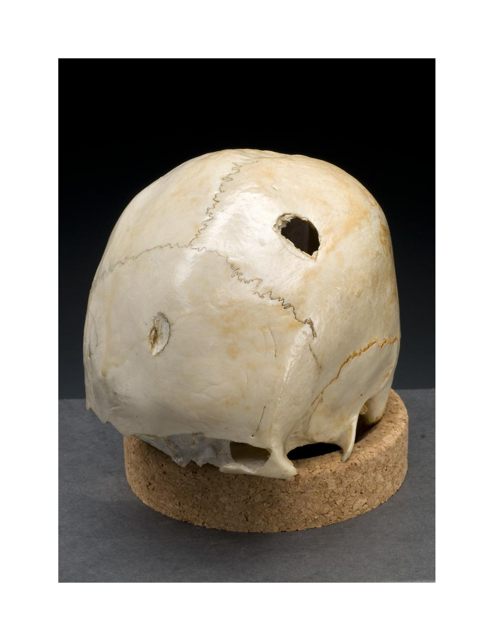

Select an answer and submit. For keyboard navigation, use the up/down arrow keys to select an
answer.
a
Complete Fracture
b
Infraction
Your answer
Explanation
The answer is B. It is an infraction.
Submitted: Answered - Correct!0 attempts left
You have reached the maximum number of attempts
You have reached the maximum number of attempts
Explanation
Explanation
The answer is B. It is an infraction.
Q3
HomeworkAnswered
How would you characterize the fracture of the radius seen in this x-ray of a child's forearm? [9]
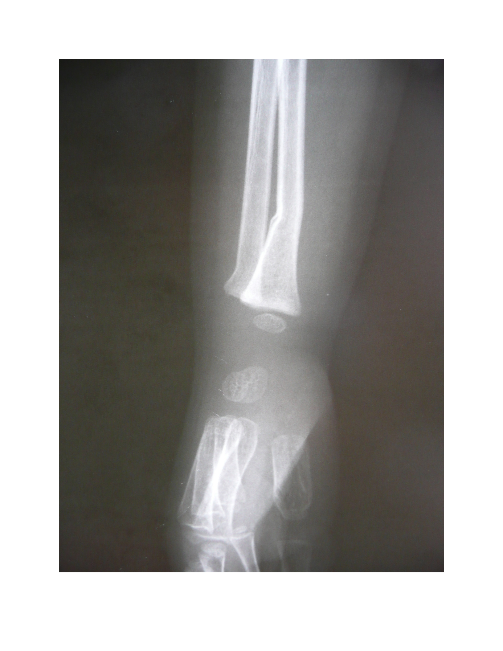

Select an answer and submit. For keyboard navigation, use the up/down arrow keys to select an
answer.
a
Complete Fracture
b
Infraction
Your answer
Explanation
The answer is B.
Submitted: Answered - Correct!0 attempts left
You have reached the maximum number of attempts
You have reached the maximum number of attempts
Explanation
Explanation
The answer is B.
Steeping some tea...
Secondary Fracture Lines
The preceding discussion focused on the formation of a primary fracture associated with the
direct area of impact. However, discontinuities are often associated with secondary fracture
lines, which usually originate from the point of impact or circumscribe the impact site.
Secondary fracture lines result when the bone directly at the point of contact (e.g., a bullet entry
point) cannot absorb all of the energy imparted into the body through the formation of the
primary fracture (e.g., a bullet hole). There are two types of secondary fracture lines.
Radiating Fractures disperse outward from the site of impact and can usually be traced back to
it. This fact is useful for identifying the site of impact if the damage to the body has result in
extreme fragmentation. Radiating fractures represent the energy traveling through the bone along
the lines of least resistance - areas of low bone density, low bone thickness, high proportions of
trabecular bone, cranial sutures, and the numerous foramina located throughout the body.
Concentric Fractures or Hoop Fractures form concentric fracture lines that surround the site

of impact. These lines are usually associated with high velocity projectile and low velocity blunt
force trauma. Concentric fractures are associated with high levels of energy impacting the body.
Figure 11 shows both types of fractures resulting from a gunshot wound to the distal femur. Note
the single large vertical radiating fracture (blue arrows) that begins at the gunshot entry point
(red arrow) and proceeds parallel along the shaft of the long bone, and the smaller concentric
fracture (green arrow) located distal to the entry wound that affects the superior portion of the
joint surface. The concentric fracture is curvilinear and has a contour that follows that of the
primary impact site.

Gunshot to the distal femur. [11]
Figure 12 depicts another example exhibiting a more typical secondary fracture pattern on the
skull. The red arrow indicates the point of bullet impact and the green arrows identify two
concentric fractures. This figure nicely demonstrates what is meant by the term "concentric" - the
fracture lines form like rings in a pond when a pebble is thrown into the water. These fracture
lines represent the effects of the force of the bullet and the energy produced when it impacts the
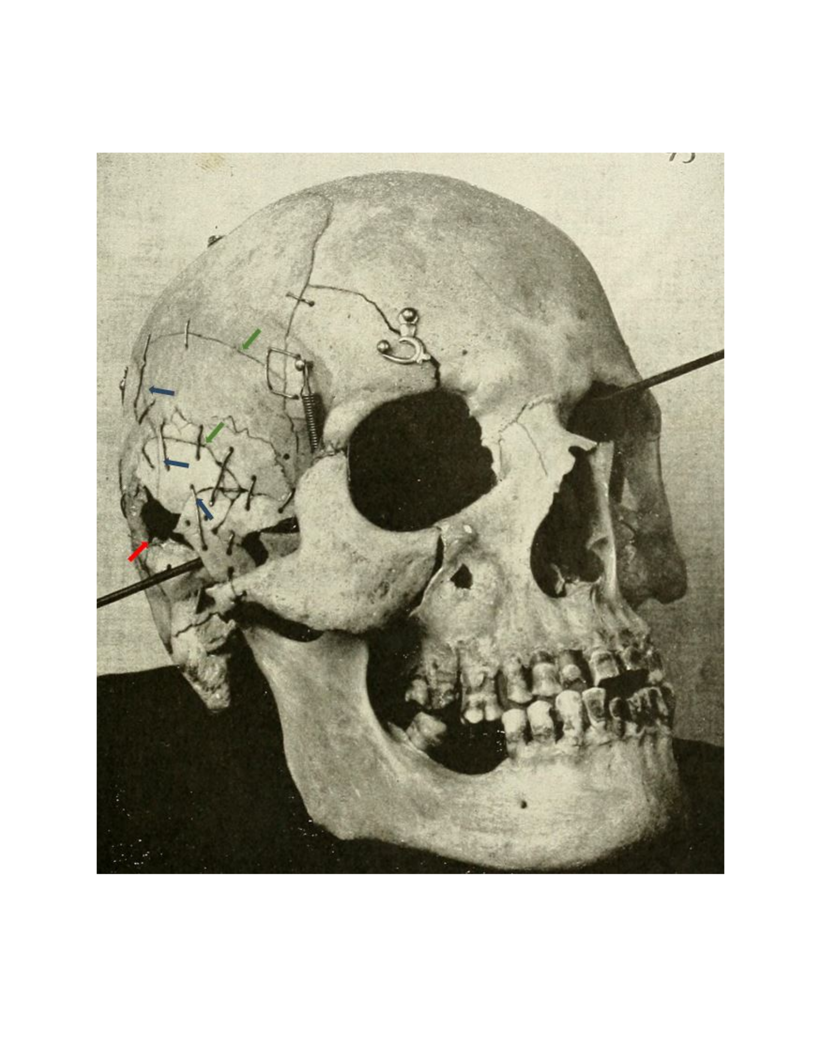
skull. These are also known as hoop fractures. The blue arrows indicate multiple radiating
fractures that originate from the primary site of bullet impact.
Skull with multiple gunshot wounds. [12]
The image below depicts a gunshot exit wound located on the posterior aspect of the left
squamosal suture (where the temporal bone meets the parietal bone). You can use the slider to

quiz yourself on identifying the different kinds of secondary fractures that were discussed above.
The red line outlines the exit defect, the blue lines mark the paths of radiating fractures, and the
green lines mark concentric fractures. Note how the radiating fractures can be traced back to the
exit defect and the concentric fractures more or less encircle it. Both kinds of fractures are used
to identify the point of impact (in this case, the exit defect). This image is also a good
demonstration of the fact that concentric fractures are often incomplete.
Steeping some tea...
Steeping some tea...
Steeping some tea...
It is important to consider that radiating fractures have a single point of origin. This is a critical
feature for distinguishing perimortem trauma with radiating fractures from damage that occurs to
a cranium from natural processes once buried in the ground. Consider the cranium in Figure 15.
At first glance the damage and breakage to the side of this cranium may seem similar to blunt
force trauma with extensive radiating fractures. However, note how haphazard the fracture lines
are. There is no clear impact site that is the point of origin for these different fracture lines. The
fractures lines intersect at right angles to each other and the broken fragments of bone are square
and rectangular vs more triangular in shape. This damage is caused by soil compression and
erosion and is not the result of trauma.
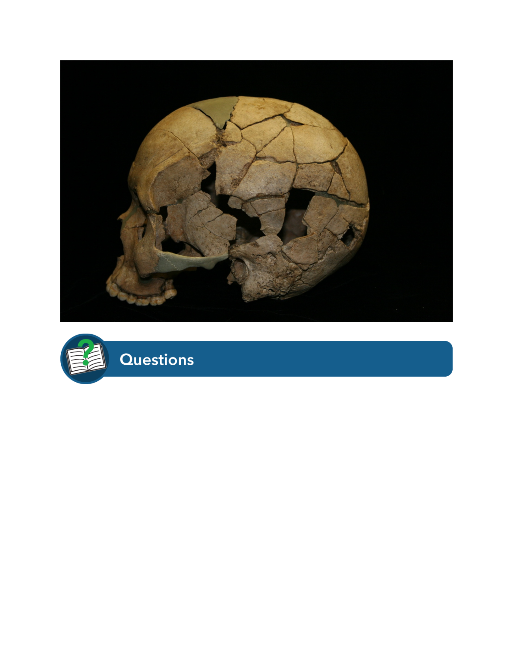
Lateral view of cranium depicting crushing due to soil compression. [15]
Steeping some tea...
Timing of Bone Injury
One of the most important considerations when assessing trauma is determining when a fracture
occurred with respect to the time of the decedent's death. Forensic anthropologists typically
recognize three time periods. Antemortem trauma occurred before death. Perimortem trauma
occurred around the time of death. Postmortem breakage (not really a fracture) occurred after the
individual died. The perimortem period is likely of greatest forensic relevance. However, it is
important to stress that in forensic pathology the perimortem interval refers to the time within
seconds or minutes of a decedent dying. In forensic anthropology the time interval is much more
coarsely defined and is usually measured in weeks, not minutes or seconds.
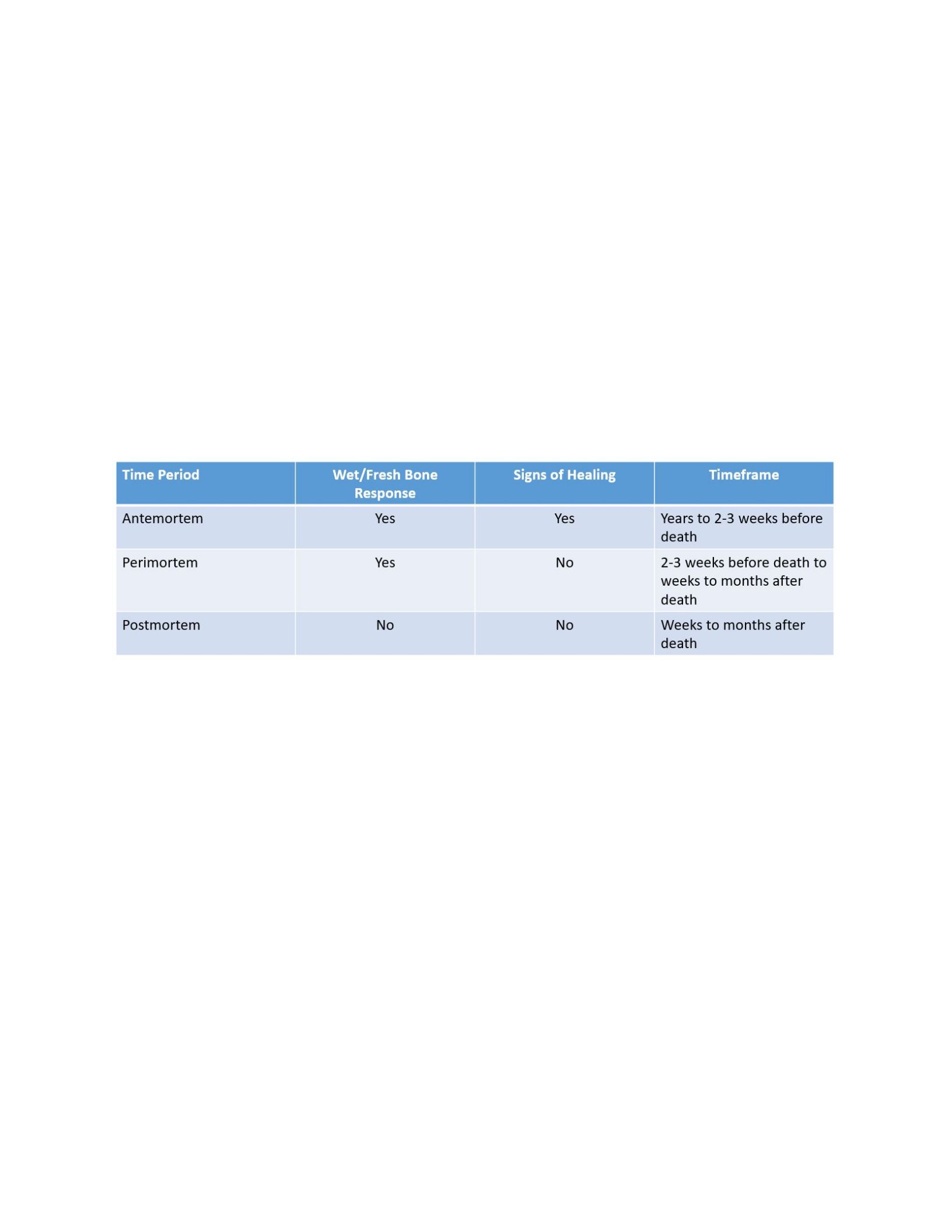
Antemortem trauma contributes to an investigation in two ways. First, if the trauma occurred
well before the time of death there may be a medical history that can be matched to trauma that
is observed on the body of an unknown decedent. That is, a fracture with a distinct pattern of
healing could be compared to x-rays of an individual for whom a missing persons report has
been filed. Second, trauma identified in various stages of healing may indicate a pattern of injury
consistent with torture or domestic abuse. For child decedents, observation of antemortem
trauma may indicate a history of child abuse and neglect.
The challenge with identifying antemortem trauma is that it is entirely based on observations of
bone healing or signs of secondary infection in response to the traumatic insult. However, bone
does not begin healing at the level of gross, macroscopic observation (i.e., with the naked eye)
for days to weeks after an injury occurs. A decedent could have received a blow to the head
causing blunt force trauma 1-2 weeks prior to being shot and killed. However, if the healing of
the first incident had not begun at the time of the second incident then the timelines will not be
distinguishable through forensic analysis.
Table 1. Diagnostic criteria for differentiating antemortem, perimortem, and postmortem bone
fracture/breakage.
Perimortem trauma is likely of greatest interest to the forensic anthropologist because it may
directly relate to the cause of death. Observations of perimortem gunshot wounds, stab marks, or
blunt force injuries would all be suggestive of the means by which the decedent died.
Postmortem trauma could be of forensic relevance, but it is less likely related to cause and
manner of death. The challenge is in differentiating perimortem and postmortem trauma. With
respect to traumatic injuries of bone, the difference between a perimortem fracture and a
postmortem break is simply a function of the collagen content remaining in the bone at the time
the break occurs. After death, bone loses moisture and the collagen component of bone begins to
decay. This process of decay is a roughly continuous process, but it is also dependent upon the
environment in which the body is decomposing. At a certain point after death, bone will begin to
break like a dry, brittle material. Therefore, the difference between perimortem and postmortem
trauma is whether the bone fractures with a wet/fresh bone response indicating high levels of
collagen or moisture or with a dry bone response indicating low levels of collagen or moisture.
These criteria are summarized in Table 1.
Antemortem Trauma

The first step in determining the timing of bone injury is observing whether there is any evidence
of osteogenic response at the site of fracture. The term "osteogenic" means "bone formation"
and reflects the body's response to an injury. If the cellular processes were no longer functioning
at the time the injury was sustained (i.e., the individual was not alive), then the healing process
will not be evident at the site of fracture. Antemortem trauma can also be identified if the wound
shows evidence of secondary infection. The healing process and the body's response to infection
involve both osteoclasts (cells that remove bone) and osteoblasts (cells that make new bone). A
general description of the healing process is as follows:
Soon after the traumatic event, blood from broken blood vessels will pool at the site of
the injury resulting in visible bruising and swelling. The pooling of blood forms a
hematoma, which is the body's initial attempt to stabilize the injury. Within 5-10 days, the
bone itself begins to heal as evidenced by increased porosity or pitting (small holes in
the surface of the bone) surrounding the fracture margins. Osteoclasts will begin
removing sharp fracture edges and resorbing dead pieces of bone within the fracture site.
This results in the rounding of the broken margins. As healing continues, osteoblasts
will begin producing new bone to mend the fracture ends. Initially a bony callus forms
that is composed of woven bone, which is less structurally sound and more disorganized
in appearance. This initial fracture callus will be replaced with mature bone that is more
structurally sound as the wound is remodeled for months after the injury occurred.
In summary, antemortem trauma is evidenced by three macroscopic features of bone healing:
increased porosity or pitting surrounding the fracture site, rounding of broken fracture margins,
and the deposition of new bone (bony callus) to completely mend the fracture.
Antemortem Cranial Gunshot Trauma
Figure 17 is a high-resolution close-up showing the external surface of the skull with active
infection. The small holes represent porosity, which covers the entire area of the image. Also
note how the surface looks somewhat uneven.
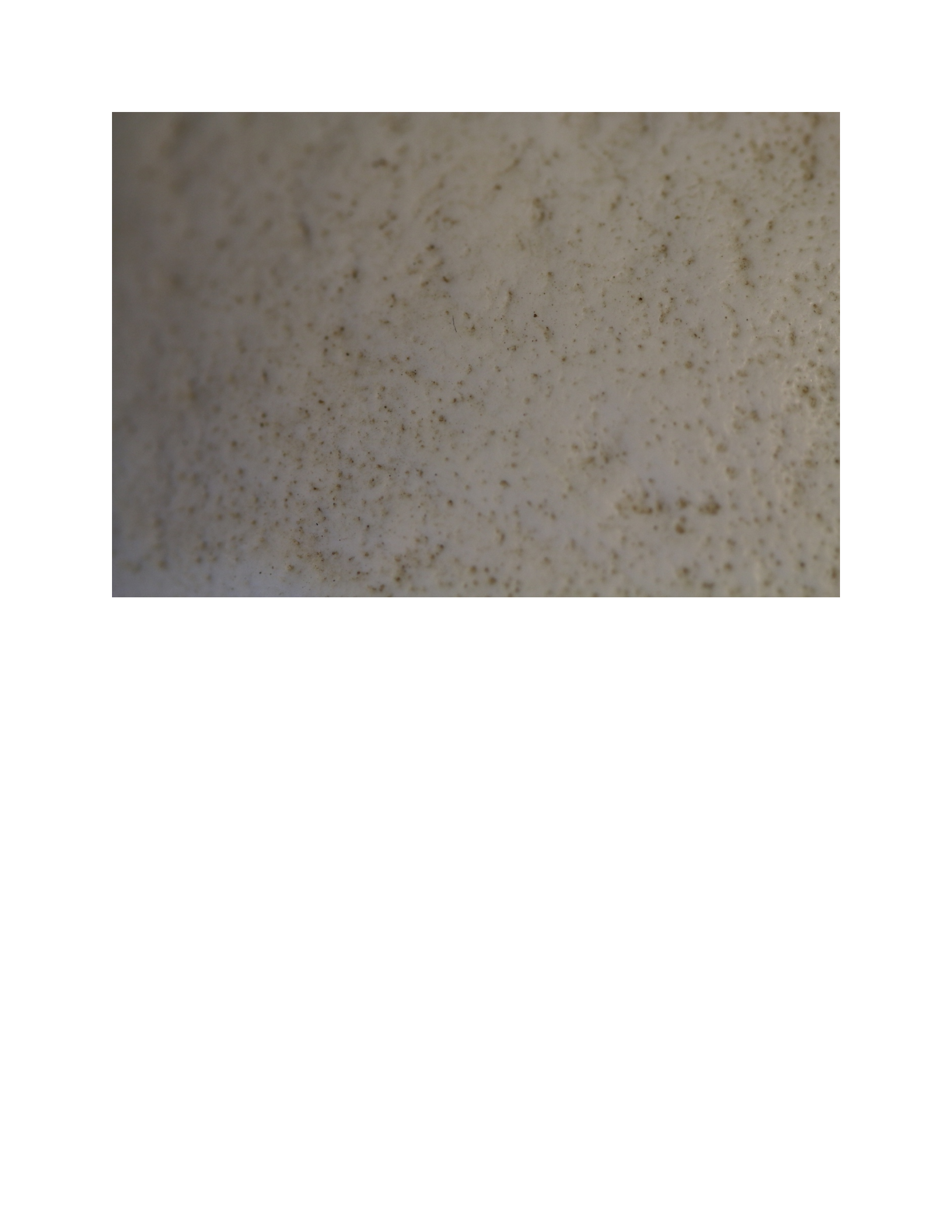
Close-up image of a bone surface showing porosity. [17]
Figure 18 shows the same specimen from a different angle. Note the left side of the image
exhibits porosity and an uneven surface that is not smooth, while the right side of the image
shows no porosity and is much smoother in texture. This image nicely demonstrates the
difference between bone that is actively responding to a localized infection (left of the dashed
line) and bone that is not (right side of the dashed line).

Active bone response to infection (left of line) and smooth, non-porous bone that is not actively
responding to infection (right of line). [18]
Figure 19 shows a single cranial gunshot wound that was fatal. The individual lived for 5 days
after the trauma occurred but there is no signs of healing or infection. There is no porosity
surrounding the gunshot wound. The edges of the bullet hole are sharp and crisp. The bone
surrounding the bullet hole is smooth and even indicating there is no remodeling of the margin.
Even though the individual lived for 5 days with this injury this would be considered a
perimortem trauma because no evidence of healing or active bony response is evident.
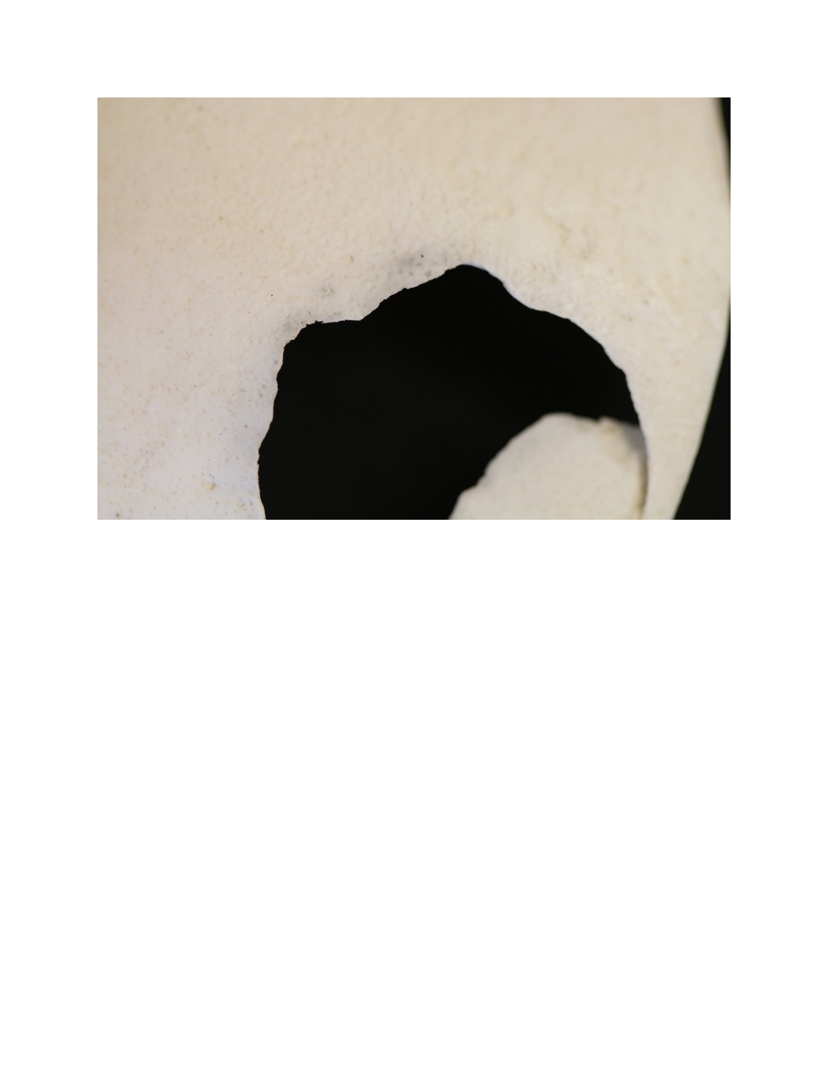
Perimortem gunshot wound showing no evidence of healing or remodeling of the fracture
margins. [19]
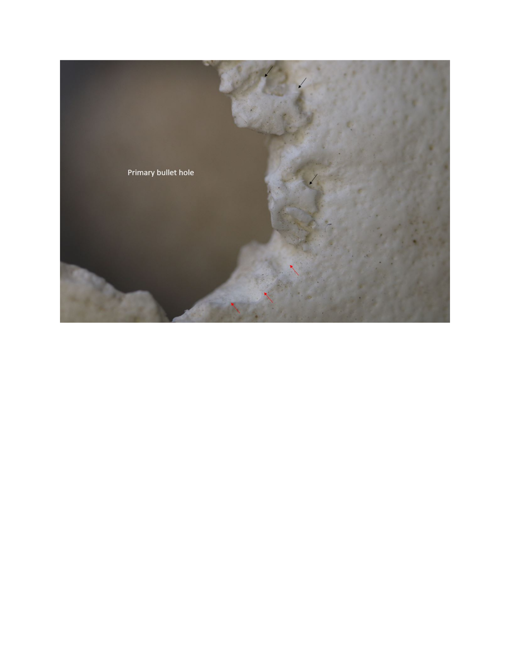
Compare Figure 19 with Figure 20. Figure 20 shows a bullet hole that has begun to heal. The
black arrows indicate areas of active bone removal via osteoclastic activity. The red arrow
indicates a fracture margin that looks smoothed. Compare the sharpness of the fracture margin in
both figures. Note also the surrounding bone looks more porous and uneven in Figure 20,
another indication of active bone response. This individual lived for 20 days after being shot in
the head with a single bullet.

Bullet hole showing rounding of fracture margins. Black arrows indicate active areas of bone
removal. Red arrow indicated a smoothed fracture margin. [20]
Figure 21 shows evidence of even further healing. This individual survived for 37 days after
being shot in the head. The black arrows indicate areas of new bone formation via osteoblastic
activity. The body is attempting to close the hole by adding new bone to the fracture margins.
The red arrows indicate an area that has been smoothed via osteoclastic activity, but no new bone
has been laid down yet.
Combined osteoblastic and osteoclastic activity on a bullet wound. [21]
Given enough time, the body will attempt to completely close the bullet wound via osteoblastic
activity. The individual in Figure 22 survived for 10 years after being shot in the head. The lack
of porosity indicates the healing process has stopped. Note that much of the wound has been
filled in with new bone and note how rounded the edges are.

Healed cranial gunshot wound. The fracture margins are rounded with much new bone added to
attempt to fill in the hole created by the bullet impact. Note the lack of porosity around the
impact site, indicating the healing process has stopped. [22]
Antemortem Postcranial Callus Formation
For most postcranial fractures the body will begin to lay down new bone quickly to mend the
two halves of fractured bone. The result is a bony callus, initially composed of disorganized
woven bone and eventually remodeled and replaced with mature bone. Woven bone can be laid
down very quickly and it is used by the body to stabilize the fracture as soon as possible. Mature
bone is better organized at the microscopic level and is structurally stronger than woven, thus
providing a longer-term solution to the healing process. The fracture callus in Figure 23 depicts
the fibrous woven bone callus of initial fracture repair. This is a severe example, but it
demonstrates the disorganized appearance of a woven bone callus.

Large fracture callus. [23]
Figure 24 is subtler. This is a minor rib fracture that shows the early stages of fracture repair.
The lighter colored bone indicated by the black arrow is a small woven bone fracture callus. The
color difference results in the difference in bone density between the fibrous woven bone callus
and the surrounding compact bone of the rib shaft.
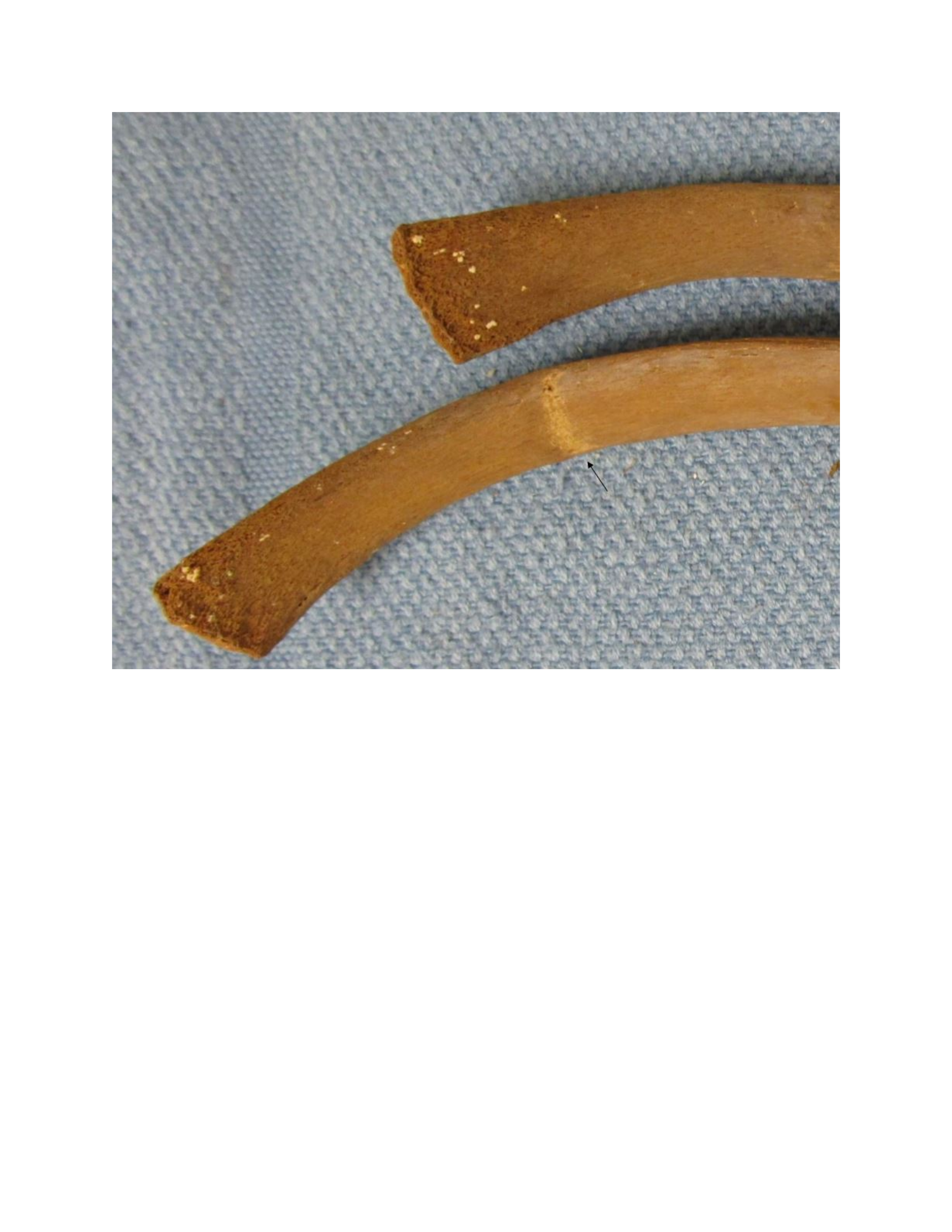
Early fracture callus formation of a rib. [24]
Figures 25-27 show examples of mature bone fracture calluses. Note how smooth the surfaces of
the healing fractures are in comparison with Figure 23. Figures 25-27 are examples of fracture
calluses during the remodeling stages of healing and likely the body has stopped attempting to
mend these fractures any further.
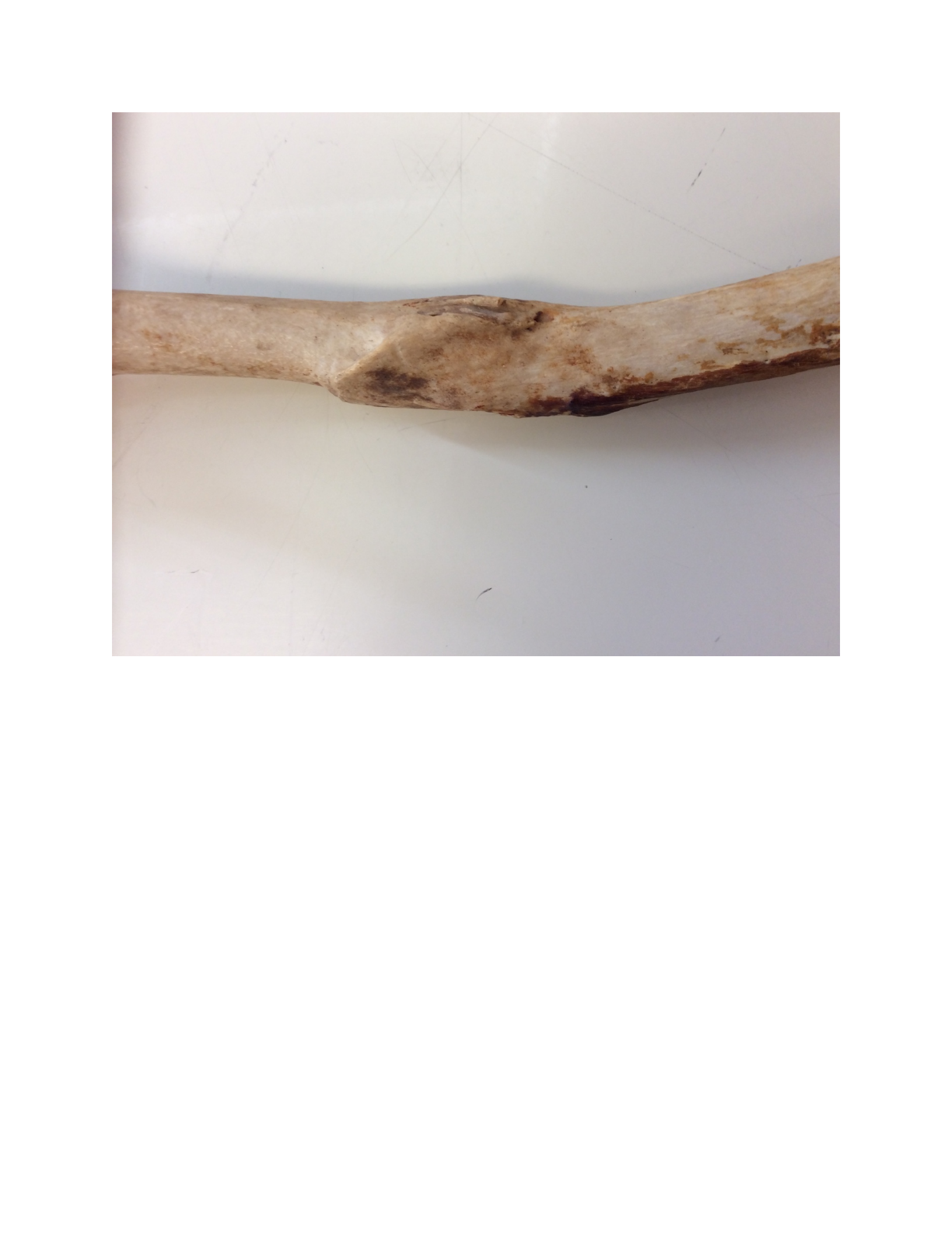
Well-healed fracture callus in a humerus. [25]
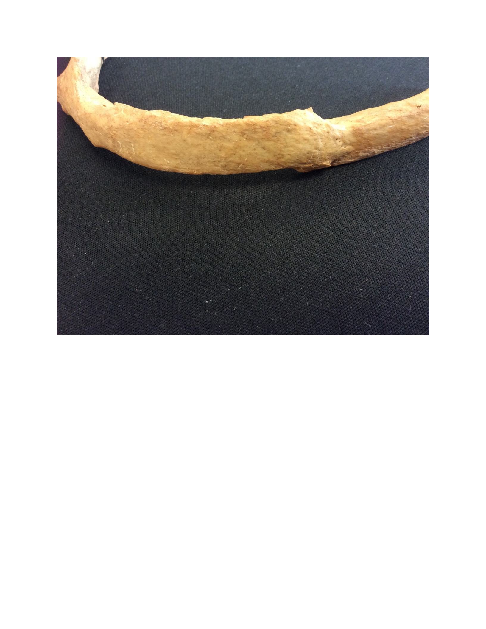
Well-healed fracture callus in a rib. [26]
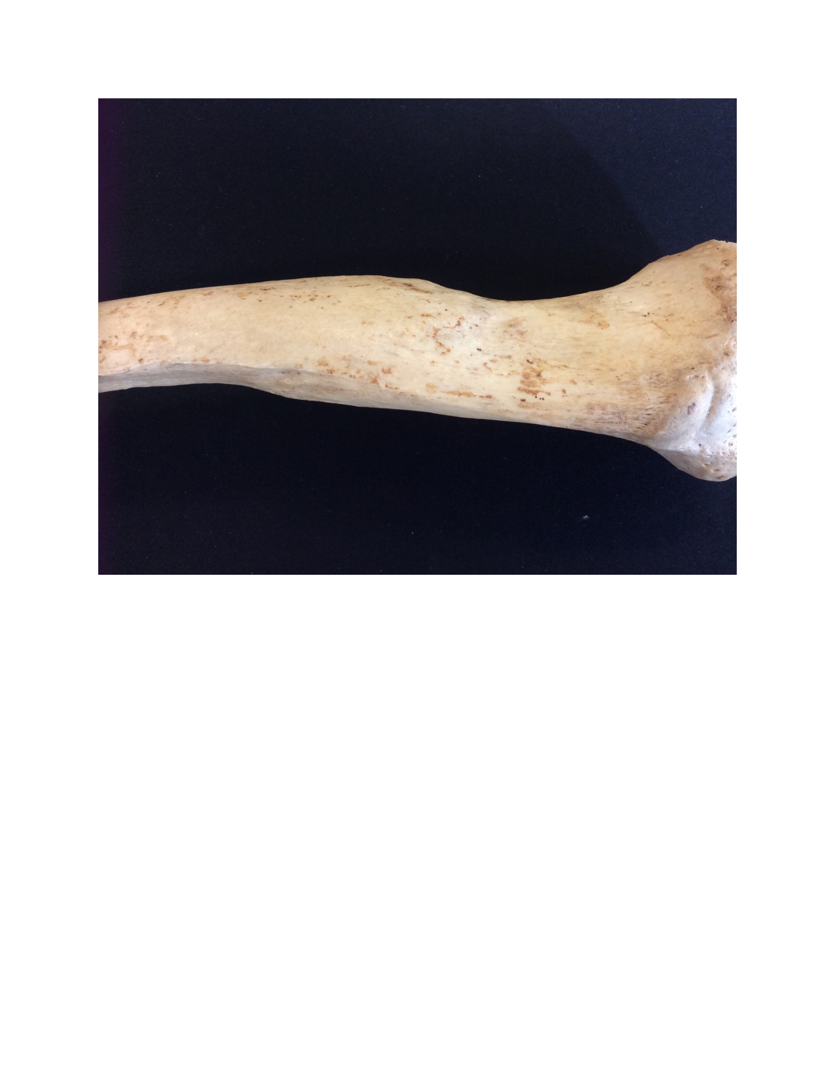
Well-healed fracture callus in a tibia. [27]
Perimortem Trauma and Postmortem Alterations
If there is no evidence of healing, then the fracture could either be perimortem or postmortem in
timing. Recall that perimortem wounds are those in which the fracture occurred while the bone
was in "a biomechanically fresh state (Christensen et al., 2014:349)." This is called a wet bone,
green bone, or greasy bone response. The high collagen content of wet bone provides elasticity;
the bone can better withstand stress and will deform prior to breaking. Postmorterm fractures are
those in which the break occurred when the bone was in a biomechanically brittle state. Dry bone
is weaker than wet bone. It will not deform prior to fracturing, breaks under lower loads, and will
tend to shatter into multiple pieces. Differentiating the two is one of the most difficult tasks in
forensic anthropology (Cattaneo and Cappella, 2017; Galloway et al., 2014; Maples, 1986;
Symes et al., 2012, 2014; Ubelaker, 1991; Ubelaker and Adams, 1995; Wheatley, 2008; Wieberg
and Wescott, 2008). Research on this topic is ongoing and the following discussion is meant to
provide general guidelines and not hard and fast rules. The postmortem loss of elasticity in bone
is a continuous process and not one that can easily be divided into discrete either/or categories.
Still, experimental studies have identified a number of indicators that allow the forensic
anthropologist to identify perimortem trauma with some consistency (Table 2).
Appearance of the fracture line on the external surface of the bone
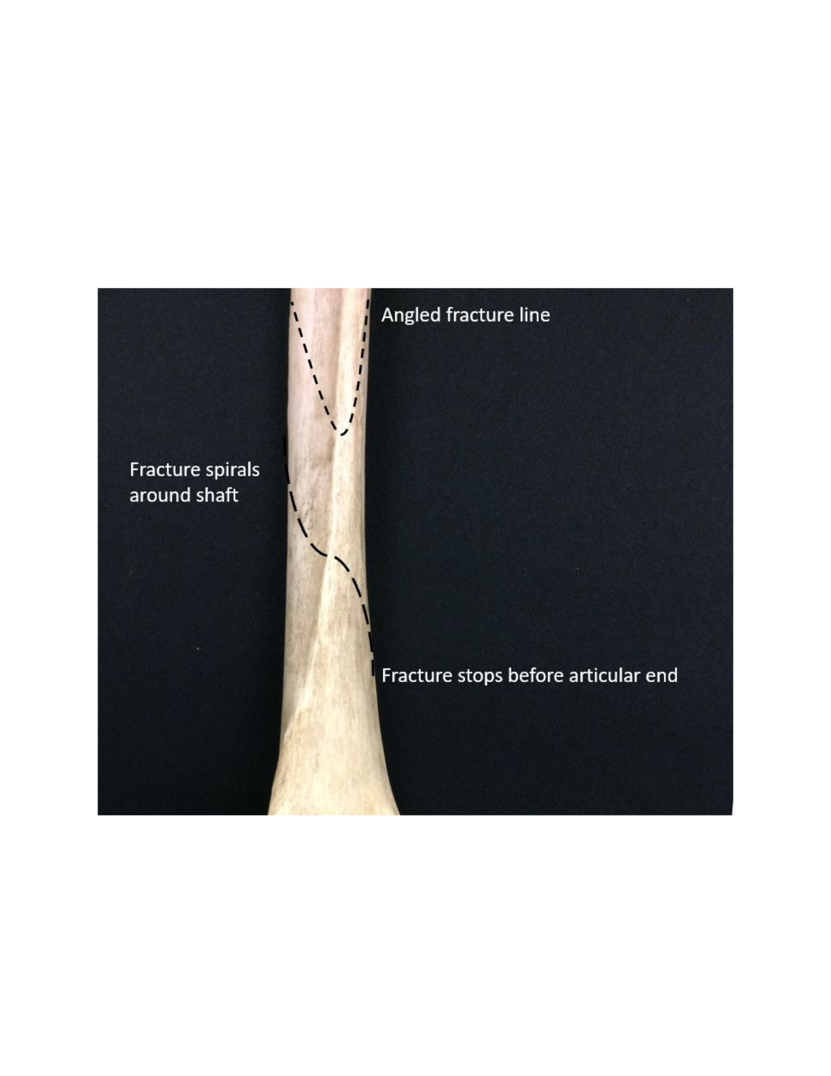
One consideration is the way the fracture line propagates with respect to the overall shape of the
bone. Perimortem fractures can exhibit a variety of appearances. Some fractures have a zig-zag
pattern with sharp projections and acute changes in direction, like a bolt of lightning. Others can
be curvilinear in their path and encircle the shaft in a radial pattern. Perimortem fracture lines
also tend to stop at the articular ends of long bones. This is because the articular ends of a long
bone contain a high proportion of trabecular bone, which can dissipate the energy of the trauma
more effectively without producing a fracture line (Figure 28).
Examples of fracture line pathways consistent with perimortem trauma. [28]
Postmortem fractures tend to be aligned either perpendicular to the bone shaft or longitudinally
along the bone shaft. These fractures tend not to have a curved pathway around the bone and
look more regular in their appearance. Postmortem fractures can also have a stepped pathway
along the bone length. Postmortem fracture lines will continue through the articular ends of
bones (Figure 29).
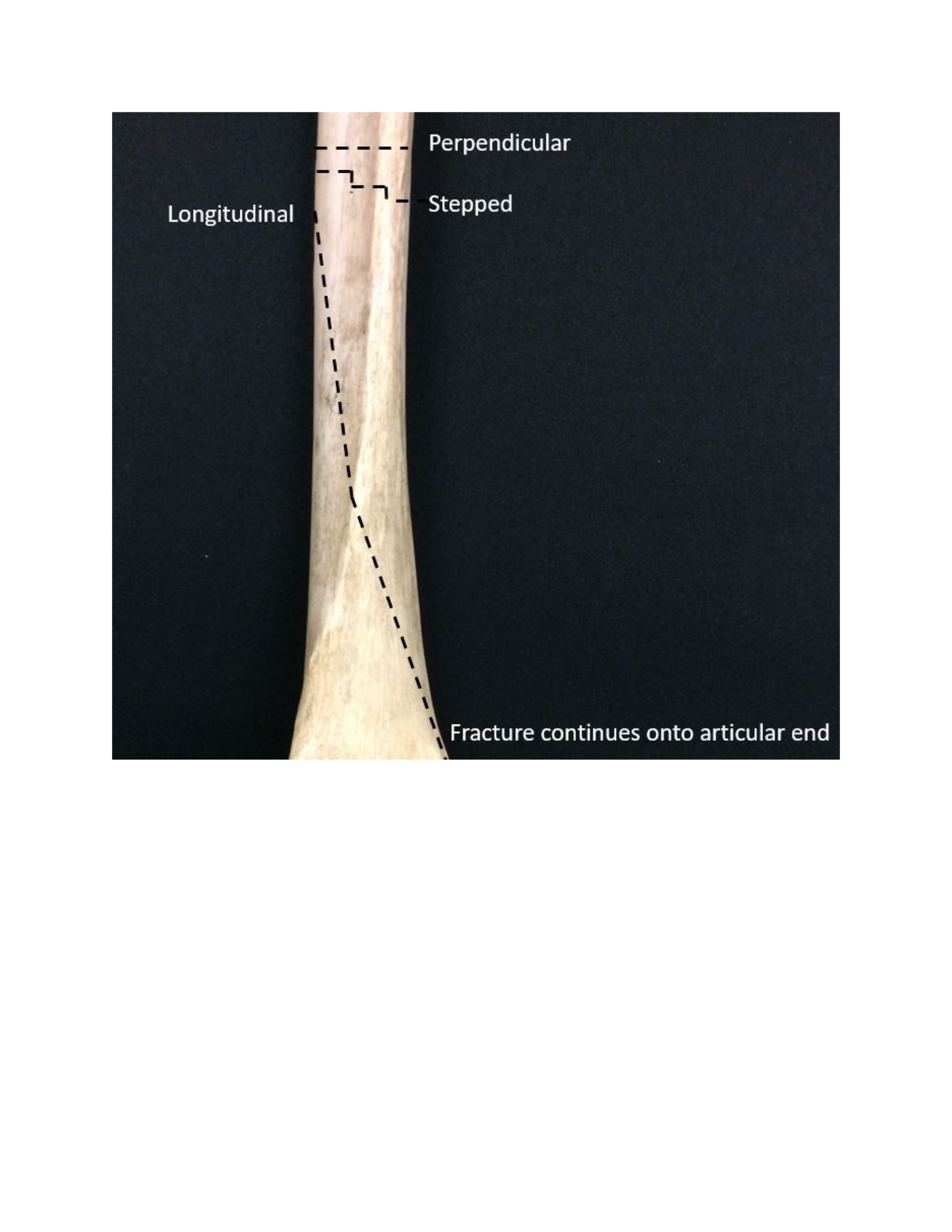
Examples of fracture line pathways consistent with postmortem trauma. [29]
In perimortem fractures the line itself tends to be sharp, clear and well defined. The bone tends
not to shatter into multiple pieces. When fragmentation does occur perimortem wounds are much
more likely to have partially adhering bone and hinge fractures (Figure 30) in which the
smaller fragments do not completely separate (i.e., they are infractions).
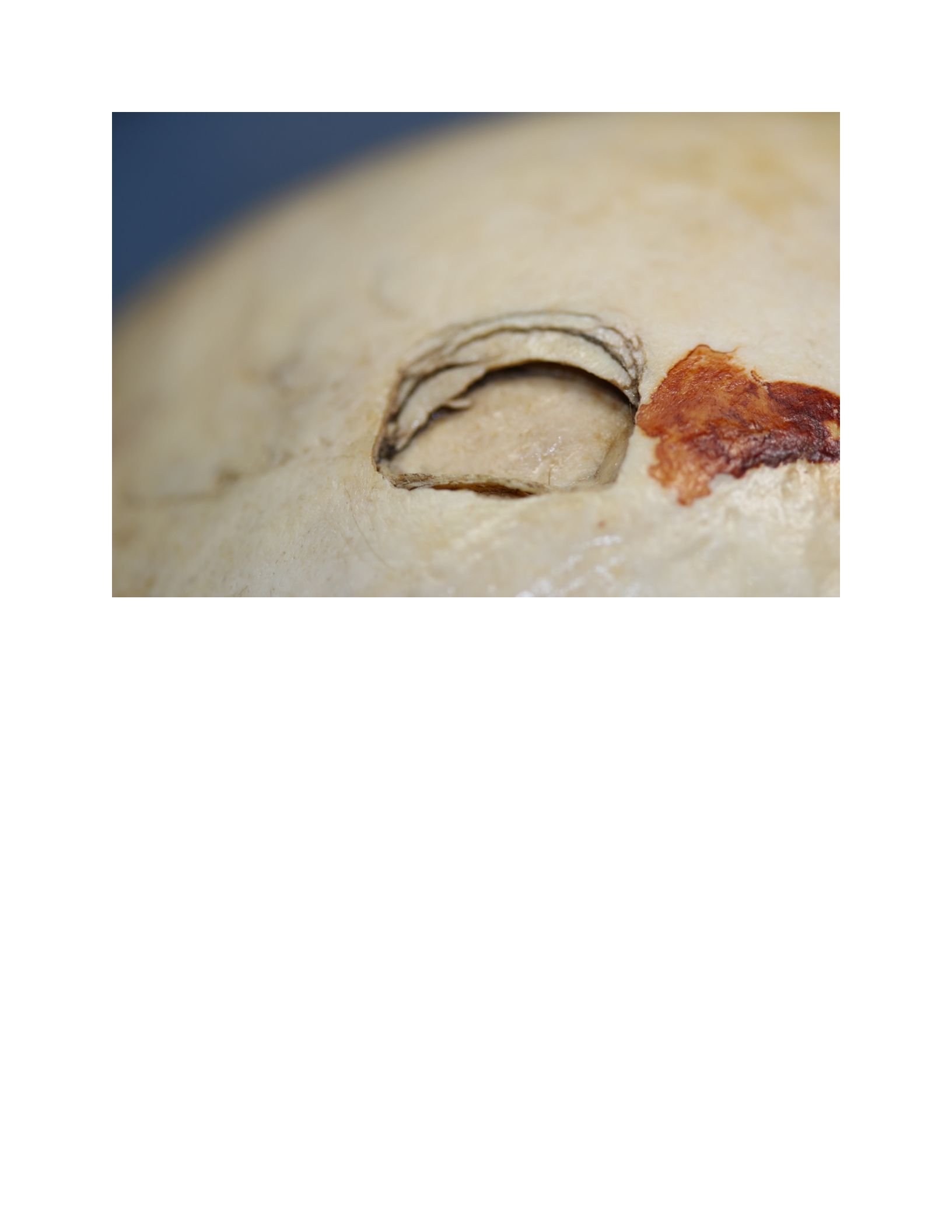
Hinge fractures of a cranial vault wound. Note that multiple pieces of bone are adhering around
the trauma site. [30]
In postmortem fractures the line tends to be ragged in appearance. There is a greater tendency for
the bone to shatter into multiple pieces, but these pieces will completely separate from the main
body of the bone and not adhere. Hinge fractures are not seen with postmortem fractures.
Appearance of the exposed fracture edge
Thus far we have discussed the appearance of fractures on the external surface of the bone.
However, when a complete discontinuity results from trauma the margin of the broken edge
(Figure 31) also exhibits diagnostic criteria of peri- and postmortem trauma. Three observations
are noteworthy: fracture profile angle, fracture profile texture, and the fracture profile color.
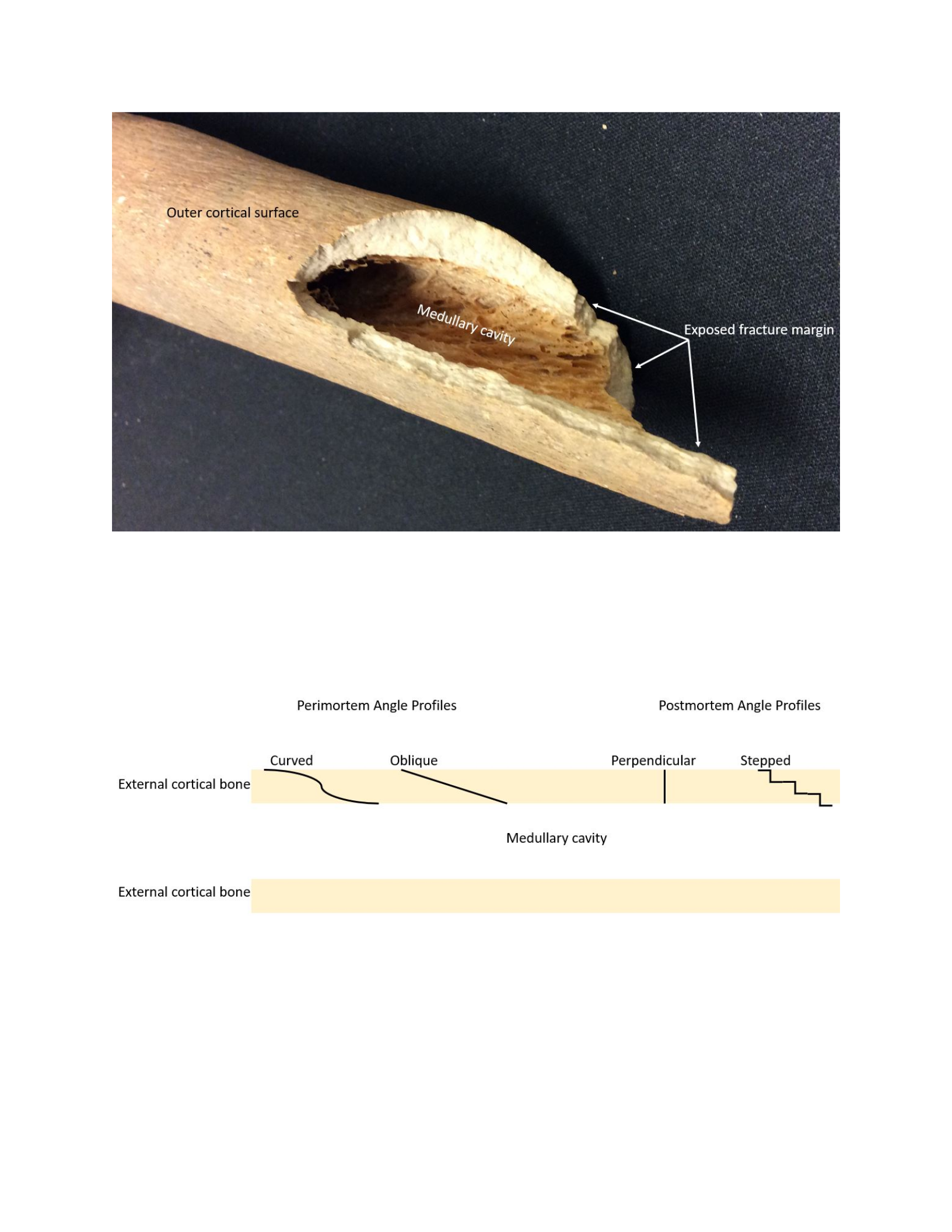
Broken fracture profile on a human long bone. [31]
The fracture profile angle refers to the shape of the broken edge. In perimortem trauma the
broken edge is obliquely angled or curved. In postmortem trauma the bone breaks at a right angle
to the shaft and the fracture line is straight or perpendicular with respect to the outer surface of
the bone. Postmortem breaks can also exhibit a stepped appearance (Figure 32).
Fracture profile angle in perimortem and postmortem trauma. [32]
The fracture profile texture refers to the texture of the broken edge itself. In perimortem trauma
the broken edge is smooth. In postmortem trauma the broken edge is rough and ragged.
Postmortem fractures have the appearance of being roughly torn, reflecting the lack of elastic
properties in the bone. Although difficult to photograph the differences between a smooth and
rough profile texture are best indicated by the black arrows in Figure 33.

Smooth (left) and rough (right) profile textures. [33]
The fracture profile color refers to whether the color of the fractured edge is the same color as
the exterior cortical surface. In perimortem trauma, the edge and exterior surface of the bone are
exposed to the same burial environment. As such, any changes in bone color that occurs due to
soil contact will be shared by both surfaces. In postmortem trauma the exterior surface is
exposed to burial color changes for a longer period of time than the fractured margin. Therefore,
you expect the margin to be lighter in color than the exterior bone surface. This is demonstrated
in Figure 33 (above). The perimortem trauma has a consistent color throughout the bone. The
postmortem trauma is tan on the exterior and nearly white along the fracture edge margin. Note
that it is possible for postmortem breaks to assume the same color as the exterior bone surface
depending on when the break happened and how long the body has been in the soil.

Table 2. Summary of differences between perimortem and postmortem trauma.
Given how difficult it can be to determine the timing of trauma please answer these practice
questions and consider the feedback before moving on to the graded component of the lab.
Practice 1
HomeworkAnswered
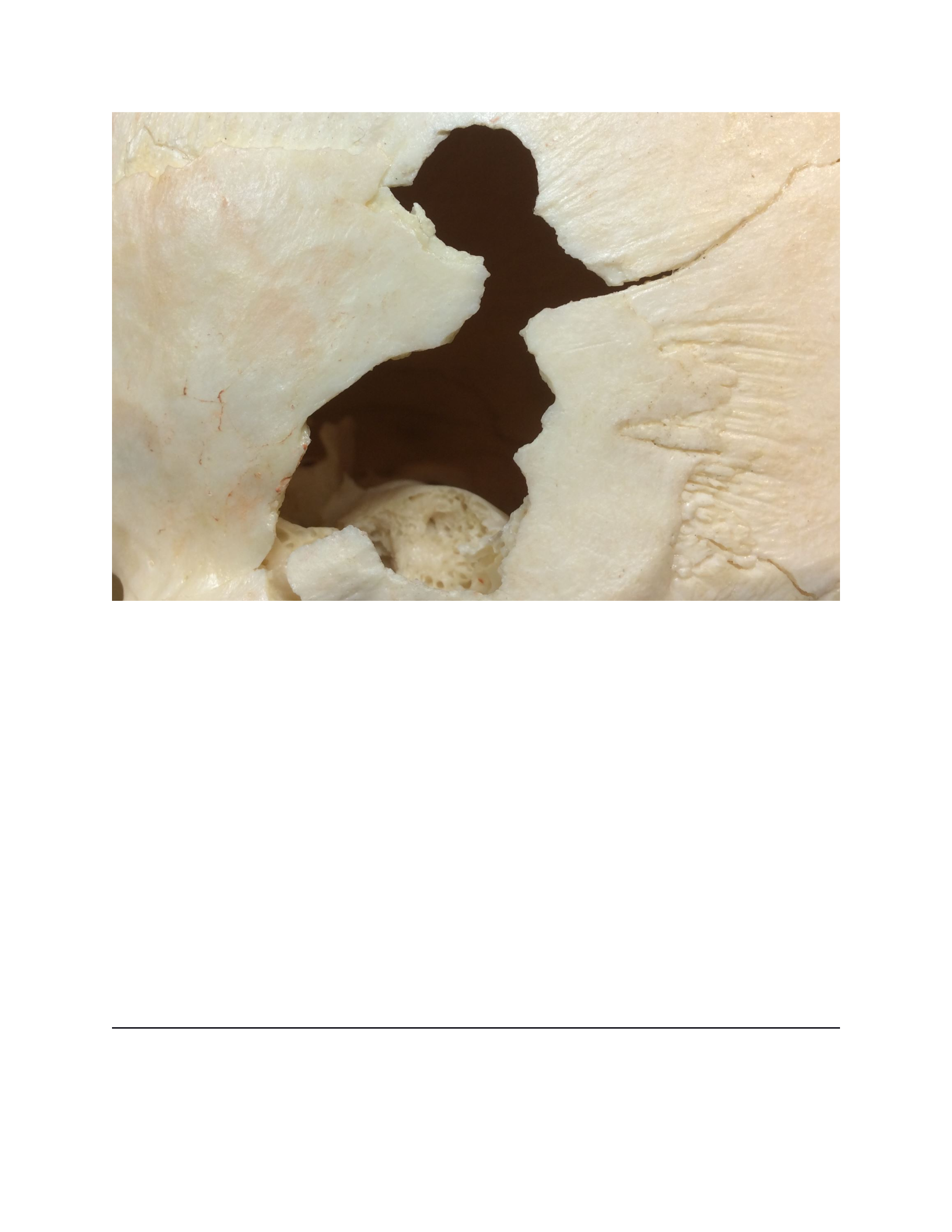
Describe the timing of this gunshot wound to the head. [34]
Select an answer and submit. For keyboard navigation, use the up/down arrow keys to select an
answer.
a
Antemortem
b
Perimortem
Your answer
c
Postmortem
Explanation
This is a perimortem wound. There are no signs of healing indicating it is not antemortem. The
fracture lines are crisp and there is a clear bullet impact site.
Submitted: Answered - Correct!0 attempts left
You have reached the maximum number of attempts
You have reached the maximum number of attempts
Explanation
Explanation
This is a perimortem wound. There are no signs of healing indicating it is not antemortem. The
fracture lines are crisp and there is a clear bullet impact site.
Steeping some tea...
Practice 3
HomeworkAnswered
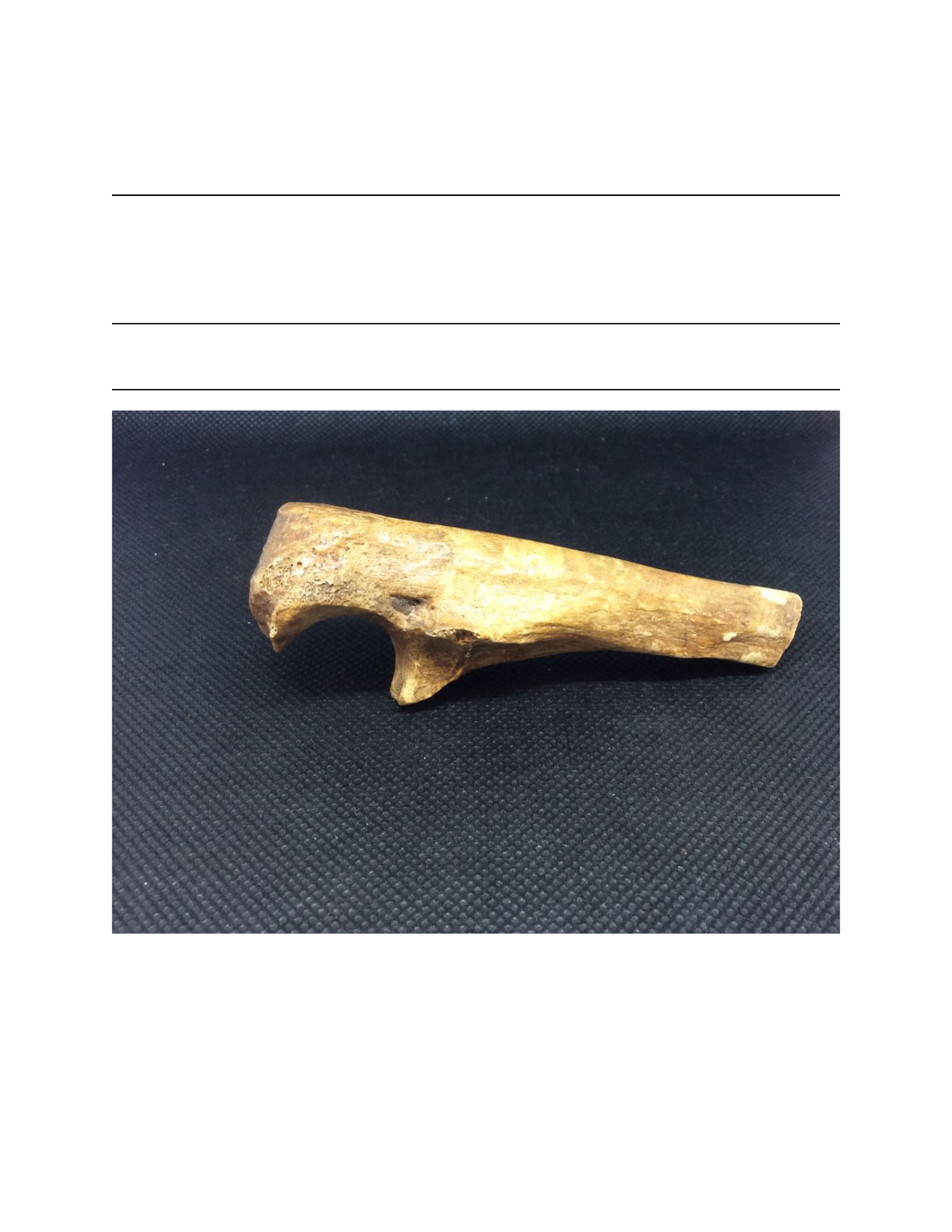
Describe the timing of the fracture to the end of this ulna. [36]
Select an answer and submit. For keyboard navigation, use the up/down arrow keys to select an
answer.
a
Antemortem
b
Perimortem
c
Postmortem

Your answer
Explanation
This is a classic postmortem long bone fracture. Even though you cannot see the profile, the bone
is broken perpendicular to the shaft.
Submitted: Answered - Correct!0 attempts left
You have reached the maximum number of attempts
You have reached the maximum number of attempts
Explanation
Explanation
This is a classic postmortem long bone fracture. Even though you cannot see the profile, the bone
is broken perpendicular to the shaft.
Practice 4
HomeworkAnswered
Describe the timing of this trauma to the cranial vault. [37]

Select an answer and submit. For keyboard navigation, use the up/down arrow keys to select an
answer.
a
Antemortem
b
Perimortem
Your answer
c
Postmortem
Explanation
This is a perimortem wound to the skull in the form of a depressed infraction. There is a hinge
fracture present and no signs of healing. That is, there is no porosity around the impact site and
the edges appear sharp. A break like this could never happen in the postmortem interval.
Submitted: Answered - Correct!0 attempts left

You have reached the maximum number of attempts
You have reached the maximum number of attempts
Explanation
Explanation
This is a perimortem wound to the skull in the form of a depressed infraction. There is a hinge
fracture present and no signs of healing. That is, there is no porosity around the impact site and
the edges appear sharp. A break like this could never happen in the postmortem interval.
Steeping some tea...
Steeping some tea...
Steeping some tea...
Practice 8
HomeworkAnswered
Describe the timing of this cranial wound. [41]

Select an answer and submit. For keyboard navigation, use the up/down arrow keys to select an
answer.
a
Antemortem
b
Perimortem
Your answer
c
Postmortem
Hint
This is a well healed antemortem wound to the skull. This is indicated by the smoothed fracture
profile edge. Although the hole has not been filled with new bone, there is no porosity around the
fracture margins, indicating that the healing process has ended. Osteoclasts have remodeled the
fracture margins to create a smooth appearance.
Submitted: Answered - Incorrect0 attempts left
You have reached the maximum number of attempts
You have reached the maximum number of attempts

Hint
Hint
This is a well healed antemortem wound to the skull. This is indicated by the smoothed fracture
profile edge. Although the hole has not been filled with new bone, there is no porosity around the
fracture margins, indicating that the healing process has ended. Osteoclasts have remodeled the
fracture margins to create a smooth appearance.
Practice 9
HomeworkAnswered
Describe the timing of this fracture on the cranial vault. [42]
Select an answer and submit. For keyboard navigation, use the up/down arrow keys to select an
answer.
a
Antemortem
b
Perimortem
c
Postmortem

Your answer
Explanation
This one is a bit more challenging. There is no real evidence of healing around this oddly shaped
hole in the skull. The primary evidence for this being postmortem is the difference in color in the
fracture wall versus the outer surface of the cranium. The lack of hinge fractures or secondary
fracture lines also suggests this is not a perimortem wound.
Submitted: Answered - Correct!0 attempts left
You have reached the maximum number of attempts
You have reached the maximum number of attempts
Explanation
Explanation
This one is a bit more challenging. There is no real evidence of healing around this oddly shaped
hole in the skull. The primary evidence for this being postmortem is the difference in color in the
fracture wall versus the outer surface of the cranium. The lack of hinge fractures or secondary
fracture lines also suggests this is not a perimortem wound.
Steeping some tea...
The following questions are graded.
Steeping some tea...
Q9
HomeworkAnswered

What type of fracture is shown at the end of this long bone? Note the fracture profile as well as
how the fracture relates to the long bone shaft (ignore the small fracture at the bottom of the
image that runs longitudinally along the shaft). Ignore the color of the broken bone surface here
and focus on the profile primarily in making your decision. [45]
Select an answer and submit. For keyboard navigation, use the up/down arrow keys to select an
answer.
a
Antemortem
b
Perimortem
c
Postmortem
Your answer
Explanation
The answer is C. The fracture lines are perpendicular to the shaft.
Submitted: Answered - Correct!0 attempts left
You have reached the maximum number of attempts
You have reached the maximum number of attempts
Explanation
Explanation
The answer is C. The fracture lines are perpendicular to the shaft.
Q10
HomeworkAnswered
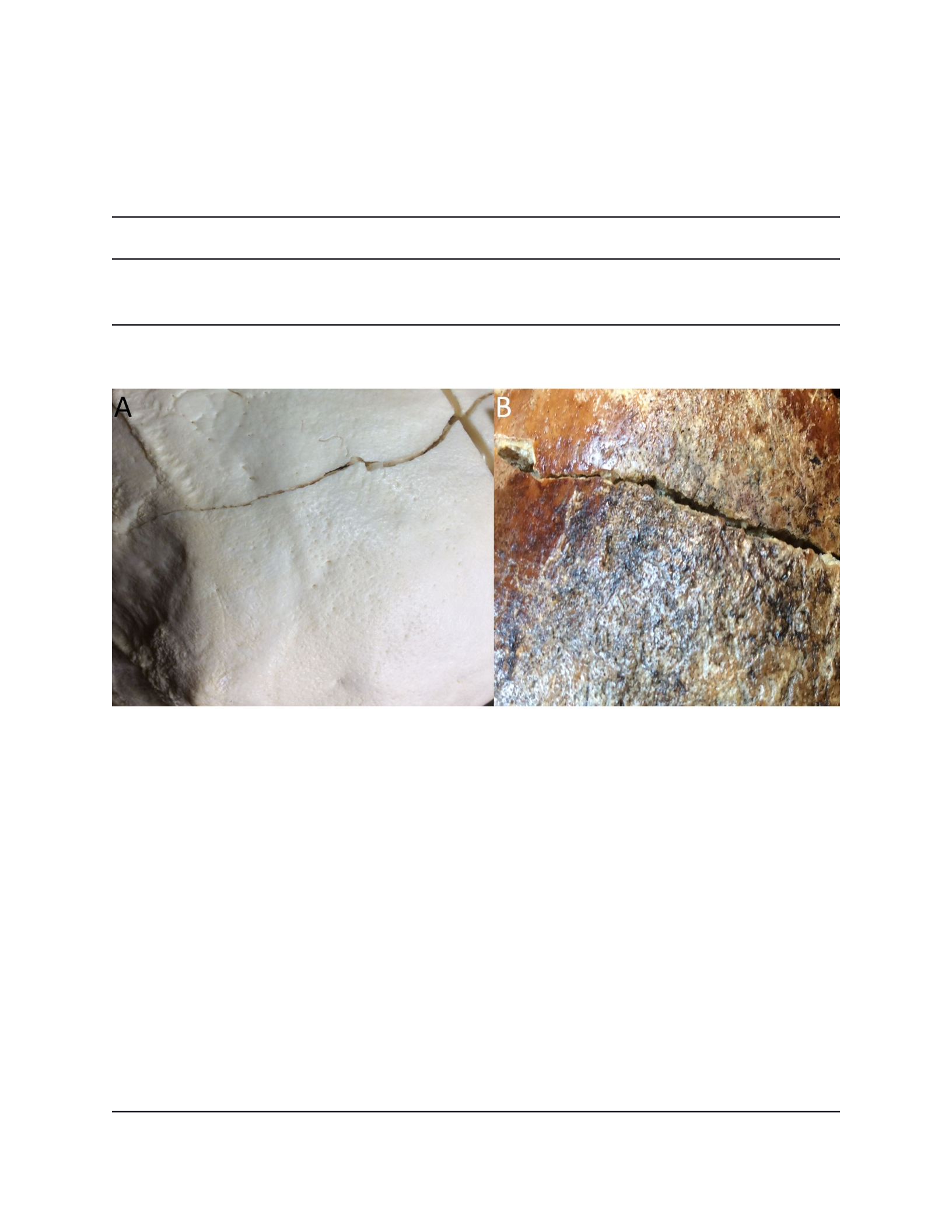
Consider the two images shown here. Based on the fracture patterns shown on these skull
fragments and what we have learned in this lab, which shows evidence of PERIMORTEM
trauma? [46]
Select an answer and submit. For keyboard navigation, use the up/down arrow keys to select an
answer.
a
A
Your answer
b
B
Explanation
The answer is A. The fracture line is clean and crisp, while on specimen B the fracture line is
eroded and rounded at the edges. The sharpness of the edges of A is indicative of a perimortem
injury.
Submitted: Answered - Correct!0 attempts left
You have reached the maximum number of attempts

You have reached the maximum number of attempts
Explanation
Explanation
The answer is A. The fracture line is clean and crisp, while on specimen B the fracture line is
eroded and rounded at the edges. The sharpness of the edges of A is indicative of a perimortem
injury.
Steeping some tea...
Steeping some tea...
Steeping some tea...
Q14
HomeworkAnswered
Consider this x-ray of a fractured humerus. Note the large red arrow is pointing to the early
stages of healing in the form of a fracture callus (that wispy, translucent bone trying to make a
connection between the bone ends). In an x-ray, the white areas are denser (radiopaque). In
looking at this image please select ALL of the characteristics that apply to describe this fracture.
Note there are multiple correct answers that must be selected to receive credit. [50]


Multiple answers: Multiple answers are accepted for this question
Select one or more answers and submit. For keyboard navigation...
a
Fracture
Your answer
b
Infraction
c
Comminuted
d
Displaced
Your answer
Hint
Incorrect, this is a fracture that is comminuted and displaced. The answer is A, C, and D.
Submitted: Sorry, you didn't select ALL the correct answers - Incorrect0 attempts left
You have reached the maximum number of attempts
You have reached the maximum number of attempts
Hint
Hint
Incorrect, this is a fracture that is comminuted and displaced. The answer is A, C, and D.
Q15
HomeworkAnswered
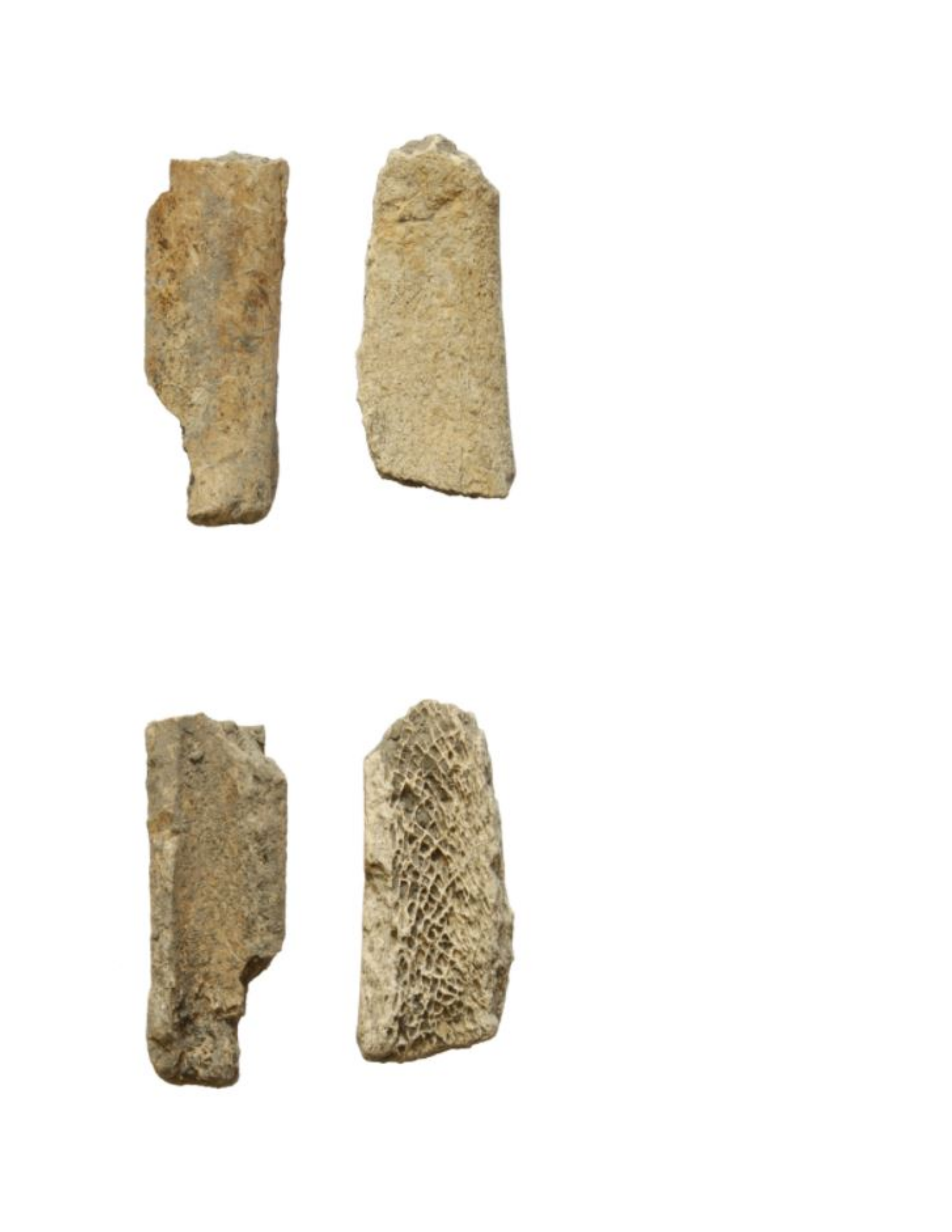
How would you characterize the fractures to these long bone ends? [51]

Select an answer and submit. For keyboard navigation, use the up/down arrow keys to select an
answer.
a
Antemortem
b
Perimortem
c
Postmortem
Your answer
Explanation
The answer is C. The perpendicular breaks with respect to the shaft indicate a postmortem
interval.
Submitted: Answered - Correct!0 attempts left
You have reached the maximum number of attempts
You have reached the maximum number of attempts
Explanation
Explanation
The answer is C. The perpendicular breaks with respect to the shaft indicate a postmortem
interval.
Q16
HomeworkAnswered
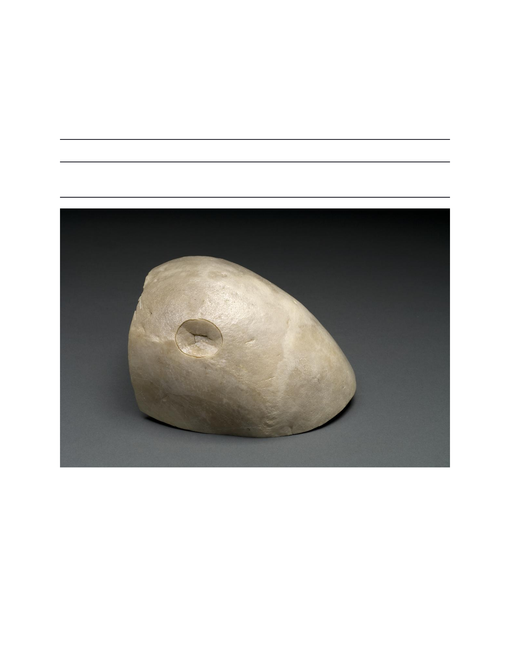
How would you characterize the fractures to this long bone end? Look closely at the shape of the
fracture line and the appearance of the fracture margins. [52]

Select an answer and submit. For keyboard navigation, use the up/down arrow keys to select an
answer.
a
Antemortem
b
Perimortem
Your answer
c
Postmortem
Explanation
The answer is B. The appearance of these fractures is consistent with perimortem trauma.
Submitted: Answered - Correct!0 attempts left
You have reached the maximum number of attempts
You have reached the maximum number of attempts
Explanation
Explanation
The answer is B. The appearance of these fractures is consistent with perimortem trauma.
Q17
HomeworkAnswered
Please characterize the timing of this infraction. [53]
Select an answer and submit. For keyboard navigation, use the up/down arrow keys to select an
answer.
a
Antemortem
b
Perimortem
Your answer
c
Postmortem

Explanation
The answer is B. A hinge fracture with no signs of healing suggests a perimortem time frame.
Submitted: Answered - Correct!0 attempts left
You have reached the maximum number of attempts
You have reached the maximum number of attempts
Explanation
Explanation
The answer is B. A hinge fracture with no signs of healing suggests a perimortem time frame.
Q18
HomeworkAnswered
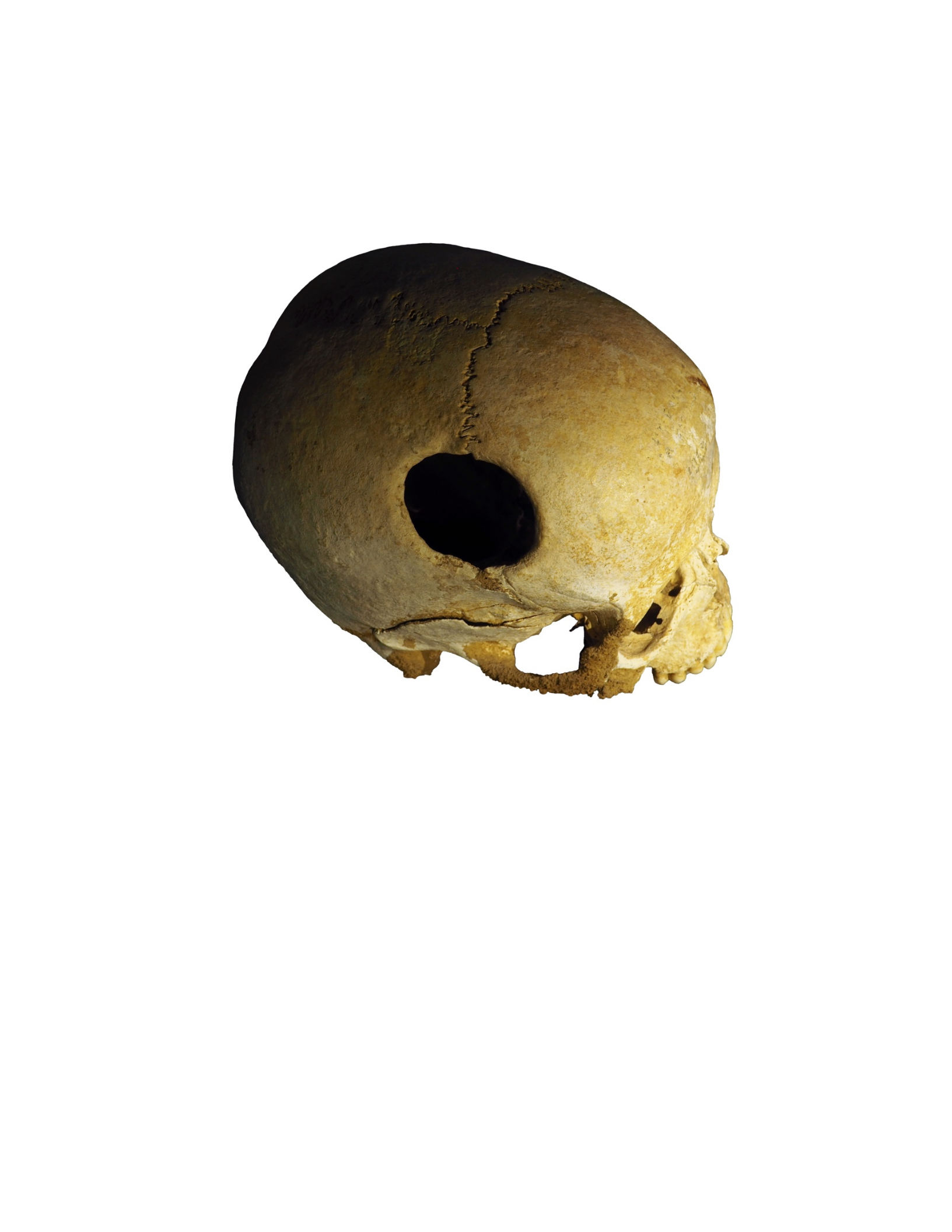
Look carefully at this image of a woman who experienced trepanation, the intentional cutting of
holes in the skull. This is an ancient medical practice throughout the world (with some modern-
day proponents - I do NOT recommend), and believe it or not people DID survive this procedure
in many cases. Looking closely at the margins of the hole removed from this skull, would you
say this woman survived or not? [54]
Select an answer and submit. For keyboard navigation, use the up/down arrow keys to select an
answer.
a
She did survive
Your answer
b
She did not survive
Explanation
The answer is A. The edges are well rounded indicating she did survive the event.

Submitted: Answered - Correct!0 attempts left
You have reached the maximum number of attempts
You have reached the maximum number of attempts
Explanation
Explanation
The answer is A. The edges are well rounded indicating she did survive the event.
Q19
HomeworkAnswered
Look closely at this attempt at surgical repair of the skull. Did this individual survive, and if so
for how long? [55]
Select an answer and submit. For keyboard navigation, use the up/down arrow keys to select an
answer.
a

This individual did not survive, the edges of the bone are sharp
b
This individual survived for a few weeks, there is porosity present and rounding of the edges
c
This individual survived for an extended period of time, the edges are rounded, porosity is
lacking and some bony bridging is present.
Your answer
Explanation
The answer is C. The edges are well rounded, there is no porosity, and there is a bony bridge
connecting the fragment of bone to the rest of the skull
Submitted: Answered - Correct!0 attempts left
You have reached the maximum number of attempts
You have reached the maximum number of attempts
Explanation
Explanation
The answer is C. The edges are well rounded, there is no porosity, and there is a bony bridge
connecting the fragment of bone to the rest of the skull
Q20
HomeworkAnswered
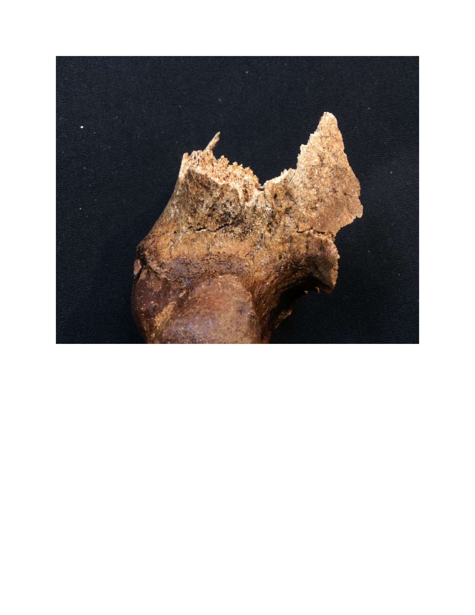
How would you characterize the damage to this long bone? This is the distal end of a femur.
Look closely at clues from coloration and the appearance of the fractured edge. Does this look
clean and sharp consistent with perimortem trauma? Or does the fractured bone look rough and
uneven? Consider color differences between the fracture margin and the surface of the bone?
[56]
Select an answer and submit. For keyboard navigation, use the up/down arrow keys to select an
answer.
a
Antemortem
b
Perimortem
c
Postmortem
Your answer
Explanation
The answer is C. These are postmortem breaks. The fracture margin is lighter than the
surrounding bone indicating these breaks occurred AFTER the bone was removed from the
ground. Also the fracture lines are not sharp and clean in appearance. They look more nibbled.
Submitted: Answered - Correct!0 attempts left

